Transcription
4E x e r c i s eThe Cell: Anatomy and DivisionThe Anatomy of the Composite Cell section can be given as an out-of-class assignment to save time.This might be necessary if audiovisual material is used.Time Allotment: 2 hours.Multimedia Resources: See Appendix B for Guide to Multimedia Resource Distributors.Inside the Living Cell (WNS: 50 minutes, DVD)An Introduction to the Living Cell (CBS: 30 minutes, DVD)A Journey Through the Cell (FHS: DVD)Part One: Cells: An Introduction (20 minutes)Part Two: Cell Functions: A Closer Look (20 minutes)Mitosis and Meiosis (DE: 23 minutes, VHS, DVD)Practice Anatomy Lab 3.0 (PAL) (PE: DVD, Website)Laboratory MaterialsOrdering information is based on a lab size of 24 students, working in groups of 4. A list of supplyhouse addresses appears in Appendix A.3-D model of composite cell or chartof cell anatomy24 slides of simple squamousepithelium24 slides of teased smooth muscle24 slides of human blood cell smear24 slides of sperm24 slides of whitefish blastulae24 compound microscopes, lens paper,lens cleaning solution, immersionoil3-D models of mitotic stagesVideo or animation of mitosisChenille sticks (pipe cleaners), twodifferent colors, cut into 3-inchpiecesAdvance Preparation1. Set out slides (one per student) of simple squamous epithelium, teased smooth muscle, human blood cell smear, sperm, andwhitefish blastulae. Students will also need lens paper, lens cleaning solution, immersion oil, and compound microscopes.2. Set out a model or a lab chart of a composite cell, and models of mitotic stages.3. Obtain the chenille sticks (pipe cleaners) in two different colors, and cut each into 3-inch pieces. Set out 8pieces per group, 4 of each color.Comments and Pitfalls1. Observing differences and similarities in cell structure often gives students trouble, as many of them havenever seen any cells other than epithelial cells. Slides or pictures of these cell types might help.18MARI1702 11 C04 pp018-023.indd 18Copyright 2014 Pearson Education, Inc.2/20/13 10:10 AM
Answers to Pre-Lab Quiz (p. 39)1. The structural and functional unitof all living things.2. a, chromatin3. d, selective permeability4. Ribosomes5. c, mitochondria6.7.8.9.10.interphasefalseFourb, interphasefalseAnswers to Activity QuestionsActivity 5: Observing Various Cell Structures (pp. 44–45)4. Simple squamous epithelial cells are relatively large and irregularly (“fried-egg”) shaped. Smooth musclecells are also relatively large, but are long and spindle shaped. Red blood cells and sperm are both examples of small cells. Red blood cells appear round, while sperm cells are streamlined with long flagella.Cell shape is often directly related to function. Epithelial cells fit tightly together and cover large areas.Elongated muscle cells are capable of shortening during contraction. The red blood cells are small enoughto fit through capillaries, and are actually biconcave in shape, which makes them flexible and increasessurface area (not obvious to the students at this point). Sperm cells’ streamlined shape and flagella aredirectly related to efficient locomotion.The sperm cells have visible projections (flagella), which are necessary for sperm motility.The function of sperm is to travel through the female reproductive system to reach the ovum in the uterinetubes. This requires motility, provided by the flagella.None of the cells lacks a plasma membrane.Mature red blood cells have no nucleus.Nucleoli will probably be clearly visible in the epithelial cells, and possibly visible in the other nuclei.No. Identifiable organelles are not visible in most of these cells. Filaments may be visible in the smoothmuscle preparations. The details of organelle structure are usually below the limit of resolution of the lightmicroscope. Unless special stains are used, there is no way to see or distinguish the organelles at this level.Activity 7: “Chenille Stick” Mitosis (p. 48)2. The centromere3. The mitotic spindle4. KinetochoresThe nuclear envelope5. The metaphase plate6. Pulling apart of the sister chromatids.Each sister chromatid is now a chromosome.7. The events of telophase cause the chromosomes and cell structures to revert to their interphase appearance: the chromosomes uncoil, a nuclear envelope forms around each chromatin mass, nucleoli appear inthe nucleus, and the spindle breaks down and disappears.Copyright 2014 Pearson Education, Inc.MARI1702 11 C04 pp018-023.indd 19Exercise 4192/20/13 10:10 AM
S h ee t4NameLab Time/Datee x e r c i s eThe Cell:Anatomy and DivisionRe v i e wAnatomy of the Composite Cell1. Define the following terms:organelle: A highly organized intracellular structure that performs a specific (metabolic) function for the cell. cell: The basic structural and functional unit of living organisms 2. Although cells have differences that reflect their specific functions in the body, what functions do they have in common?Ability to metabolize, to reproduce, to grow (increase in mass), to respond to a stimulus, and to move 3. Identify the following cell parts:plasma membrane1. external boundary of cell; regulates flow of materials into and out of the cell; site ofcell signalinglysosome2. contains digestive enzymes of many varieties; “suicide sac” of the cellmitochondria3. scattered throughout the cell; major site of ATP synthesismicrovilli4. slender extensions of the plasma membrane that increase its surface areainclusions5. stored glycogen granules, crystals, pigments, and so onGolgi apparatus6. membranous system consisting of flattened sacs and vesicles; packages proteins forexportnucleus7. control center of the cell; necessary for cell division and cell lifecentrioles8. two rod-shaped bodies near the nucleus; associated with the formation of the mitoticspindlenucleolus9. dense, darkly staining nuclear body; packaging site for ribosomesmicrofilamentsrough ER or endoplasmicreticulum10. contractile elements of the cytoskeleton11. membranous system; involved in intracellular transport of proteins and synthesis of membrane lipidsribosomes12. attached to membrane systems or scattered in the cytoplasm; site of protein synthesischromatin or chromatinthreads13. threadlike structures in the nucleus; contain genetic material (DNA)peroxisome14. site of free radical detoxification20MARI1702 11 C04 pp018-023.indd 20Copyright 2014 Pearson Education, Inc.2/20/13 10:10 AM
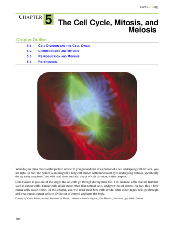
4. In the following diagram, label all parts provided with a leader line.NucleusNuclear envelopeNucleolusSmoothendoplasmicreticulumNuclear tochondrionIntermediate filamentsPeroxisomeDifferences and Similarities in Cell Structure5. For each of the following cell types, list (a) one important structural characteristic observed in the laboratory, and (b) thefunction that the structure complements or ensures.squamous epitheliuma. cells fit closely together like floor tiles b. often a lining or covering tissue sperma. has a tail or flagellum b. allows sperm to propel itself to an egg smooth musclea. cells have an elongated shape b. a long axis allows a greater degree of shortening red blood cellsa. anucleate (no nucleus); disc shaped b. more “room” to carry hemoglobin or oxygen; large surface area Copyright 2014 Pearson Education, Inc.MARI1702 11 C04 pp018-023.indd 21Review Sheet 4212/20/13 10:10 AM
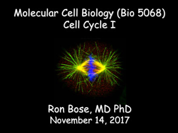
6. What is the significance of the red blood cell being anucleate (without a nucleus)? Limited life span. Does not reproduce. The nucleus is gone; therefore, the cell cannot manufacture new proteins, etc. Did it ever have a nucleus? (Use an appropriate reference.) Yes If so, when? Before its release into the bloodstream 7. Of the four cells observed microscopically (squamous epithelial cells, red blood cells, smooth muscle cells, and sperm),which has the smallest diameter? sperm Which is longest? smooth muscle or sperm (variable) Cell Division: Mitosis and Cytokinesis8. Identify the three phases of mitosis in the following photomicrographs.a. metaphase b. anaphase c. prophase 9. What is the importance of mitotic cell division? Provides cells for body growth and for repair of damaged tissue or provides additional cells with the same genetic makeup 10. Draw the phases of mitosis for a cell that contains four chromosomes as its diploid or 2n number.22Review Sheet 4MARI1702 11 C04 pp018-023.indd 22Copyright 2014 Pearson Education, Inc.2/20/13 10:10 AM
11. Complete or respond to the following statements:Division of the 1 is referred to as mitosis. Cytokinesis is division of the2. The major structural difference between chromatin and chromosomesis that the latter are 3 . Chromosomes attach to the spindle fibers by undivided structures called 4 . If a cell undergoes mitosis but not cytokinesis,the product is 5 . The structure that acts as a scaffolding for chromosomalattachment and movement is called the 6 . 7 is the period of cell lifewhen the cell is not involved in division. Two cell populations in the bodythat do not routinely undergo cell division are 8 and 9 .1.nucleus 2.cytoplasm 3.coiled/condensed/shortened 4.centromeres 5.a binucleate cell or multinucleated cell 6.spindle 7.interphase 8.neurons 9.skeletal and cardiac muscle cells 12. Using the key, categorize each of the events described below according to the phase in which it occurs.Key: a.anaphaseb.interphasec.metaphased.prophased1. Chromatin coils and condenses, forming chromosomes.a2. The chromosomes are V shaped.e3. The nuclear envelope re-forms.e4. Chromosomes stop moving toward the poles.c5. Chromosomes line up in the center of the cell.d6. The nuclear envelope fragments.d7. The mitotic spindle forms.b8. DNA synthesis occurs.b9. Centrioles replicate.dd (or a, c, and d)e.telophase10. Chromosomes first appear to be duplex structures.11. Chromosomal centromeres are attached to the kinetochore fibers.e12. Cleavage furrow forms.aandc (possibly d)13. The nuclear envelope(s) is absent.13. What is the physical advantage of the chromatin’s coiling and condensing to form short chromosomes at the onset of mitosis?Short, compact bodies are mechanically much easier to manipulate during mitosis than are long, thin chromatin threads. Copyright 2014 Pearson Education, Inc.MARI1702 11 C04 pp018-023.indd 23Review Sheet 4232/20/13 10:10 AM
The Cell: Anatomy and Division 3-D model of composite cell or chart of cell anatomy 24 slides of simple squamous epithelium 24 slides of teased smooth muscle 24 slides of human blood cell smear 24 slides of sperm 24 slides of whitefish blastulae 24 compound microscopes, lens paper, lens cleaning solution, immersion oil 3-D models of mitotic stages