Transcription
Neuroscience 146 (2007) 1536 –1545CHRONIC COCAINE TREATMENT ALTERS DENDRITICARBORIZATION IN THE ADULT MOTOR CORTEX THROUGHA CB1 CANNABINOID RECEPTOR– DEPENDENT MECHANISMI. BALLESTEROS-YÁÑEZ,a O. VALVERDE,b C. LEDENT,cR. MALDONADOb* AND J. DEFELIPEa*(Soria et al., 2005). Furthermore, the CB1 antagonistrimonabant attenuates the relapse due to cocaine-associated cues or cocaine re-exposure (De Vries et al., 2001;De Vries and Schoffelmeer, 2005). The endocannabinoidsystem participates in a variety of physiological phenomena including cognitive functions, locomotor activity andreward (Zimmer et al., 1999; Lichtman et al., 2002;Valverde, 2005; Maldonado et al., 2006) and, recently, ithas been proposed to regulate the addictive processesinduced by different drugs of abuse (Maldonado et al.,2006).The cerebral cortex participates in many physiologicalfunctions including cognition and emotion, and it is particularly relevant in the consolidation of the addictive processes (Jentsch et al., 2000; Kalivas and Volkow, 2005;Kringelbach, 2005). Numerous studies in rats have revealed that repeated cocaine administration induces electrophysiological and morphological adaptations in layers IIIand V pyramidal neurons of rat prefrontal, parietal andoccipital cortex (Robinson and Kolb, 1999; Robinson et al.,2001; Nasif et al., 2005; Huang et al., 2006). Recently, ithas been shown that layer III pyramidal neurons of ratmotor cortex are also sensitive to the administration ofdrugs (Ballesteros-Yáñez et al., 2007). However, there areno data available regarding the involvement of CB1 cannabinoid receptors in the microanatomical cortical changesinduced by chronic cocaine exposure. For this purpose, weanalyze the effect of cocaine administration in layer IIIpyramidal neurons of wild type and CB1 knockout mice.The motor cortex is essential for the translation ofbiological relevant environmental and pharmacologicalstimuli into adaptive motor responses, including thoseseen during behavioral sensitization (Pierce and Kalivas,1997; Zimmer et al., 1999). Furthermore, numerousGABAergic neurons containing CB1 mRNA and receptorsare present in layers II–III of the motor cortex (Marsicanoand Lutz, 1999; Freund et al., 2003; Harkany et al., 2005).The CB1 receptor is involved in the presynaptic regulationof GABA and dopamine release, modulating cortical pyramidal neuron activity (Trettel et al., 2004; Laviolette andGrace, 2006).In the present study, we have analyzed the structure oflayer III pyramidal neurons of the motor cortex followingexposure to cocaine in both wild type and CB1 knockoutmice. Specifically, we analyzed the size, branching characteristics, density and total number of spines in completely reconstructed basal dendritic arbors of intracellularly injected pyramidal cells. In this way we were able toaCajal Institute, CSIC, Avda Doctor Arce 37, 28002 Madrid, SpainbDepartament de Ciències Experimentals i de la Salut, UniversitatPompeu Fabra, C/ Doctor Aiguader 88, 08003 Barcelona, SpaincIRIBHM, Université libre de Bruxelles, B-1070 Bruxelles, BelgiumAbstract—The CB1 cannabinoid receptors modulate the addictive processes associated with different drugs of abuse,including psychostimulants. Mice lacking CB1 receptors exhibit an important attenuation of the reinforcing responsesproduced by cocaine in an operant self-administration paradigm. We have investigated the effect of chronic cocainetreatment on dendrite structure and spine density of theprincipal cortical neuron, the pyramidal neuron, in CB1knockout mice and wild type littermates. Layer III pyramidalcells of the motor cortex were injected intracellularly in fixedcortical slices and their morphometric parameters analyzed.Under basal conditions, the field area of the dendritic arborswas more extensive and dendritic spine density was higher inwild type mice than in CB1 knockout mice. Chronic treatmentof cocaine diminished the size and length of the basal dendrites and spine density on pyramidal cells from wild typemice. However, the total number of spines in the pyramidalcells of CB1 knockout mice augmented slightly followingchronic cocaine treatment, although no changes in the morphology of the dendritic arbor were observed. Our data demonstrate that microanatomy and synaptic connectivity areaffected by cocaine, the magnitude and nature of thesechanges depend on the presence of CB1 receptors. 2007IBRO. Published by Elsevier Ltd. All rights reserved.Key words: basal dendritic arbor, cannabinoids, CB1 knockout mice, cocaine, intracellular injection, morphology.Cocaine is a widely used drug of abuse that induces euphoria and increases locomotor activity (e.g. Woolvertonand Johnson, 1992), and enhances dopaminergic activityby acting on the monoamine reuptake system, binding toone of the multiple monoamine transporters (Rothman andBaumann, 2003). Recent studies in CB1 receptor deficientmice and pharmacological assays have demonstrated thatthe endocannabinoid system participates in the addictionrelated to drugs of abuse (Ledent et al., 1999; Maldonadoet al., 2006). In this sense, acquisition of cocaine selfadministration behavior is impaired in CB1 knockout mice*Corresponding author. Tel: 34-91-585-4735; fax: 34-91-585-4754(J. DeFelipe); Tel: 34-93-542-28-45; fax: 34-93-542-28-02 (R.Maldonado).E-mail address: defelipe@cajal.csic.es(J. DeFelipe), rafael.maldonado@upf.edu (R. Maldonado).Abbreviations: PB, phosphate buffer.0306-4522/07 30.00 0.00 2007 IBRO. Published by Elsevier Ltd. All rights 6
I. Ballesteros-Yáñez et al. / Neuroscience 146 (2007) 1536 –1545consistently analyze the same population of cells and dendritic compartment.EXPERIMENTAL PROCEDURESAnimalsMice lacking CB1 cannabinoid receptors were generated as reported previously (Ledent et al., 1999). In order to homogenize thebackground of the mice, the first generation of heterozygotes wasbackcrossed for 30 generations onto a CD1 background (CharlesRivers Laboratories, L’Arbresle, France), selecting for the mutantCB1 gene in each generation. Heterozygote crosses producedwild type and knockout littermates for use in subsequent experiments. Breeding pairs were periodically renovated by crossingheterozygous mice with wild type CD1 females (Charles RiversLaboratories) in order to maintain the genetic diversity of theoutbred background. Male, 3 month old CB1 cannabinoid receptorknockout mice (n 8) and their wild type littermates (n 9) weremaintained in controlled conditions (21 1 C and 55 10% humidity), with a 12-h light/dark cycle. Food and water were availablead libitum during all experiments. All animals were handled inaccordance with the guidelines set out in the European Community Directive 86/609/EEC regulating animal research and all theprocedures were approved by the local ethical committee (CEEAIMAS-UPF). All efforts were made to minimize the number of miceused and their suffering.Cocaine administrationCocaine hydrochloride was obtained from the “Ministerio deSanidad y Consumo” (Spain), it was dissolved in sterile 0.9%physiological saline and administered by the i.p. route. Animalsreceived an injection of cocaine (10 mg/kg) or saline on themorning of 13 consecutive days (9:00 a.m.) and 24 h after the lastcocaine injection, the animals were killed and their brains quicklyremoved. This treatment schedule was that used previously toevaluate cocaine-induced behavioral sensitization in mice lackingCB1 cannabinoid receptors (Martin et al., 2000). The animals werekilled 24 h after the last cocaine injection in order to avoid any biasintroduced by the effects of the acute cocaine injection.PerfusionMice were deeply anesthetized with a mixture of ketamine (50 mg/kg, Rhone Merieux, France), absolute ethanol 5%, xylazine(10 mg/kg, Sigma Chemical Co., Madrid, Spain), and sterile distilled water 95%. Subsequently, they were transcardially perfusedwith 4% paraformaldehyde in 0.1 mol/l phosphate buffer, pH 7.4(PB) and their brains were carefully removed. The cortex of theright hemisphere was gently flattened between two glass slides(e.g. Welker and Woolsey, 1974) and it was further immersed in4% paraformaldehyde for 24 h. Sections (150 m) were cut parallel to the cortical surface with a vibratome (The Vibratome Company, St. Louis, MO, USA). We identified the section containinglayer III through its cytoarchitectural features, facilitating the injection of cells at the base of layer III (Elston and Rosa, 1997).Intracellular injectionsCell morphology was studied in motor cortex (secondary motorcortex, area M2 according to Franklin and Paxinos, 1997 (Fig. 1).The methods employed for cell injection and cell reconstructionhave been described in detail previously (Elston and Rosa, 1997;Benavides-Piccione et al., 2005). Briefly, sections were pre-labeled with 4,6-diamidino-2-phenylindole (D9542; Sigma, St.Louis, MO, USA) and the cells were injected with Lucifer Yellow(8% in 0.1 M Tris buffer, pH 7.4) by continuous current. Afterinjection of neurons, the sections were first immunostained with a1537rabbit antibody against Lucifer Yellow produced at the Cajal Institute (diluted 1:400,000 in stock solution: 2% bovine serum albumin[A3425, Sigma]; 1% Triton X-100 [30632, BDH Chemicals, Poole,UK]; and 5% sucrose in PB). This antibody was detected with abiotinylated donkey anti-rabbit secondary antibody (diluted 1:200in stock solution; RPN1004; Amersham Pharmacia Biotech, LittleChalfont, UK) and visualized with a streptavidin– horseradish peroxidase complex (diluted 1:200 in PB; RPN1051; Amersham)using 3,3 -diaminobenzidine as the chromogen (D8001; SigmaChemical Co., Madrid, Spain: Fig. 1).Cell reconstructionIn this analysis, we only included cells identified as pyramidalneurons through the labeling of the proximal portion of the apicaldendrite, and whose entire basal dendritic arbor was completelyfilled and contained within the section. An equivalent number ofneurons was retrieved from each animal. These neurons werereconstructed in three dimensions using Neurolucida (MicroBrightField, Williston, VT, USA; Fig. 2), and the following morphological parameters and features of the basal dendritic tree weremeasured:Basal dendritic field area: considered as the area enclosed bya polygon that joins the most distal points of dendritic processes(convex area).Branching complexity (Sholl analysis): number of dendriticbranches that intersect concentric spheres of increasing 25 mradii (centered on the cell body) as a function of the distance fromthe soma.Number of branches by branch order.Total dendrite length (per cell) of the basal dendritic arbor.The tortuosity of branches is the ratio of the actual length ofthe segment divided by the distance between the endings of thesegment. A straight segment is the smallest tortuosity possible 1.These values are expressed per cell.Spine densityThe density of dendritic spines on the labeled pyramidal cells wasdetermined by counting the number of spines in each 10 m segment ( 100 oil objective) starting from the soma and continuing tothe distal tips of 156 randomly selected dendrites (39 per experimental group) that projected horizontally from different cells (e.g. seeEayrs and Goodhead, 1959; Valverde, 1967). No distinction wasmade between sessile and pedunculated spines. Correction factorsused in other studies when quantifying spines (e.g. Feldman, 1984)were not used here as the DAB reaction product is more transparentthan the Golgi precipitate, permitting the spines that grow from theunderside of the dendrites to be visualized (see Fig. 2 in BallesterosYáñez et al., 2006). The estimation of the total number of spinesfound in the basal dendritic arbor of an “average” pyramidal cell wascalculated by multiplying the average number of spines of a givenportion of dendrite by the average number of branches for the corresponding region. This analysis was carried out over the entiredendritic arbor.Statistical analysisIn order to study the contribution of each animal to the variance ofthe dataset, every morphometric parameter was previously analyzed within each experimental group. We found that there wereno substantial differences among the animals within any specificgroup. One-way ANOVA was used for the statistical comparisonsof basal dendritic area, dendritic length and tortuosity. Repeatedmeasures ANOVA was used to analyze the branching pattern,number of branches per order and density of spines. When, theyrevealed significant differences in the data set, a post hoc Bonferroni test was performed. All statistical comparisons were madeusing the SPSS statistical package (SPSS Science, Chicago, IL,USA).
1538I. Ballesteros-Yáñez et al. / Neuroscience 146 (2007) 1536 –1545
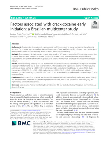
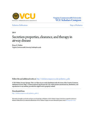
I. Ballesteros-Yáñez et al. / Neuroscience 146 (2007) 1536 –1545RESULTSIn this study, we have completely reconstructed 245 layerIII pyramidal cells in the motor area (Figs. 1 and 2). Thesecells were subject to morphometric analyses and theycorresponded to 138 neurons from animals administeredsaline (80 neurons from wild type and 58 from CB1 knockout mice) and 107 neurons from the cocaine-treated group(62 neurons from wild type and 45 from CB1 knockoutmice). We compared the structure of the pyramidal neurons in wild type and CB1 knockout mice and analyzed theeffects of chronic cocaine treatment in both groups ofanimals.Cocaine alters the morphometric parameters ofpyramidal neurons in wild type miceChronic cocaine administration reduced the extension ofthe basal dendritic arbor in wild type mice by approximately24%, from 3.54 0.09 104 m2 in saline-treated animalsgroup to 2.70 0.11 104 m2 in mice that consumed cocaine (F3,241 14.8, P 0.0001). Similarly, the dendriticlength diminished by approximately 13% from 2.11 0.04 103 m in animals that received saline to 1.83 0.05 103 min mice that consumed cocaine (F3,241 5.78, P 0.001). Incontrast, the tortuosity of dendrites was augmented inanimals that received cocaine (1.42 0.02) when compared with those that received saline (1.33 0.01;F3,241 5.801, P 0.01). However, the density of spines onpyramidal neurons was lower in wild type mice afterchronic cocaine treatment (16.4 0.9) than in salinetreated animals (18.0 0.7; mean S.E.M. of the peak ofspine density). In conjunction, these two morphometricvalues indicated that the total number of spines decreasedby 25% in the wild type mice subjected to chronic cocainetreatment (1587 spines) when compared with salinetreated wild type mice (2076 spines; Fig. 3, supplementarymaterial 1–3).Differential basal morphometric parameters in CB1knockout mice and wild type littermatesUnder basal conditions, the basal dendritic arbor of pyramidalcells in the motor cortex was more extensive in wild type micethan in the CB1 knockout mice (F3,241 14.8, P 0.0001: Fig.4). The dendritic spine density was also significantly higher inwild type (18.0 0.7 spines/10 m) than in CB1 knockoutmice (16.4 0.9 spines/10 m, mean S.E.M. of the peakspine density; F3,153 15.1, P 0.0001: Fig. 4; supplementarymaterial 1–3). A total of 2076 spines per cell were estimatedfor the wild type animals whereas in the CB1 knockout animals this number was 1858 (Fig. 4). No significant differences between these two groups of animalswere found in the other morphological parameters examined.1539The effects of cocaine on the morphometricparameters of pyramidal cells in CB1 knockout miceThe chronic administration of cocaine to CB1 knockoutmice induced only minor changes in the structure of thedendritic arbor of pyramidal neurons. However, CB1knockout mice that were administered cocaine exhibited ahigher spine density (20.0 0.9, maximum value S.E.M)than the CB1 knockout mice that received saline(14.6 1.1, F3,153 15.1, P 0.0001: supplementary material 3). In addition chronic cocaine treatment produced aslight increase in the total number of spines of around 7%in CB1 knockout mice per basal dendritic arbor. While, themean of 1858 spines was calculated for CB1 knockoutmice that received saline, the number of spines for thosethat were administered cocaine was calculated as 2003(Fig. 5: supplementary material 1–3).Together, our data show that chronic cocaine treatment induced changes in the microanatomy of pyramidalcells in both wild type and CB1 knockout mice, although themagnitude of these modifications is modulated by the CB1receptors.DISCUSSIONThe current study reveals that the lack of CB1 cannabinoidreceptors induces changes in the morphology of pyramidalneurons in the mouse motor cortex. Moreover, the morphological changes of the basal dendritic arbor induced bychronic cocaine administration are dependent on the expression of CB1 cannabinoid receptors. The size of dendritic arborof pyramidal cells is related to the topographic sampling areaand mixing of inputs, while branching complexity determinesthe degree to which the integration of inputs is compartmentalized within the arbors (Poirazi and Mel, 2001; Elston,2003). Each dendritic spine forms an excitatory synapse andsuch spines represent the main target of these synapses(DeFelipe and Fariñas, 1992). Thus, the differences in thesize, branching pattern and spine density produce variationsin the total number of excitatory inputs to each pyramidal cell,and in their integrative properties.Genotype differences under basal conditionsThe basal dendritic arbor of pyramidal cells in CB1 knockout mice is smaller and there were fewer dendritic spinesthan in wild type littermates. This implies changes in thenumber of excitatory inputs to pyramidal neurons and inthe processing of information in CB1 knockout mice. Theseresults are relevant to the mechanisms underlying thebehavioral responses observed in CB1 knockout mice.Indeed, anatomical, neurochemical and behavioral evidence indicates that endocannabinoids play an importantFig. 1. (A, B) Low-power photomicrographs of the sections of the mouse cerebral cortex parallel to the cortical surface, showing the region where thecells where injected in the motor cortex (lateral view of the right hemisphere). (A) Wild type animals; B, knockout mouse. These layer III neurons wereinjected with Lucifer Yellow and then processed with a light-stable diaminobenzidine. (C, E) High magnification of the boxed areas in A and B,respectively. (D, F) Pyramidal neurons injected in cocaine wild type animal (D) and in cocaine knockout animal (F). (G–J) High-power photomicrographs of horizontally projecting dendrites from layer III pyramidal neurons in the motor cortex of wild type mouse (G, I) and of knockout mouse (H,J) cortex in control (G, H) and cocaine-treated mouse (I, J). Scale bar 1.36 mm (A, B); 475 m (C–F); 16.4 m (G–J).
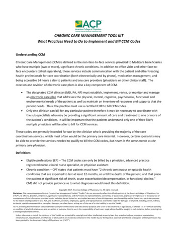
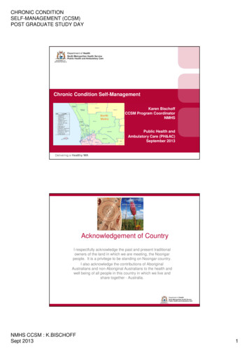
1540I. Ballesteros-Yáñez et al. / Neuroscience 146 (2007) 1536 –1545Fig. 2. Neurolucida drawings of layer III pyramidal neurons, as seen in the plane of a section parallel to the cortical layers, from control and treatedmice in motor cortex. The cells illustrated have basal dendritic arbors which approximated to the average size for each group. Scale bar 125 m.role in cognition and motor activity through CB1 receptors(Terranova et al., 1996; Varvel and Lichtman, 2002; BilkeiGorzo et al., 2005).CB1 knockout mice of the age used here perform similarly to wild type mice in an operant behavior paradigmreinforced by natural rewards (food and water: Soria et al.,2005). Moreover, they display better learning in the activeavoidance paradigm (Martin et al., 2002). The endocannabinoid system has also been implicated in emotional-likeresponses (Valverde, 2005), including the extinction ofaversive memories (Marsicano et al., 2002). Deletion ofthe CB1 cannabinoid receptors augments the sensitivity todevelop an anhedonic-like state after exposure to achronic unpredictable mild stress (Martin et al., 2002).Furthermore, CB1 cannabinoid receptor gene deletion alterslocomotor activity, displaying hypoactivity (Zimmer et al.,1999). Thus, the morphological modifications observed couldparticipate in the changes in motor and emotional-like responses, or in addictive vulnerability in these knockout mice.The changes in dendritic arborization in knockout miceunder basal conditions seem to be developmental rather thanrelated to adult dendritic remodeling. However, remodelingcannot be ruled out when considering the age-relatedchanges in hippocampal neurons in CB1 knockout mice(Bilkei-Gorzo et al., 2005). The cannabinoid system plays animportant role in developmental processes such as neurogenesis, axonal elongation and synaptogenesis (FernandezRuiz et al., 2000; Jin et al., 2004). Furthermore, CB1 cannabinoid receptors are important modulators of dopaminergicactivity (Melis et al., 2004b), and D1 dopamine receptormediated signaling contributes to the growth of dendrites ofpyramidal neurons in certain cortical areas (Stanwood et al.,2005). Therefore, CB1 receptors could contribute to thegrowth of pyramidal neurons by mediating dopamine activity.The effects of cocaine in wild type and CB1knockout miceThe size and length of the pyramidal cells basal dendriticarbors were reduced in wild type mice treated with cocaine, the tortuosity was increased, and the spine densitywas lower than in the control mice. In contrast, the number
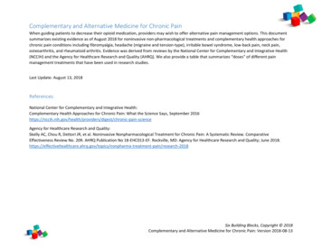
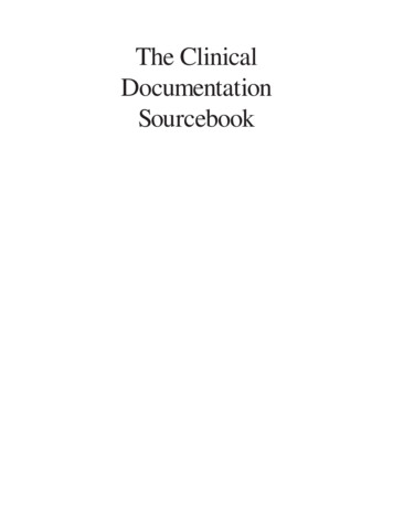
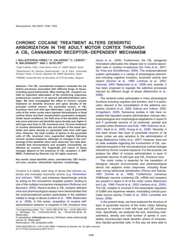
I. Ballesteros-Yáñez et al. / Neuroscience 146 (2007) 1536 –15451541Fig. 3. Graphs representing the statistically different morphological parameters analyzed in this study. (A) Basal dendritic field area; (B) total basaldendritic length; (C) tortuosity per cell; (D) estimation of the total number of spines; (E) Sholl analysis; (F) quantity of branches per branch order;(G) number of spines per distance from the soma. Morphological parameters of pyramidal neurons from control (white) or cocaine-treated (black)wild-type mice.of spines increased in CB1 knockout mice that weretreated with cocaine, while no change was observed in themorphology of the dendritic arbor in these animals.We do not know whether the effects observed here aredue to a direct effect on pyramidal neurons or whether theyare secondary to changes in cortical networks due to theinteractions of cannabinoids with neurotransmitters. Although located in pyramidal and GABAergic neurons, thehighest level of CB1 receptors in the cortex is found incertain GABAergic neurons and axon terminals that inner-Fig. 4. Graphs representing the statistically different morphological parameters analyzed in this study. (A) Basal dendritic field area; (B) total basaldendritic length; (C) tortuosity per cell; (D) estimation of the total number of spines; (E) Sholl analysis; (F) quantity of branches per branch order;(G) number of spines per distance from the soma. Morphological parameters of pyramidal neurons from wild-type (white) or CB1 knockout (black) mice.
1542I. Ballesteros-Yáñez et al. / Neuroscience 146 (2007) 1536 –1545Fig. 5. Graphs representing the statistically different morphological parameters analyzed in this study. (A) Basal dendritic field area; (B) total basaldendritic length; (C) tortuosity per cell; (D) estimation of the total number of spines; (E) Sholl analysis; (F) quantity of branches per branch order;(G) number of spines per distance from the soma. Morphological parameters of pyramidal neurons from control (white) or cocaine-treated (black) CB1knockout mice.vate both pyramidal cells and GABAergic interneurons(Marsicano and Lutz, 1999; Harkany et al., 2005). Furthermore, CB1 receptors coexist with the 5-HT3A serotoninreceptor in GABAergic neurons of the superficial neocortical layers in rodents, and GABA release by these cellsdepends on the functional state of both receptors (Moraleset al., 2004). The functional balance between these tworeceptors might be altered by cocaine administration,which would modify the levels of cortical serotonin (Steketee, 2003; Arnold, 2005). Moreover, cocaine administrationinhibits monoamine transporters, increasing extracellulardopamine and producing functional changes in the dopaminergic innervation of cortical neurons (Steketee, 2003;Arnold, 2005). Manipulation of the synaptic inputs to pyramidal cells induces morphological changes in their dendritic arbors and spines (e.g. Elston, 2002; Lewis et al.,2003). Thus, the functional changes produced by cocainetreatment in dopamine transmission could at least be partially responsible for the morphological changes foundhere. These changes could be implicated in the enhancedlocomotor activity induced by cocaine treatment (e.g.Woolverton and Johnson, 1992; Uhl et al., 2002). Endocannabinoids also play an important role in the control ofreward circuits (Maldonado et al., 2006). Moreover,through activating presynaptic CB1 receptors and inhibitingthe glutamatergic inputs from the prefrontal cortex, theyfacilitate the release of dopamine in the nucleus accumbens (Robbe et al., 2002; Melis et al., 2004a). Therefore,the endocannabinoid system might regulate the dopaminemodulation of cortical information processing, supporting acorrelation between unbalanced endocannabinoid signal-ing and dopamine-dependent processes under conditionssuch as drug addiction or stress (Melis et al., 2004b).Finally, the differential effects of cocaine on pyramidalcell morphology in CB1 knockouts might be attributed tothe disappearance of the cross-talk between the cannabinoid and monoamine systems in these mutant mice (Glassand Felder, 1997; Schlicker and Kathmann, 2001; Van derStelt and Di Marzo, 2003; Jarrahian et al., 2004). Indeed,the possible participation of an endocannabinoid-monoamine interaction is supported by different findings. Cannabinoid agonists increased the synthesis of dopamine/norepinephrine in brainstem slices containing the locuscoeruleus or the hippocampus (Moranta et al., 2006),whereas the CB1 antagonist rimonabant was reported toincrease monoamine efflux from the prefrontal cortex inrats following “in vivo” microdialysis (Tzavara et al., 2003).Behavioral and neurochemical evidence indicates that theactivation or blockage of the CB1 cannabinoid receptorsmodulates the responses elicited by the stimulation of5HT1A and 5HT2A receptors (Cheer et al., 1999; Darmaniet al., 2003). In addition, chronic antidepressant treatmentmodifies the expression of the CB1 cannabinoid receptorsin the rat brain (Hill et al., 2006) and the inhibitor of anandamide hydrolysis, URB597, enhances the firing activity ofserotonergic neurons in the dorsal raphe nuclei of rats(Gobbi et al., 2005). In contrast, CB1 blockage exacerbates normal reactions to acute stress (Martin et al., 2002).Furthermore, the endocannabinoid system has been reported to be involved in the behavioral responses inducedby psychostimulants. Thus, changes in pyramidal cellscould contribute to the impaired reinforcement of cocaine
I. Ballesteros-Yáñez et al. / Neuroscience 146 (2007) 1536 –1545observed in CB1 knockout mice (Soria et al., 2005) or afterthe administration of the CB1 antagonist rimonabant (DeVries et al., 2001). Moreover, it has been reported that thereinforcing effects of other drugs of abuse (morphine, nicotine and ethanol) were also impaired in CB1 knockoutmice (Maldonado et al., 2006). It might therefore be interesting to evaluate whether other drugs of abuse producesimilar effects on pyramidal cells to cocaine in the CB1mutant mice. However, we cannot rule out that some adaptive changes brought about by the genetic deletion of theCB1 receptor could contribute to the morphological changesin the pyramidal cells.Comparison with previous studies ofthe effects of cocainePrevious studies in the anterior cingulate cortex of rats andrabbits have shown that cocaine increases the number ofdendritic branches, tortuosity and/or spine density in pyramidal cells (Robinson and Kolb, 1999, 2004; Robinson etal., 2001; Stanwood et al., 2001; Kolb et al., 2003).Whereas our results agree with increase of branch tortuosity after cocaine exposure, we found that the drug reduces the extension of the basal dendritic trees and thedensity of spines in motor cortex pyramidal cells in wildtype mice. This discrepancy could be due to the differenttreatment schedules, the different animal species used(rabbits and rats versus mice) and the techniques appliedin each study (Golgi staining versus labeling with intracellular injections: e.g. Robinson and Kolb, 1999; BallesterosYáñez et al., 2007). For example, the changes observedpreviously might be attributed to the long-lasting effects ofcocaine or to re-adaptation to an environment without cocaine (cocaine treatment over 4 weeks and the animalswere kill 24 –25 days after the last injection). The differences we observed here are more readily attributable tothe chronic effects of this drug since cocaine was administered for 13 days and the mice were killed 24 h after thelast dose of cocaine. This procedure was used to comparethe results with those from a previous study evaluating thebehavioral sensitization induced by cocaine in CB1 knockout mice (Martin et al., 2000). Interestingly, CB1 knockoutmice developed similar behavioral sensitization and cocaine-induced conditioned place preference as wild-typelittermates (Martin et al., 2000). However, mutant mice didnot acquire a reliable operant behavior to self-administercocaine (Soria et al., 2005). Moreover, cocaine does notappear to equally affect all cortical areas. Thus, cocaineadministration increased dendritic branches and spinedensity in the cingulate cortex, but it did not change themorphology of pyramidal neurons in the rat parietal oroccipital cortex (Robinson et al., 2001). We have identifiedspecific morphological changes in neurons of the motorcortex induced by cocaine treatment that depend on thepresence of CB1 receptors. The labeling technique usedhere validates the reliability and accuracy of these findings.Further studies using this technique will be necessary toclarify the possible reversibility of these changes after thelong-term disruption of the cocaine treatment.1543CaveatsThere are two limi
CHRONIC COCAINE TREATMENT ALTERS DENDRITIC ARBORIZATION IN THE ADULT MOTOR CORTEX THROUGH ACB 1 CANNABINOID RECEPTOR–DEPENDENT MECHANISM I. BALLESTEROS-YÁÑEZ,a O. VALVERDE,b C. LEDENT,c R. MALDONADOb* AND J. DEFELIPEa* aCajal Institute