Transcription
Biomed Microdevices (2009) 11:547–555DOI 10.1007/s10544-008-9260-xIntegrated microfluidic devices for combinatorial cell-basedassaysZeta Tak For Yu & Ken-ichiro Kamei &Hiroko Takahashi & Chengyi Jenny Shu &Xiaopu Wang & George Wenfu He & Robert Silverman &Caius G. Radu & Owen N. Witte & Ki-Bum Lee &Hsian-Rong TsengPublished online: 9 January 2009# Springer Science Business Media, LLC 2008Abstract The development of miniaturized cell culture platforms for performing parallel cultures and combinatorialassays is important in cell biology from the single-cell levelto the system level. In this paper we developed an integratedmicrofluidic cell-culture platform, Cell-microChip (CellμChip), for parallel analyses of the effects of microenvironmental cues (i.e., culture scaffolds) on different mammaliancells and their cellular responses to external stimuli. As amodel study, we demonstrated the ability of culturing andassaying several mammalian cells, such as NIH 3T3 fibroblast, B16 melanoma and HeLa cell lines, in a parallel way.For functional assays, first we tested drug-induced apoptoticresponses from different cell lines. As a second functionalassay, we performed “on-chip” transfection of a reporter geneencoding an enhanced green fluorescent protein (EGFP)followed by live-cell imaging of transcriptional activation ofcyclooxygenase 2 (Cox-2) expression. Collectively, our CellμChip approach demonstrated the capability to carry outparallel operations and the potential to further integrateadvanced functions and applications in the broader space ofcombinatorial chemistry and biology.Keywords Microfluidic devices . Cell-based assay .Apoptosis . Transfection . Cell cultureElectronic supplementary material The online version of this article(doi:10.1007/s10544-008-9260-x) contains supplementary material,which is available to authorized users.Zeta Tak For Yu and Ken-ichiro Kamei contributed equally to this work.Z. T. F. YuDepartment of Mechanical and Aerospace Engineering,University of California,Los Angeles, CA 90095, USAC. J. Shu : O. N. WitteDepartment of Microbiology, Immunology, and MolecularGenetics, University of California,Los Angeles, CA 90095, USAKameiTakahashi·: X.X. WangWang :· G.G.W.W.HeHe: ·Z. T. F. Yu : K.-i.Kamei· :H.H.TakahashiC. G. Radu (*) : H.-R. Tseng (*)Crump Institute for Molecular Imaging, University of California,Los Angeles, CA 90095, USAe-mail: caiusradu@ucla.edue-mail: hrtseng@mednet.ucla.eduO. N. WitteThe Howard Hughes Medical Institute, University of California,Los Angeles, CA 90095, USAR.SilvermanSilverman: ·KameiKameiWang·: G.G. W.W. HeHe :· R.Z. T. F. Yu : K.-i.· :X.X.WangC. G. Radu : O. N. Witte (*) : H.-R. TsengDepartment of Molecular and Medical Pharmacology,University of California,Los Angeles, CA 90095, USAe-mail: owenw@microbio.ucla.eduK.-B. Lee (*)Department of Chemistry and Chemical Biology,Institute for Advanced Materials, Devices and Nanotechnology,The Rutgers Stem Cell Research Center, Rutgers,The State University of New Jersey,Piscataway, NJ 08854, USAe-mail: kblee@rci.rutgers.eduDO09260; No of Pages
5481 IntroductionConventional cell-based experiments are typically performed on a large cell population. Researchers have begunto appreciate that there are local variables associated withthe heterogeneous microenvironment in the macroscopicculture setting, which often leads to experimental inconsistency. On the other hand, differences among individualcells are often ignored since the conventional assays (e.g.,Weston blots and microarray analysis) are conducted at acollective fashion. As a result, it is challenging to elucidatecomplex cellular systems and analyze dynamic signalingpathways (Irish et al. 2006) using the conventionalexperiment systems. To overcome these challenges, it isessential to develop a new technology platform to enable(1) improved control on the cell culture microenvironment,(2) precise cell assays with the single-cell resolution, and(3) sequential and parallel operation by combining thosementioned in (1) and (2). We envision such a technologycan be applied for screening drug candidates (Dittrich andManz 2006; Padron et al. 2000), evaluating biologicalpathways (Minor 2003), and understanding pharmacologicaleffects (Hill et al. 1998; Umezawa 2005), thus constitutingcritical technological foundations for a broad spectrum ofbiomedical research.Microfluidic devices (Auroux et al. 2002; Dittrich andManz 2006; Dittrich et al. 2006; Reyes et al. 2002) offer arobust analytical tool that allows rapid analysis of cellularresponses to external stimuli in a parallel way. Moreover,microfluidics, with its intrinsic advantages of sample/reagenteconomy, precise control over physical and chemical microenvironments, high throughput, scalability and digital controllability, whose features allow microfluidics to investigatecomplex and dynamic biological processes at the single-celllevel. In addition, microscale cell culture using microfluidicsallows investigating the function of microenvironmental cues atthe single-cell level. Especially, compared to static microfluidiccell-culture systems, integrated microfluidics allows for thecontrol of adding and/or removing of biochemical cues atspecific temporal as well as spatial points. This uniqueadvantage makes it possible to do novel microenvironmentalexperiments, such as cell–cell interactions and extracellularmatrix (ECM)–cell interactions. However, there are severalissues associated with the fabrication and control of integratedmicrofluidic cell culture systems in order to achieve routinecellular assays of mammalian cells. Even though microfluidiccell culture systems have been developed extensively, they canmainly be described into two ways: what kinds of cell cultureplatforms were used, and what types of cells and applicationswere tested. Many different cell culture platforms have beendeveloped, including two-dimensional, three-dimensional(Cartmell et al. 2003; Toh et al. 2007) and co-culture platformsBiomed Microdevices (2009) 11:547–555(Sin et al. 2004). Those microfluidic approaches enabled toculture and assay many different cells, such as liver (Kaneet al. 2006; Sin et al. 2004; Zhang et al. 2008), muscle(Tourovskaia et al. 2005; Tourovskaia et al. 2006), neural(Millet et al. 2007; Park et al. 2006), and stem cells (Chunget al. 2005; Gomez-Sjoberg et al. 2007; Kim et al. 2006).Despite these recent advances, several critical questions andseveral challenges should be addressed more explicitly, andoptimized to fully achieve the potential of microfluidic cellculture and assays. It requires the abilities: (1) to test a robustand flexible fluidic configuration (e.g., flow through vs.circulation) for cell culture and (2) to fabricate microfluidicnetwork capable of performing sequential and paralleloperations such as parallel culture of multiple cell typesand subsequent phenotypic and functional assays in the samemicrofluidic chip. In this context, incorporation of isolationvalves (Unger et al. 2000) and peristaltic pumps (Chou et al.2001) should allow individual addressability and digitalcontrollability (Lee et al. 2005; Wang et al. 2006) of eachcell culture chamber embedded on a microfluidic device,which in turn should enable complex manipulations of theculture microenvironments as well as multiple analyticalmeasurements.Over the years, there have been a variety of microfluidicchips directed for functional biological assays. Theseinclude differentiation of cell through different flow rates(Gu et al. 2004), fully automated cell culture system bytwo-layer PDMS chips (Gomez-Sjoberg et al. 2007),optimization of drug cocktail to regulate cell activitiesthrough closed-loop control algorithm and microfluidicplatform (Wong et al. 2008), and modelling of galactosepathway in an alternating culture environment (Bennett etal. 2008). All these devices or systems have providedadditional modules and thus are superior to the conventional setting on conducting biological research or routineoperations.In this paper, we describe the design and operation ofpolydimethylsiloxane (PDMS)-based Cell-microChip (CellμChip, Fig. 1) envisioned as a digitally controlled platformfor performing parallel cell culture and sequential cellularassays. The potential of the Cell-μChip to support theoptimal culture and assays of human and murine cell lineswas demonstrated. To determine the optimal cultureconditions, these cells were cultured in six cell culturechambers embedded on a Cell-μChip under two differentmedium supply modes in parallel (i.e., circulation anddirect feeding). The growth and viability of the microcultures were monitored and quantified in real time in theCell-μChip using an integrated CCD camera. Sequentialstaining with acridine orange (AO) and propidium iodide(PI) (Hudson et al. 1969; Traganos et al. 1977) allowedthe identification of viable and dead cells, respectively. To
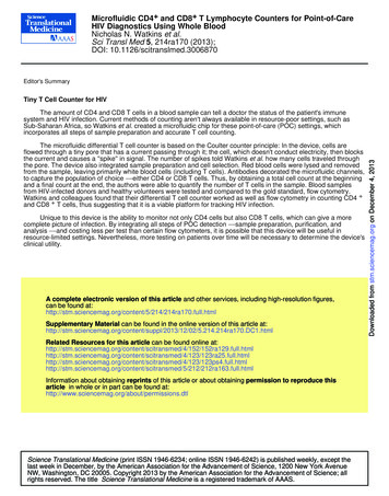
Biomed Microdevices (2009) 11:547–555Fig. 1 (a) Schematic representation of an integrated CellmicroChip (Cell-μChip) forperforming multiple cell cultureand assays under a digitally controlled interface. Three pairs ofparallel-oriented cell culturechambers are incorporated in aCell-μChip, where multiple celltypes can be cultured under twodifferent modes of medium supply, i.e., circulatory (channels i, iiiand v) and direct feeding (channels ii, iv and vi). The operation ofthis microchip is controlled bypressure driven valves with theirdelegated functions indicated bytheir colors: red for regular valve(for isolation and gating) andyellow for pumping valve (forfluid transport and circulation).(b) Optical image of the actualdevice. The microchip was loadedwith various colors of food dyesto enhance the visualization ofdifferent parts in the entire system: red and yellow as in (a); blueindicates the flow channel and themedium reservoir549(a)OutletportInletportiMediumtubing iiiiiivvvi(1) Cell culturechambers i vi:100– 150 nL(2) lar valveValve for pump(b)demonstrate the ability to perform “on chip” functional assays,we analyzed drug-induced apoptotic responses. Furthermore,we showed that the Cell-μChip is amenable to complexsequential operations such as genetic manipulation andmonitoring of transcriptional activation of gene expression.2 mm2 Experimentaloutside the culture chambers were removed by washing withfresh DMEM. The Cell-μChip was placed in an incubator for6 h. The pump was turned on to introduce the DMEM in acirculating or feed through fashion in the respective culturechambers. The flow rate of the medium was controlled in therange of 0.1–4 nL s 1. Cell growth was monitored bycollecting bright field micrographs of cells inside theCell-μChip at 12-h intervals.2.1 Microfluidic cell culture2.2 Immunoassay for fibronectinUsing the integrated valves and pumps, bovine fibronectin(FN) (Sigma) solution (1.0 mg mL 1) filled in Teflon tubingwas introduced into the six cell culture chambers of theCell-μChip. The Cell-μChip was kept at 37 C for 30 minfor FN coating. DMEM was then introduced into the cellculture chambers to extrude the FN solution. The mediumreservoirs and medium tubings were then dead-end filledwith DMEM at 10 psi for 60 min (Song et al. 2008). Then,individual cell suspensions (NIH 3T3, HeLa and B16) with 2 106 cells mL 1 were sequentially introduced by gravitationinto the cell culture chambers. After cell loading, cells locatedTo confirm FN coating efficiency in cell culture chambers,immunoassay for FN was performed. After FN coating ina Cell-μChip, blocking solution containing 5% BSA and0.1% N-dodecyl-β-D-maltoside (DDM) (Pierce) was introduced in a Cell-μChip, and then kept at roomtemperature for 1 h. Mouse anti-FN (BD Biosciences)was loaded into cell culture chambers, and incubated atroom temperature for 2 h. Excess antibody in cell culturechambers were rinsed with PBS with 0.1% Tween 20(PBS-T) twice. Then secondary goat anti-mouse IgGconjugated with Alexa555 (Invitrogen) were introduced in
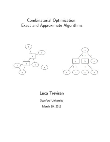
550cell culture chambers and incubated at room temperature for1 h. After washing with PBS-T twice, fluorescent intensitywas measured with a fluorescent microscope, and quantifiedwith MetaMorph.2.3 AO/PI fluorescence stainingAfter culturing the cells in the Cell-μChip for 4 days, asolution composed of DMEM, AO and PI in a ratio of10:1:1 was introduced into the cell culture chambers. After5 min incubation at 37 C, the cells were imaged under afluorescence microscope.Biomed Microdevices (2009) 11:547–5553 Results and discussion3.1 Design of the Cell-μChipThe PDMS-based Cell-μChips (Fig. 1) were fabricated bymultilayer soft lithography approach (see supplementaryinformation) (Unger et al. 2000; Xia and Whitesides 1998).It is important to note that the biocompatible and gaspermeable properties (Kim et al. 2007; Korin et al. 2007) ofPDMS matrices help to retain proper physiological(a)30 min2h1 mg mL-1NIH 3T3, HeLa and B16 cells were cultured in the cellculture chambers of the Cell-μChip for 24 h. Staurosporine(0, 0.1, 1 and 10 μM as final concentrations) oractinomycin D (0, 0.1, 1 and 10 μM as final concentrations)in DMEM culture medium were loaded into the cell culturechambers. After 2 h incubation, the medium in all cellculture chambers was replaced by the MitoTracker Reddye (Invitrogen) for staining of viable cells. The CellμChip was incubated for 30 min at 5% CO2, 37 C.Sequentially, the MitoTracker Red solution was replacedby a solution containing 100 μL of Annexin bindingbuffer and 5 μL of Annexin V-Alexa488 for staining theapoptotic cells. Following 15 min incubation at RT, cellculture chambers were flushed with Annexin bindingbuffer and the cells were imaged using a fluorescencemicroscope.0.01 mg mL-12.4 Apoptosis assay(b)NIH 3T3 cells were loaded into the six culturechambers of the Cell-μChip and were allowed to settlefor 24 h. Cells were transfected with the pCox2-EGFPplasmid (a kind gift from Prof. Harvey R. Herschman,UCLA), encoding an enhanced green fluorescent protein(EGFP) under a murine Cox-2 promoter (Liang et al.2004). The transfection mixture containing plasmid(0.5 μg), medium (30 μL) and transfection reagent(2.5 μL, Superfect reagent, Qiagen) was incubated at RTfor 10 min. The mixture was further diluted with 150 μL ofDMEM and loaded into all culture chambers of the CellμChip. After 3-h incubation at 5% CO2, 37 C, the mixturewas replaced with serum-free DMEM and cells wereincubated for an additional day. To activate the Cox-2promoter the culture media in three out of six chamberswas replaced with media containing the induction agentTPA (50 ng mL 1). Following 7 h incubation at 5% CO2,37 C, EGFP expression was imaged using a fluorescencemicroscope.Fluorescence Intensity (A.U.)2.5 On-chip transfection and reporter gene imaging4000350030 min2h3000250020001500100050000.01 mg mL-11 mg mL-1Fibronectin ConcentrationFig. 2 Fibronectin coating efficiency on the PDMS surface in a CellμChip determined by immunofluorescence assay. (a) Fluorescenceimages of immunostained FN on the PDMS surface. (b) Quantitativeanalysis of FN coating efficiency determined with fluorescenceimages shown in (a)
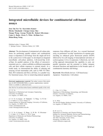
Biomed Microdevices (2009) 11:547–555551(a)Day 0Day 1Day 2Day 7Day 8200 µmDay 3matic valves and peristaltic pumps. This design enabled todigitally control sequential operations. The chip consistedof three identical pairs of parallel-oriented culture chamberswith identical dimensions (3 0.5 0.1 mm3, correspondingto a volume of 150 nL). To allow synchronized pumping,six internally connected peristaltic pumps were incorporatedat the ends of the six cell culture chambers.Each pair of culture chambers was configured to havetwo types of medium supplies: one allowing mediarecirculation through the culture chamber for cellularauto-conditioning and the other enabling direct feeding ofcells with fresh media. The size of medium reservoir couldaccommodate about 10 μL of culture media, a volumesufficient to sustain continuous on-chip cell culture for8 days. In contrast, supply Teflon tubings were utilized to(a)(b)(b)(c)(c)200 µmNIH 3T3HeLaB16(d)20(e)100 µmFig. 3 Long term culture of NIH 3T3 cells in a Cell-μChip. (a) Timelapse images of the NIH 3T3 cell proliferation in the microchip in aduration of 8 days. (b)–(e) Dead (PI)/live (AO) staining of NIH 3T3cells cultured in a Cell-μChip for 4 days. (b) A bright fieldmicrograph of NIH 3T3 cell morphologies. (c) A green fluorescencemicrograph of the live-stained cells. (d) A red fluorescence micrograph of the dead-stained cells. (e) A merged fluorescence image of(b) and (c)conditions for a wide range of mammalian cells suitable fordifferent screening applications. A fluidic network forindividually addressable cell culture chambers and solution/reagent transport was integrated with embedded pneu-Fold Changes of Cell NumberNIH 3T3(d)HeLa15with mediumre-circulationB16NIH 3T3 with directmedium feeding10500246DaysFig. 4 Demonstration of six parallel cell cultures in a closely relatedmicroenvironment. After 3 days culture, the cell morphologies wereshown in (a) NIH 3T3, (b) HeLa and (c) B16. (d) Growth curves ofchip-cultured NIH 3T3, HeLa and B16 cells were quantified bymonitoring the number of cells inside the cell culture chambers overtime. After 5 days culture, we could not count cell number preciselydue to cell confluence and multiple cell layers in the cell culturechambers
552Biomed Microdevices (2009) 11:547–555Staurosporine LivingApoptosisB16Living0.1ActinomycinD (µM)Fig. 5 Multiparametric apoptosis assays performed in the CellμChips. NIH 3T3, HeLa and B16 cells were treated with eitherstaurosporine or actinomycin D to induce apoptosis. Apoptotic cellswere stained with Annexin V conjugated with Alexa488 (green), andliving cells were stained with MitoTracker (red)store and deliver fresh culture media. This design allowedus to perform six cell culture experiments in a closelyrelated microenvironment under two different culture mediasupply modes.searching optimal ECMs, FN is effectively coated on thePDMS surface in our Cell-μChip (Fig. 2). Several differentconditions, such as different FN concentrations andincubation time, were tested to optimize the FN coatingcondition. We confirm the efficiency and homogeneity ofFN coating on PDMS by immunoassay. As a result, 1 mgmL 1 of FN for 30 min incubation at 37 C is the optimalcondition in a Cell-μChip.3.2 Surface modification with fibronectinWe initially used bare PDMS surface to seed cells,however, we found that cells either could not attach to thesurface well or they detached so easily when new freshmedia were supplied. We reasoned that this problem wasdue to the inherent hydrophobicity of PDMS materials.Thus, we tested to use ECMs in order to make the surfacehydrophilic and biocompatible for cell adhesion. In our3.3 Cell culture in the Cell-μChipWe initially demonstrated to culture NIH 3T3 cells in allculture chambers of a Cell-μChip (Fig. 3, SupplementaryInformation and Fig. S1). Generally, following loading on
Biomed Microdevices (2009) 11:547–555(a)(b)20µm(c)(d)553numbers increased significantly for all three cell lines. Toexamine cell viability in the three reservoir-attached cellculture chambers, we performed sequential PI/AO staining.As a result, most of the cells are viable, and few dead cellswere observed (data not shown). To quantify the cultureparameters in the Cell-μChip we sought to determinegrowth rate for the three cell types (Fig. 4(d)). We observedthat the curves reflected a linear growth phase until the cellsreached confluence followed by a stationary phase. Comparing the cell growth with medium-recirculation or directfeeding setting, first we used NIH 3T3 cells and continuously monitored them. There was no clear difference inNIH 3T3 cell growth between the two settings (Fig. 4(d)).In the case of HeLa and B16 cells, we obtained similarresults as NIH 3T3 cells (data not shown). This resultindicates that a Cell-μChip serves a platform to performmultiple cell culture in a single device.3.5 On-chip apoptosis assayFig. 6 On-chip transfection and EGFP induction in NIH 3T3 cells.The plasmid vector which encodes EGFP driven by a Cox-2 promoterwas transfected with NIH 3T3 cells. (a) Bright field and (b)fluorescence images of NIH 3T3 cells stimulated with TPA for 7 h.(c) Bright field and (d) fluorescence images of NIH 3T3 cells withoutstimulationthe chip, it took about 1 h for cells to attach and spread ontothe FN-coated culture chamber surfaces. NIH 3T3 cellsgrew to confluence, and the whole microchannel was fullyoccupied at day 8. During this period, we continuouslymonitored cell growth inside the Cell-μChip using a CCDcamera (see ESM Movie 1). Interestingly, cells in a CellμChip grew on the bottom as well as ceiling. Thisphenomenon is not allowed under conventional cultureconditions using Petri dishes. This observation indicatedthat a Cell-μChip could provide unique and intrinsiccharacteristics of cell culture manipulations. To determinecell viability we performed AO/PI fluorescence staining(Fig. 3(b)–(e)). AO staining (Fig. 3(c)) indicated that themajority of cells in the chamber were viable. PI staining(Fig. 3(d)) showed a small percentage of dead cells.3.4 Parallel culture of multiple cell lines in a singleCell-μChipTo demonstrate this concept, we cultured NIH 3T3, HeLaand B16 cells in a single Cell-μChip (Fig. 4). NIH 3T3,HeLa and B16 cells were sequentially loaded into the pairsof culture chambers; i–ii, iii–iv and v–vi accordingly. Aftercell adhesion, the six peristaltic pumps were turned on tofeed cells with the medium, in either recirculation or directfeeding setting. The results were consistent with thoseobserved for the parallel culture of NIH 3T3 cells. CellApoptosis is not only fundamentally involved in developing cells and maintaining tissue homeostasis, but also canbe closely related to several diseases including cancer,autoimmune, and neurodegeneration. Even though, extensive studies have been reported to dissect apoptosis’smolecular basis, more system level as well as single-celllevel analysis by using integrated microfluidics would bringnew insights for the underlying mechanisms of thesebiological processes. To demonstrate the capability of “onchip” functional analyses with the Cell-μChip, we performed a drug-induced apoptosis assay (Fig. 5). We usedtwo kinds of apoptosis inducers, staurosporine (ST)(Rajotte et al. 1992; Tafani et al. 2001; Wang et al. 1996)and actinomycin D (AD) (Martin et al. 1990). NIH 3T3,HeLa and B16 cells were treated with apoptosis inducers atfour concentrations (0, 0.1, 1 and 10 μM) in the CellμChips. ST or AD treated cells increased apoptotic cellpopulation with a dose-dependent manner of apoptosisinducers. Although cell viability among untreated andtreated cells appeared to be the same based on theMitoTracker staining, the cells underwent the apoptosisprocess at various speeds according to the drug concentration, as demonstrated by the Annexin V staining. Weconclude that the Cell-μChip served as a platform toperform multiparametric functional assays.3.6 On-chip transfection and monitoring of reporter geneexpressionPlasmid DNA transfection is one of the common methodsto manipulate gene expression in mammalian cells.However, the detailed conditions of optimal transfectionfor different cell lines are variable. Combinatorial approach
554from parallel microfluidic cellular assays can help toidentify the optimal condition. To demonstrate the feasibilityof performing the other assay in the Cell-μChip, genetransfection experiments were carried out (Fig. 6). Forproof-of-concept, we used a plasmid vector with anenhanced green fluorescent protein (EGFP) driven by acyclooxygenase-2 (Cox-2) promoter. Therefore, EGFPexpression serves as a reporter of Cox-2 transcription. Thebasal expression level of Cox-2 in NIH 3T3 cells is very low.Tetradecanoylphorbol acetate (TPA) can activate Cox-2transcription. EGFP expression in these cells was monitoredunder a fluorescence microscope. As shown in Fig. 6, eventhough weak EGFP signals were detected in the negativecontrol chambers, strong EGFP signals were observed inTPA-induced transfected cells in the corresponding cellculture chambers.4 ConclusionIn summary, we developed a fully digitally controlledmicrofluidic cell culture and assay platform that couldsupport parallel cell culture and sequential cell assays.Through the integration of isolation valves, murine andhuman cells lines could be cultured in different cell culturechambers and tested for different conditions of cell cultureand assays in a single Cell-μChip. Real-time monitoring ofcell morphology and numbers, viability assay, apoptosisassay and transfection to monitor expression of a reportergene vector were also performed using the same platform.Our results indicate that intrinsic advantages of microfluidicdevices enable the execution of complicated and integratedbiological operations in stand-alone devices such as theCell-μChip. We envision that this platform will be furtherintegrated with advanced functions and utilities for moresophisticated cell culture applications.Acknowledgements This research was supported by the NIHNanoSystems Biology Cancer Center, the DOE-UCLA Institute ofMolecular Medicine and the NIH-UCLA Center for In Vivo Imagingin Cancer Biology and Siemens Medical Solutions USA Inc. We thankStephanie M. Shelly, Dan Rohle, Shirley Quan and Mireille Riedinger forthe outstanding technical support with conventional cell culture conditions. ONW is an Investigator of the Howard Hughes Medical Institute.CGR was supported by a Developmental Project Award (ICMIC, NIH/NCI grant no. CA08630). C.J.S. was supported by a National Institutes ofHealth (NIH) Research Training in Pharmacological Sciences traininggrant PHS T32 CM008652.ReferencesP.A. Auroux, D. Iossifidis, D.R. Reyes, A. Manz, Micro total analysissystems. 2. Analytical standard operations and applications Anal.Chem. 74, 2637–2652 (2002). doi:10.1021/ac020239tBiomed Microdevices (2009) 11:547–555M.R. Bennett, W.L. Pang, N.A. Ostroff, B.L. Baumgartner, S. Nayak,L.S. Tsimring, J. Hasty, Metabolic gene regulation in adynamically changing environment Nature 454, 1119–1122(2008). doi:10.1038/nature07211S.H. Cartmell, B.D. Porter, A.J. Garcia, R.E. Guldberg, Effects ofmedium perfusion rate on cell-seeded three-dimensional boneconstructs in vitro Tissue Eng. 9, 1197–1203 (2003).doi:10.1089/10763270360728107H.P. Chou, M.A. Unger, S.R. Quake, A microfabricated rotary pumpBiomed. Microdevices 3(4), 323–330 (2001). doi:10.1023/A:1012412916446B.G. Chung, L.A. Flanagan, S.W. Rhee, P.H. Schwartz, A.P. Lee,E.S. Monuki, N.L. Jeon, Human neural stem cell growth anddifferentiation in a gradient-generating microfluidic device LabChip 5, 401–406 (2005). doi:10.1039/b417651kP.S. Dittrich, A. Manz, Lab-on-a-chip: microfluidics in drugdiscovery Nat. Rev. Drug Discov. 5, 210–218 (2006).doi:10.1038/nrd1985P.S. Dittrich, K. Tachikawa, A. Manz, Micro total analysis systems.Latest advancements and trends Anal. Chem. 78, 3887–3907(2006). doi:10.1021/ac0605602R. Gomez-Sjoberg, A.A. Leyrat, D.M. Pirone, C.S. Chen, S.R. Quake,Versatile, fully automated, microfluidic cell culture system Anal.Chem. 79, 8557–8563 (2007). doi:10.1021/ac071311wW. Gu, X. Zhu, N. Futai, B.S. Cho, S. Takayama, Computerizedmicrofluidic cell culture using elastomeric channels and Brailledisplays Proc. Natl. Acad. Sci. U. S. A. 101, 15861–15866(2004). doi:10.1073/pnas.0404353101D.C. Hill, S.K. Wrigley, L.J. Nisbet, Novel screen methodologies foridentification of new microbial metabolites with pharmacologicalactivity Adv. Biochem. Eng. Biotechnol. 59, 73–121 (1998).doi:10.1007/BFb0102297B. Hudson, W.B. Upholt, J. Devinny, J. Vinograd, The use of anethidium analogue in the dye-buoyant density procedure for theisolation of closed circular DNA: the variation of the superhelixdensity of mitochondrial DNA Proc. Natl. Acad. Sci. U. S. A. 62,813–820 (1969). doi:10.1073/pnas.62.3.813J.M. Irish, N. Kotecha, G.P. Nolan, Mapping normal and cancer cellsignalling networks: towards single-cell proteomics Nat. Rev.Cancer 6, 146–155 (2006). doi:10.1038/nrc1804B.J. Kane, M.J. Zinner, M.L. Yarmush, M. Toner, Liver-specificfunctional studies in a microfluidic array of primary mammalianhepatocytes Anal. Chem. 78, 4291–4298 (2006). doi:10.1021/ac051856vL. Kim, M.D. Vahey, H.Y. Lee, J. Voldman, Microfluidic arrays forlogarithmically perfused embryonic stem cell culture Lab Chip 6,394–406 (2006). doi:10.1039/b511718fL. Kim, Y.C. Toh, J. Voldman, H. Yu, A practical guide tomicrofluidic perfusion culture of adherent mammalian cells LabChip 7, 681–694 (2007). doi:10.1039/b704602bN. Korin, A. Bransky, U. Dinnar, S. Levenberg, A parametric study ofhuman fibroblasts culture in a microchannel bioreactor Lab Chip7, 611–617 (2007). doi:10.1039/b702392hC.C. Lee, G. Sui, A. Elizarov, C.J. Shu, Y.S. Shin, A.N. Dooley, J.Huang, A. Daridon, P. Wyatt, D. Stout, H.C. Kolb, O.N. Witte,N. Satyamurthy, J.R. Heath, M.E. Phelps, S.R. Quake, H.R.Tseng, Multistep synthesis of a radiolabeled imaging probe usingintegrated microfluidics Science 310, 1793–1796 (2005).doi:10.1126/science.1118919Q. Liang, M. Yamamoto, D.T. Curiel, H.R. Herschman, Noninvasiveimaging of transcriptionally restricted transgene expressionfollowing intratumoral injection of an adenovirus in which theCOX-2 promoter drives a reporter gene Mol. Imaging Biol. 6,395–404 (2004). doi:10.1016/j.mibio.2004.09.002S.J. Martin, S.V. Lennon, A.M. Bonham, T.G. Cotter, Induction ofapoptosis (programmed cell death) in human leukemic HL-60
Biomed Microdevices (2009) 11:547–555cells by inhibition of RNA or protein synthesis J. Immunol. 145,1859–1867 (1990)L.J. Millet, M.E. Stewart, J.V. Sweedler, R.G. Nuzzo, M.U. Gillette,Microfluidic devices for culturing primary mammalian neurons atlow densities Lab Chip 7, 987–994 (2007). doi:10.1039/b705266aL.K. M
monitoring of transcriptional activation of gene expression. 2 Experimental 2.1 Microfluidic cell culture Using the integrated valves and pumps, bovine fibronectin (FN) (Sigma) solution (1.0 mg mL 1) filled in Teflon tubing was introduced into the six cell culture chambers of the Cell-μChip. The Cell-μChip was kept at 37 C for 30 min for .