Transcription
Pan et al. Journal of Hematology & 2019) 12:124RESEARCHOpen AccessSingle-cell RNA sequencing revealscompartmental remodeling of tumorinfiltrating immune cells induced by antiCD47 targeting in pancreatic cancerYu Pan1†, Fengchun Lu1†, Qinglin Fei1, Xingxing Yu1, Ping Xiong2, Xunbin Yu3, Yuan Dang4, Zelin Hou1, Wenji Lin5,Xianchao Lin1, Zheyang Zhang6, Minggui Pan7 and Heguang Huang1*AbstractBackground: Human pancreatic ductal adenocarcinoma (PDAC) responds poorly to immune checkpoint inhibitor(ICPi). While the mechanism is not completely clear, it has been recognized that tumor microenvironment (TME)plays key roles. We investigated if targeting CD47 with a monoclonal antibody could enhance the response ofPDAC to ICPi by altering the TME.Methods: Using immunohistochemistry, we examined tumor-infiltrating CD68 pan-macrophages (CD68 M) andCD163 M2 macrophages (CD163 M2) and tumor expression of CD47 and PD-L1 proteins in 106 cases of PDAC.The efficacy of CD47 blockade was examined in xenograft models. CD45 immune cells from syngeneic tumormodels were subjected to single-cell RNA-sequencing (scRNA-seq) by using the 10x Genomics pipeline.Results: We found that CD47 expression correlated with the level of CD68 M but not CD163 M2. High levels oftumor-infiltrating CD68 M, CD163 M2, and CD47 expression were significantly associated with worse survival.CD47high/CD68 Mhigh and CD47high/CD163 M2high correlated significantly with shorter survival, whereas CD47low/CD68 Mlow and CD47low/CD163 M2low correlated with longer survival. Intriguingly, CD47 blockade decreased thetumor burden in the Panc02 but not in the MPC-83 syngeneic mouse model. Using scRNA-seq, we showed thatanti-CD47 treatment significantly remodeled the intratumoral lymphocyte and macrophage compartments inPanc02 tumor-bearing mice by increasing the pro-inflammatory macrophages that exhibit anti-tumor function,while reducing the anti-inflammatory macrophages. Moreover, CD47 blockade not only increased the number ofintratumoral CD8 T cells, but also remodeled the T cell cluster toward a more activated one. Further, combinationtherapy targeting both CD47 and PD-L1 resulted in synergistic inhibition of PDAC growth in the MPC-83 but not inPanc02 model. MPC-83 but not Panc02 mice treated with both anti-CD47 and anti-PD-L1 showed increasednumber of PD-1 CD8 T cells and enhanced expression of key immune activating genes.Conclusion: Our data indicate that CD47 targeting induces compartmental remodeling of tumor-infiltratingimmune cells of the TME in PDAC. Different PDAC mouse models exhibited differential response to the anti-CD47and anti-PD-L1 blockade due to the differential effect of this combination treatment on the infiltrating immunecells and key immune activating genes in the TME established by the different PDAC cell lines.Keywords: CD47, PD-L1, Immunotherapy, Pancreatic cancer, Immune checkpoint inhibitor* Correspondence: heguanghuang22@163.com†Yu Pan and Fengchun Lu contributed equally to this work.1Department of General Surgery, Fujian Medical University Union Hospital,No. 29 Xinquan Road, Fuzhou 350001, ChinaFull list of author information is available at the end of the article The Author(s). 2019 Open Access This article is distributed under the terms of the Creative Commons Attribution 4.0International License (http://creativecommons.org/licenses/by/4.0/), which permits unrestricted use, distribution, andreproduction in any medium, provided you give appropriate credit to the original author(s) and the source, provide a link tothe Creative Commons license, and indicate if changes were made. The Creative Commons Public Domain Dedication o/1.0/) applies to the data made available in this article, unless otherwise stated.
Pan et al. Journal of Hematology & Oncology(2019) 12:124IntroductionPancreatic ductal adenocarcinoma (PDAC) is a highlyaggressive malignancy with 5-year survival rate of approximately 9% [1]. Immune checkpoint inhibitors(ICPis) have shown little activity in PDAC despite theirbroad efficacy in many other malignancies [2–5], likelyrelated to the nature of the tumor microenvironment(TME) in PDAC [6, 7]. Some previous studies [8–10]showed that PDAC TME often contained a wide rangeof CD4 T cells, CD8 T cells, regulatory T cells, neutrophils, and macrophages infiltration.Tumor-associated macrophages (TAM) are the mostabundant tumor-infiltrating immune cells in PDAC [11].They can be divided into two subsets: immunestimulatory macrophages (or M1 macrophages, M1) andimmune-regulatory macrophages (or M2 macrophages,M2). M1 secretes gamma interferon (IFN- ) and otherinflammatory cytokines, whereas M2 produces immunosuppressive cytokines such as interleukin 10 (IL-10) thatparticipates in the tumor immune escape in the TMEand promotes tumor cell proliferation [12, 13]. Moreover, TAM was associated with poor survival in patientswith PDAC [10]; thus, TAM may be a valid therapeutictarget of PDAC. Some recent studies [14–16] showedthat CD47, a “don’t eat me” signal that binds to its receptor signal regulatory protein α (SIRPα) on phagocytesto suppress macrophage phagocytosis, was widelyexpressed on the surface of malignant cells. Evidencehas accumulated that anti-CD47 targeting can inducemacrophage phagocytosis of tumor cells and may improve cell-mediated immune response [11, 16, 17].Blocking CD47-SIRPα pathway has been shown to be effective in inhibiting several malignancies in preclinicalstudies [15, 18]. However, the expression of CD47 inPDAC has not been comprehensively studied. Also, therelationship between the tumor expression of CD47 andTAM in PDAC remains unclear. The impact of CD47blockade on macrophages, CD4- and CD8-positive Tcells is not understood.In this study, we explored the effect of targeting CD47on the TME of PDAC and if targeting both CD47 andPD-L1 could enhance the inhibitory effect on PDACgrowth. We investigated the effect of anti-CD47 inpatient-derived PDAC xenografts and studied the mechanism of such effect using single-cell RNA-sequencing(scRNA-seq), a high dimensional profiling to evaluatefunctional and genetic changes of tumor-infiltrating immune cell populations of syngeneic mouse models following CD47 targeting.Materials and methodsPatients and tissue samplesHuman pancreatic cancer tumor samples were collected from the patients who received surgery atPage 2 of 18Fujian Medical University Union Hospital, Fuzhou,China, from November 2010 to January 2019. All patients had histologically confirmed PDAC. Patientswith neoadjuvant treatment, inflammatory diseases,or active infection were excluded. A total of 106 patients who had been diagnosed with PDAC were enrolled in the study. The stage of each patient wasassessed based on the American Joint Committee onCancer version 8 (AJCC 8). Informed consent wasobtained before sample collection. The study was approved by the Committee for the Ethical Review ofResearch, Fujian Medical University Union Hospital.Formalin-fixed paraffin-embedded samples were obtained for immunohistochemistry analysis.Cell linesThe murine PDAC cell lines Panc02 and MPC-83, syngeneic to C57BL/6 mice, and Kunming (KM) mice wereobtained from Shanghai Aolu Biological Technology Co.Ltd (Shanghai, China). Human pancreatic cancer celllines including PANC-1, BxPC-3, SW1990, CFPAC-1,and AsPC-1 were obtained from the Cell Bank, ChineseAcademy of Sciences (Shanghai, China). All cell lineswere genotyped for identification by the Cell Bank,Chinese Academy of Sciences, and were tested to ruleout mycoplasma contamination.MiceMale athymic nude (BALB/c-nu) mice, 4–5 weeks ofage, male C57BL/6 mice, 5 weeks of age, and male KMmice, 5 weeks of age, were obtained from Beijing VitalRiver Laboratory Animal Technology Co., Ltd. (Beijing,China). Male NCG (NOD-Prkdcem26Cd52Il2rgem26Cd22/NjuCrl) mice, 4–5 weeks of age, were obtained fromNanjing Biomedical Research Institute of NanjingUniversity (Nanjing, China).AntibodiesMonoclonal rabbit anti-human PD-L1 antibody (E1L3N,#13684) and monoclonal rabbit anti-human CD68 antibody (D4B9C, #76437) were obtained from Cell Signaling Technology and the polyclonal rabbit anti-human/mouse CD47 antibody (ab175388), monoclonal rabbitanti-human/mouse CD163 antibody (clone EPR19518),monoclonal rabbit anti-mouse PD-L1 antibody (cloneEPR20529), rabbit anti-CD4 antibody (EPR19514), antiCD8 antibody (YTS169.4), rabbit anti-iNOS antibody(ab15323), and rabbit Anti-CD206 antibody (ab64693)were from Abcam. Anti-mouse CD8a monoclonal antibody, PE (Clone: 53-6.7) were purchased fromeBioscience. Anti-mouse CD279 (PD-1), FITC (Clone:29F.1A12) were purchased from Biolegend.
Pan et al. Journal of Hematology & Oncology(2019) 12:124Page 3 of 18In vivo tumorigenicity assayPeripheral blood mononuclear cell isolationPatient-derived xenograft (PDX) model was performedaccording to the previous studies [19, 20]. PDAC tumorsamples P962 and P989 were collected from fresh human surgical specimens at Fujian Medical UniversityUnion Hospital. Tumors were placed in RPMI supplemented with 10% fetal bovine serum (FBS) and cut into0.3 0.3 0.3 cm pieces. The right axilla of each nudemice or NCG mice was sterilized and a small incision onthe right axilla create a subcutaneous pocket, and thenthe 0.3 0.3 0.3 cm tumor piece was inserted into thepocket (P1 generation). When tumors reach 1000 mm3,the mice were sacrificed and tumors were removed andpassed to a secondary colony of mice (P2 generation).We implanted 20 tumors in 10 nude mice and 10 NCGmice, respectively. Four weeks after tumor implantation,mice were divided into two groups (n 5 tumors pergroup): control, or an anti-human CD47 in vivo mAb(200 μg/day i.p., Clone No. B6.H12, BioXcell), for 2weeks. After treatment, mice were sacrificed and tumorswere removed and weighed.The syngeneic tumor model was established in accordance with our previously described protocol[21]. Panc02 cells or MPC-83 cells were subcutaneously implanted into 20 C57BL/6 mice or 20 KMmice. When the tumor reached 100 mm3, tumorbearing mice were randomly divided into fourgroups. Then, tumor-bearing mice were treated withmouse IgG (200 μg/day i.p., Clone No. MPC-11,BioXcell), an anti-mouse CD47 in vivo mAb (200 μg/day i.p., Clone No. MIAP301, BioXcell), an antimouse PD-L1 in vivo mAb (mAb; 200 μg/day i.p.,Clone No. 10F.9G2, BioXcell), or anti-CD47 mAb anti-PD-L1 mAb. After 2 weeks of treatment, micewere sacrificed, and tumors were removed andweighed. All experiments were approved by the Ethics Committee for Animal Research of 900 Hospitalof the Joint Logistics Team.The peripheral blood mononuclear cells (PBMCs) wereisolated from xenograft mouse models by FicollHypaque gradient centrifugation (Haoyang Biotech,Tianjin, China).Tissue digestionComplete media was prepared with RPMI-1640(Hyclone), 10% FBS (Gibco), and 1% penicillinstreptomycin (Hyclone). Tumor tissues from mousexenograft models were each minced with scissors andenzymatically digested in complete media supplementedwith 1.0 mg/ml collagenase type IV (Sigma), 30 U/mlDNase type I (Sigma), and 0.5 mg/ml HAase type V(Sigma) for 50 min at 37 C. Then the cells were filteredthrough the 70 μm cell strainers (Miltenyi Biotec),washed with phosphate-buffered saline (PBS), lysed inred blood cell buffer (BioTeke, China), and resuspendedin PBS. Tumor-infiltrating immune cells (CD45 cells)were sorted by mouse TIL (CD45) MicroBeads (MiltenyiBiotec) according to the manufacturer’s protocol.Isolation of splenocytesThe spleen was removed from xenograft mouse models,placed in the sterile plastic dish with PBS, and thenminced and ground on the 70 μm cell strainers, dispersing into a single-cell suspension. Cells were washedwith PBS, lysed in red blood cell buffer, and resuspendedin PBS.Flow cytometry analysisTo determine the proportion of PD-1 CD8 T cells inlymphocytes, the cells from the tumor, spleen, and peripheral blood of mouse syngeneic tumor models werestained with PD-1-FITC mAb and CD8a-PE mAb andperformed on the BD Accuri C6 flow cytometer (BDBiosciences) as previously described [22].ImmunoblottingWestern blotting for PD-L1 and CD47 in pancreaticcancer cells was performed using methods describedpreviously [21].Immunohistochemistry (IHC)Immunohistochemical analysis and PD-L1 status weredefined as our previously described protocol [21]. TheCD47 protein staining intensity was assessed based onthe intensity score of 0 to 3 scale with 0 for negative expression, 1 to indicate weak, 2 to indicate moderate, or 3to indicate strong. The percentage of tumor cells stainedpositive was assessed based on the score of 1 to 3 representing 30%, 30–80%, and 80% cells. The CD47 protein expression was defined as high if the score is 4.Five areas of a representative field were counted at 400 magnification for CD68 and CD163 macrophages,and the average was calculated. High infiltration ofCD68 macrophages was defined as more than 200 positive cells on average, whereas that of CD163 macrophages was defined as more than 100 positive cells, asdescribed previously [12]. All specimens were evaluatedby two pathologists who were blinded to the patients’clinical information.ImmunofluorescenceImmunofluorescence assays were performed to identifythe location of PD-L1 and CD47 in pancreatic cancercells, as previously described [21].
Pan et al. Journal of Hematology & Oncology(2019) 12:124Single-Cell RNA sequencingCell capture and cDNA synthesisUsing single-cell 5′ Library and Gel Bead Kit (10x Genomics, 1000006) and Chromium Single Cell A Chip Kit(10x Genomics, 120236), the cell suspension (300–600living cells per microliter determined by Count Star) wasloaded onto the Chromium single cell controller (10xGenomics) to generate single-cell gel beads in the emulsion according to the manufacturer’s protocol. In short,single cells were suspended in PBS containing 0.04%BSA. Then the cells were added to each channel, andthe target cell will be recovered. Captured cells werelysed and the released RNA were barcoded through reverse transcription in individual GEMs [23]. Reversetranscription was performed on a S1000TM TouchThermal Cycler (Bio Rad) at 53 C for 45 min, followedby 85 C for 5 min, and hold at 4 C. The cDNA was generated and then amplified, and quality assessed using anAgilent 4200 (performed by CapitalBio, Beijing).Single-cell RNA-Seq library preparationAccording to the manufacture’s introduction, single-cellRNA-seq libraries were constructed using Single Cell 5′Library and Gel Bead Kit. The libraries were sequencedusing an Illumina Novaseq6000 sequencer with a sequencing depth of at least 77,618 reads per cell withpair-end 150 bp (PE150) reading strategy (performed byCapitalBio, Beijing).Data preprocessing with Cell Ranger pipelineThe Cell Ranger software was obtained from 10x Genomics website pression/software/downloads/latest. Alignment, filtering, barcode counting, and UMI countingwere performed with Cell Ranger count module to generate feature-barcode matrix and determine clusters.Data preprocessing with Seurat packageThe Seurat pipeline was applied to the data [24, 25].Genes expressed in less than three cells and cellexpressed less than 400 and more than 5000 genes wereexcluded. The data were normalized and the scale factorwas 104. Most variable genes were detected by the FindVariableFeatures function and used for subsequent analysis. Principle component analysis (PCA) wasperformed on about 3000 genes with PCA function. Thefirst 40 PCA components were used for the tSNE dimension reduction of the scaling matrix (with only themajority of variable genes) to obtain a two-dimensionalrepresentation of the cell state. For clustering, we usedFindClusters function, which realized the modular andoptimized clustering algorithm of SNN (shared nearestneighbor) based on 40 PCA components, and itsPage 4 of 18resolution was 0.5–1, resulting in 19–25 clusters. A resolution of 0.6 was selected for analysis.Cluster specific gene identification and marker-basedclassificationTo confirm marker genes, the function of FindAllMarkers was combined with likelihood-ratio test of singlecell gene expression. For each cluster, only the genesexpressed in more than 25% of the cells with at least0.25-fold difference were considered. To represent clusters, ImmGen and Enrichr were used. For pathway analysis, intra clusters (e.g., T cells, macrophages) withdifferent parameters (zerofold and at least 10% of thecell threshold to express this gene in clusters) were compared. To represent heatmap, the average expression ofthe markers within each cluster was used.Lymphoid clusters analysisTo detect lymphocytes, clusters expressing Cd3e wereextracted from the collected samples. Most variablegenes, PCA, tSNE, clustering, and marker selection analysis were performed as described before [24].Enrichment analysisGO enrichment and KEGG enrichment of clustermarkers were performed using KOBAS software withBenjamini-Hochberg multiple testing adjustment, usingthe top 20 markers gene of cluster. The results were visualized using R package.Bulk RNA-seq data processingThe bulk RNA-seq data were processed using the sameSeurat pipeline as a single cell RNA-seq data.Statistical analysisQuantitative data were expressed as the mean standard deviation (SD) and analyzed based on variance andStudent’s t tests. Chi-square tests were performed tocompare PD-L1, CD47, CD68, and CD163 and clinicalfeatures. Spearman’s rank correlation was evaluated todetermine the correlation between CD47, CD68, andCD163. OS was measured from the day of death fromany cause or the last censored follow-up. Survival anddate of diagnosis analysis methods were similar to thosepreviously described [21]. Data were analyzed using theStatistical Package for Social Science version 22.0 (SPSS,IBM, Armonk, USA).ResultsPatient characteristicsAdditional file 1: Table S1 shows the clinico-pathologicalcharacteristics of 106 patients with PDAC. The medianage of patients was 61 years (35–82). Fifty-eight percent ofpatients were males, and 79.3% of patients had TNM stage
Pan et al. Journal of Hematology & Oncology(2019) 12:124II (45 cases) and III (39 cases) disease. Neoadjuvant therapy was not given to any of the patients. Median overallsurvival (OS) was 12.1 months.TAM, expression of CD47, and PD-L1 in human PDACTo understand the relationship among CD47, PD-L1, andTAM in PDAC, we stained the tumor specimen from 106PDAC patients with anti-CD47, anti-PD-L1, anti-CD68,and anti-CD163 antibodies. CD47 expression in humanplacenta was used as positive control (Fig. 1a). The representative IHC staining of CD47, PD-L1, CD68, andCD163 were shown in Fig. 1a. We used the antigen CD68for pan-macrophages (CD68 M) and CD163 for M2 macrophages (CD163 M2). IHC staining showed that CD47and PD-L1 was highly expressed in 61.3% and 30.2% ofPDAC tissues (Fig. 1b). We next investigated the expression of CD47 and PD-L1 in five human PDAC cell linesby using Western blotting. CD47 was expressed at variouslevels in all five cell lines (Fig. 1c), and three cell lines(SW1990, BxPC-3, and CFPAC-1) showed PD-L1 expression, similar to our previous study [21].In 48.1% and 42.5% of the cases, high CD68 and highCD163 macrophage populations were detected (Fig. 1b).We found that 74.5% of higher numbers of CD68 M werehigh CD163 M2. CD68 M and CD163 M2 showed a significant positive correlation with each other by using caseby-case analysis (r 0.625, p 0.001; Additional file 1:Table S1). Moreover, CD68 M closely correlated withCD47 expression (r 0.261, p 0.007; Additional file 1:Table S1), but no significant correlation was found betweenCD163 M2 and CD47 expression (r 0.055, p 0.571;Additional file 1: Table S1). Compared to low CD163 macrophage populations, the high CD163 counts were associated with higher pT-stage (p 0.015; Additional file 1:Table S1) and the trend towards larger tumor diameter (p 0.058; Additional file 1: Table S1). In contrast, CD68 Mand CD47 expression did not correlate with most of theclinico-pathological variables, such as histological grade,clinical stage, tumor diameter, vascular invasion, and postoperative chemotherapy.Page 5 of 18 1.892, 1.845; p 0.009, 0.012; Additional file 1: TableS2; Fig. 1f, g). We also performed multivariate analysisto determine if CD47 expression or TAM remains independent predictors of OS. The variables of CD47 expression, CD68 M or CD163 M2, tumor diameter, Nstage, and grade were included in the multivariate analysis. We found that tumor CD47 expression (HR 1.703; p 0.038), CD68 M (HR 1.853; p 0.012),CD163 M2 (HR 1.898; p 0.014), tumor diameter(HR 1.626; p 0.047), grade (HR 1.745; p 0.011),and N stage (HR 1.831; p 0.001) were independentfactors associated with OS (Additional file 1: Table S2).To further determine the prognostic value of CD47expression and TAM, we examined the effect of theseimmune biomarkers on the OS of PDAC patients(Fig. 1h). We found that patients whose tumor cellshad high expression of CD47 and PD-L1 (CD47high/PD-L1high) were associated with worse OS comparedto low CD47 and PD-L1 expression (CD47low/PDL1low) (p 0.003, Fig. 1i). Patients whose tumor hadCD47high and high tumor-infiltrating CD68 M(CD68 Mhigh) and the patients whose tumor hadCD47high and high tumor-infiltrating CD163 M2(CD163 M2high) were associated with worse OS (p 0.003, Fig. 1j; p 0.005, Fig. 1k), compared to patients with CD47low and to patients with low tumorinfiltrating CD68 M (CD68 Mlow), and CD47low andlow tumor-infiltrating CD163 M2 (CD163 M2low) (p 0.018, Fig. 1j; p 0.007, Fig. 1k). By multivariateanalysis with the variables including tumor diameter,TNM stage, and grade, we found that CD47high/CD68 Mhigh (HR 2.126; p 0.006), CD47high/CD163 M2high (HR 1.873; p 0.035), CD47low/CD68 Mlow (HR 0.47; p 0.01), and CD47low/CD163 M2low (HR 0.376; p 0.002) were independent prognostic factors for OS (Additional file 1:Table S3). These results reveal that the combinationof different immune markers may be of predictivevalue for OS in patients with PDAC.The effect of anti-CD47 targeting in PDAC mouse modelsTAM and tumor expression of CD47 correlated with pooroutcome in PDAC patientsUnivariate analysis showed that the variables associatedwith OS included tumor diameter [hazard ratio (HR) 1.643; p 0.038], pN-stage (HR 1.82; p 0.001), andgrade (HR 2.478; p 0.001; Additional file 1: TableS2). Patients with high tumor expression of CD47 hadworse OS (HR 1.673; p 0.037; Table 2; Fig. 1d), compared to those with low CD47 expression. Similar to ourprevious study [21], PD-L1 expression was not significantly associated with OS (Fig. 1e). Moreover, high numbers of CD68 M and CD163 M2 cells within thetumor were significantly associated with worse OS (HRTo examine the effect of anti-CD47 mAb on PDAC,we used the tumors from two patients with PDAC(P962 and P989) to create tumor implantations innude mice and in NCG mice. NCG mice lack cellmediated immunity and do not produce cytokine production and have no functional B cells, macrophages,and NK cells [26]. The mouse models were established and treated as shown in Fig. 2a. The expressionof CD47 in parental tumor and xenograft was confirmed by IHC (Fig. 2b). After 2 weeks of anti-CD47treatment, mice were sacrificed, and tumors were removed and weighed. For both P962 and P989, nudemice had the similar tumor burden when compare
Pan et al. Journal of Hematology & Oncology(2019) 12:124Page 6 of 18Fig. 1 Immunostaining of CD47, PD-L1, CD68, and CD163 in human PDAC. a Staining with an anti-CD47, anti-PD-L1, anti-CD68, and anti-CD163antibody in the human PDAC tissue samples at 100 magnification and 400 magnification. Scale bar 50 μm (red line at the bottom left). bResults of immunohistochemical staining. c Immunoblotting of CD47 and PD-L1 in PDAC cell lines. ACTB (β-actin) was used as a normalizationcontrol. d Kaplan–Meyer plot of OS in 106 PDAC patients with high or low tumor CD47 expression. e Kaplan–Meyer plot of OS in 106 PDACpatients with high or low tumor PD-L1 expression. f, g Kaplan–Meyer plot of OS in 106 PDAC patients with high or low tumor-infiltrating CD68 (f) or CD163 macrophages (g). h–k Kaplan–Meyer plot of OS among four groups of patients divided on the combinations of two variables withhigh and low expression. h is an illustration of i–k. The four groups are Xlow and Ylow in blue, Xhigh and Ylow in gold, Xlow and Yhigh in green, andXhigh and Yhigh in red. “X” and “Y” represent the two different variables (CD47, PD-L1, CD68 M, or CD163 M2). “n” represent the numbers ofpatients in four groups (blue, gold, green, and red)with NCG mice, as assessed by tumor volume (p 0.419, 0.451) and weight (p 0.398, 0.409; Fig. 2c–h).P962 and P989 nude mice treated with anti-CD47mAb had reduced tumor burden (Fig. 2c–h). However, in NCG mouse models using the same humantumor implantation, treatment with anti-CD47 didnot reduce tumor growth (Fig. 2c–h). This is likelyrelated the immunodeficiency of the NCG mice thatlacks functional T cells and innate immunity response[27, 28].We investigated if the anti-tumor effect of CD47 targeting requires both innate and adaptive immunity, by
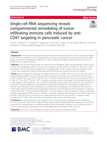
Pan et al. Journal of Hematology & OncologyFig. 2 (See legend on next page.)(2019) 12:124Page 7 of 18
Pan et al. Journal of Hematology & Oncology(2019) 12:124Page 8 of 18(See figure on previous page.)Fig. 2 Effect of CD47 blockade on the patient-derived PDAC xenograft models. a Schematic of patient-derived PDAC xenograft establishmentand tumor immunotherapy design. b Staining with an anti-CD47 antibody in two human PDAC tissue samples P962 and P989 at 100magnification and 400 magnification (upper panels). H&E and anti-CD47 antibody staining of tumor tissues from P962 and P989 patient-derivedxenograft models (P2 generation) at 100 magnification and 400 magnification (lower panels). Scale bar 50 μm (red line at the bottom left).c–h P962 and P989 xenografts from nude and NCG mice were treated intraperitoneally with IgG control or anti-CD47 mAb at 200 μg/day for 2weeks. Each group contained five animals. Tumor volume and weight were then measured (d, e, g, and h). (i–k) Panc02 cells were transplantedto C57BL/6 mice. When the tumor reached 100 mm3, tumor-bearing mice were treated with anti-CD47 antibodies for 14 days. Tumor volume andweight were then measured (j, k)implanting Panc02 cells to C57BL/6 mice. Using thismouse model, we found that treatment with anti-CD47alone resulted in significantly reduced tumor growthcompared to the untreated animals (Fig. 2i–k). Thesedata suggested that an intact immune system may be required for effective CD47 targeting to establish the immunotherapeutic effect.Composition of tumor-infiltrating immune cellsubpopulations identified by single-cell RNA-seqTo further understand the mouse immune cell subpopulations associated with anti-tumor response followinganti-CD47 treatment, we harvested the tumors on day15 following the anti-CD47 treatment and analyzed theCD45-positive immune cells by scRNA-seq with 10xGenomics pipeline (Additional file 1: Figure S1A and B).To better define the subpopulation structure of thetumor-infiltrating immune cells, we computationallypooled the data from the control and the anti-CD47group representing a total of 22,608 cells. We usedgraph-based clustering to identify transcriptional clustersconsisting of individual cell types (Fig. 3a). Comparisonwith the ImmGen database and assessment of knowncell-type markers resulted in elaboration of eight lymphoid clusters, five monocyte/macrophage clusters, threeneutrophil clusters, and three dendritic cell (DC) clusters(Fig. 3a–c). Following anti-CD47 treatment, the proportion of monocyte/macrophage populations were decreased, whereas the lymphoid populations wereincreased, including the proportions of CD4 T cells,CD8 T cells, and regulatory T cells (Tregs) (Fig. 3d–f).Using Immunohistochemical staining, we also found thatthe number and percent of CD4 T cells and CD8 Tcells were improved after anti-CD47 treatment(Additional file 1: Figure S2).To better understand and more accurately define thelymphoid clusters identified by single-cell RNA-seq, wecomputationally separated lymphoid cells (6117 cellstotal for two groups) and reanalyzed the data (Fig. 4a).This approach produced 13 distinct lymphoid clustersbroadly defined by the distribution of classical markergenes (Fig. 4b and Additional file 1: Figure S3). Clustersare named as “XXX s#”, where “XXX” represents thecell type, “s” represents the scRNA-seq, and “#” represents the different cluster.Changes in CD4 T cellsSingle-cell RNA-seq revealed two distinct clusters ofFoxP3 CD4 T cells (CD4 s1 and CD4 s2) (Fig. 4c).CD4 s1 and CD4 s2 expressed high levels of Cd4 andfunctional markers such as Lag3, Pdcd1 (PD-1), Ctla4,and Icos (Fig. 4b and Additional file 1: Figure S3). CD4s2 was distinguishable from CD4 s1 by its higher expression of CD44, Cd200, and Ccr7, and lower expression of Ccl5 and Lag3 (Additional file 1: Figure S3).KEGG revealed that both CD4 s1 and CD4 s2 displayedthe pathways associated with oxidative phosphorylationand Fc gamma R-mediated phagocytosis, and the signaling via T cell receptor, PD-1/PD-L1 checkpoint, TNF,HIF-1, and FoxO (Fig. 4g). Anti-CD47 treatment increased the percentage of total CD4 T cells (Fig. 3f),mostly by improving CD4 s1, while treatment of antiCD47 had little effect on CD4 s2 (Fig. 4d). Anti-CD47treatment not only changed the percentage of T cells inCD4 T cell clusters but also increased Pdcd1 expressionand decreased Ctla4 expression (Additional file 1: FigureS3). These data show that anti-CD47 therapy induces adramatic enhancement in the intratumoral CD4 T cells.Changes in T
Flow cytometry analysis To determine the proportion of PD-1 CD8 T cells in lymphocytes, the cells from the tumor, spleen, and per-ipheral blood of mouse syngeneic tumor models were stained with PD-1-FITC mAb and CD8a-PE mAb and performed on the BD Accuri C6 flow cytometer (BD Biosciences) as previously described [22]. Immunoblotting