Transcription
BMC PhysiologyBioMed CentralOpen AccessResearch articleRole of glucocorticoids in mediating effects of fasting and diabeteson hypothalamic gene expressionHideo Makimura1,2, Tooru M Mizuno1,2, Fumiko Isoda1,2, Joe Beasley2,3,Jeffrey H Silverstein2,3 and Charles V Mobbs*1,2Address: 1Fishberg Center for Neurobiology, Neurobiology of Aging Laboratories, Mount Sinai School of Medicine, One Gustave L. Levy Place,New York, New York, USA 10029, 2Department of Geriatrics and Adult Development, Mount Sinai School of Medicine, One Gustave L. Levy Place,New York, New York, USA 10029 and 3Department of Anesthesiology, Mount Sinai School of Medicine, One Gustave L. Levy Place, New York,New York, USA 10029Email: Hideo Makimura - Hideo.Makimura@mssm.edu; Tooru M Mizuno - Tooru.Mizuno@mssm.edu;Fumiko Isoda - Fumiko.Isoda@mssm.edu; Joe Beasley - Joseph.Beasley@msnyuhealth.org;Jeffrey H Silverstein - Jeff.Silverstein@msnyuhealth.org; Charles V Mobbs* - Charles.Mobbs@mssm.edu* Corresponding authorPublished: 09 July 2003BMC Physiology 2003, 3:5Received: 29 January 2003Accepted: 09 July 2003This article is available from: http://www.biomedcentral.com/1472-6793/3/5 2003 Makimura et al; licensee BioMed Central Ltd. This is an Open Access article: verbatim copying and redistribution of this article are permitted in allmedia for any purpose, provided this notice is preserved along with the article's original URL.AbstractBackground: Fasting and diabetes are characterized by elevated glucocorticoids and reducedinsulin, leptin, elevated hypothalamic AGRP and NPY mRNA, and reduced hypothalamic POMCmRNA. Although leptin replacement can reverse changes in hypothalamic gene expressionassociated with fasting and diabetes, leptin also normalizes corticosterone; therefore the extent towhich the elevated corticosterone contributes to the regulation of hypothalamic gene expressionin fasting and diabetes remains unclear. To address if elevated corticosterone is necessary forhypothalamic responses to fasting and diabetes, we assessed the effects of adrenalectomy onhypothalamic gene expression in 48-hour-fasted or diabetic mice. To assess if elevatedcorticosterone is sufficient for the hypothalamic responses to fasting and diabetes, we assessed theeffect of corticosterone pellets implanted for 48 hours on hypothalamic gene expression.Results: Fasting and streptozotocin-induced diabetes elevated plasma glucocorticoid levels andreduced serum insulin and leptin levels. Adrenalectomy prevented the rise in plasmaglucocorticoids associated with fasting and diabetes, but not the associated reductions in insulin orleptin. Adrenalectomy blocked the effects of fasting and diabetes on hypothalamic AGRP, NPY, andPOMC expression. Conversely, corticosterone implants induced both AGRP and POMC mRNA(with a non-significant trend toward induction of NPY mRNA), accompanied by elevated insulin andleptin (with no change in food intake or body weight).Conclusion: These data suggest that elevated plasma corticosterone mediate some effects offasting and diabetes on hypothalamic gene expression. Specifically, elevated plasma corticosteroneis necessary for the induction of NPY mRNA with fasting and diabetes; since corticosteroneimplants only produced a non-significant trend in NPY mRNA, it remains uncertain if a rise incorticosterone may be sufficient to induce NPY mRNA. A rise in corticosterone is necessary toreduce hypothalamic POMC mRNA with fasting and diabetes, but not sufficient for the reductionof hypothalamic POMC mRNA. Finally, elevated plasma corticosterone is both necessary andsufficient for the induction of hypothalamic AGRP mRNA with fasting and diabetes.Page 1 of 13(page number not for citation purposes)
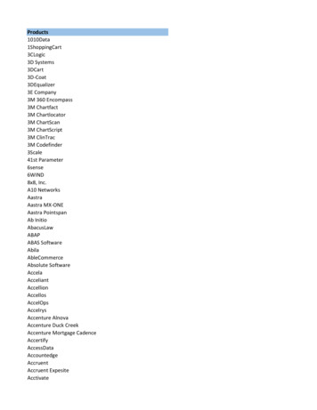
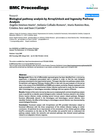
BMC Physiology 2003, undResults and DiscussionReduction in hypothalamic melanocortin tone, caused byreduction in proopiomelanocortin (POMC) mRNA andelevation in agouti-related peptide (AGRP) mRNA, isthought to play a key role in mediating nutritional effectson neuroendocrine function [1,2]. Thus fasting [3,4],insulin deficiency [5], and leptin deficiency [4] are allassociated with reduced hypothalamic melanocortintone. However, factors that regulate hypothalamicmelanocortin tone are not fully elucidated. Since bothfasting and insulin deficiency are associated with reducedplasma leptin [5], and leptin replacement reverses effectsof fasting [6] and also effects of insulin deficiency on foodintake and body weight [7], it has been suggested that thereduction in leptin accounts for the reduced hypothalamic melanocortin tone in these conditions. However,fasting can influence hypothalamic gene expression ininsulin-deficient mice independent of changes in plasmaleptin or insulin [5], as well as in leptin resistant db/dbmice [3]. These data suggest that other factors in additionto leptin may mediate effects of fasting. Since fasting,insulin deficiency, and leptin insufficiency also lead to anelevation in plasma glucocorticoids [8], and adrenalectomy of leptin-deficient ob/ob mice largely normalizeshypothalamic gene expression in these mice [9], it is plausible that elevated glucocorticoids may mediate someeffects of nutritional state on hypothalamic gene expression. This hypothesis is particularly compelling sincerecent data suggest that aberrant glucocorticoid activitymay play a key role in the development of the metabolicsyndrome [10].Study 1: Effects of adrenalectomy on hypothalamicresponses to fastingFasting reduced body weight and adipose weight in boththe sham-operated and adrenalectomized groups asexpected (Table 1). Blood glucose was also decreased byfasting in both the sham-operated group and adrenalectomized group (Table 1). Serum corticosterone concentration increased with fasting as expected in the shamoperated mice (Fig. 1A); adrenalectomy blocked this fasting-induced elevation of corticosterone (Fig. 1A). Therefore the adrenalectomized fasted mice exhibited similarlevels of corticosterone as both the adrenalectomized adlib fed mice and the sham-operated ad lib fed mice (Fig.1A). As we noted previously, a basal level of corticosterone was detectable in the adrenalectomized mice aftersurgery [9]. In contrast to corticosterone, other humoralfactors were not significantly affected by adrenalectomy.Serum insulin and serum leptin both exhibited the samedecreases in the adrenalectomized fasted and sham-operated fasted animals compared to the adrenalectomized adlib fed and sham-operated ad lib fed mice (Fig. 1B and1C); therefore the levels of these humoral factors were thesame in the sham-operated fasted mice and the adrenalectomized fasted mice.Glucocorticoids have previously been reported to increasebody weight set point, as determined by hoarding behavior in rats [11], while adrenalectomy has been reported tolower body weight set point in rats [12]. In addition, central infusion of glucocorticoids leads to increases in bodyweight and food intake with a concomitant induction ofhypothalamic NPY [13]. Nevertheless, it remains unclearif an elevation in glucocorticoid concentration contributes to the effect of fasting on hypothalamic gene expression. Adrenalectomy has been reported to blockelevations in NPY mRNA in response to fasting [14]. Onthe other hand, it has been reported that elevation ofplasma corticosterone levels is not required for the induction of NPY mRNA by fasting [15]. In addition, the extentto which hypothalamic AGRP and POMC mRNA dependson elevated glucocorticoid levels in fasting and insulindeficiency has not been determined to date. Therefore inthe present study we assessed if an elevation of plasmacorticosterone is necessary and sufficient to mediateeffects of fasting and insulin deficiency on hypothalamicmelanocortin tone, as indicated by expression of hypothalamic AGRP and POMC mRNA.Fasting increased hypothalamic AGRP mRNA as expectedin the sham-operated fasted group, and adrenalectomycompletely blocked the induction of AGRP by fasting (Fig.2A). Adrenalectomy also produced a trend to decreaseAGRP mRNA in the ad-lib fed mice but this effect was notsignificant (Fig. 2A). Fasting decreased hypothalamicPOMC mRNA as expected in the sham-operated fastedgroup, but fasting had no effect on POMC mRNA in theadrenalectomized animals (Fig. 2B). As with AGRP, NPYmRNA was also elevated after fasting, and this effect offasting on NPY mRNA was also completely blocked byadrenalectomy (Fig 2C). Adrenalectomy therefore completely blocked the effects of fasting on hypothalamicAGRP, POMC and NPY mRNAs.Study 2: Effects of adrenalectomy on hypothalamicresponses to insulin deficiency and fastingStreptozotocin injection induced diabetes in sham-operated mice (Table 2). Interestingly, adrenalectomy attenuated the rise in blood glucose in streptozotocin-injectedmice (Table 2). Fasting reduced blood glucose in the ADXSTZ-Fast group below even that of control mice (Table 2).Streptozotocin injection led to reduced body weight andadiposity in sham-operated animals as previouslydescribed [5,17] (Table 2), whereas adrenalectomy attenuated the loss of both body weight and adiposity associated with insulin deficiency (Table 2). Streptozotocininjection also led to significant hyperphagia, typical ofPage 2 of 13(page number not for citation purposes)
BMC Physiology 2003, costerone(ng/ml)5004003002001000Fed FastIntactFed FastADXB5Insulin(ng/mL)4321*0Fed FastIntact*Fed FastADXCLeptin peptide(ng/ml)4.03.02.01.0*0Fed FastIntact*Fed FastADXFigureof1 fasting on blood hormones in intact and adrenalectomized miceEffectsEffects of fasting on blood hormones in intact and adrenalectomized mice. (A) Fasting induces serum corticosterone in intact mice but not in adrenalectomized mice. Fasting decreases both serum insulin (B), and serum leptin (C) to thesame levels in intact and adrenalectomized mice. Data are expressed as mean SEM. Groups with different letters are statistically different (p 0.05), reflecting ANOVA followed by Tukey-Kramer post hoc tests comparing every group to every othergroup. Thus groups with the same letter do not differ from each other at a p 0.05 level.Page 3 of 13(page number not for citation purposes)
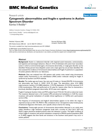
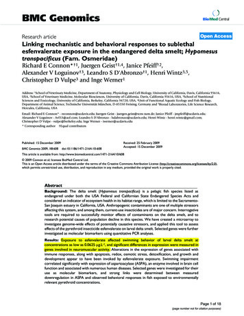
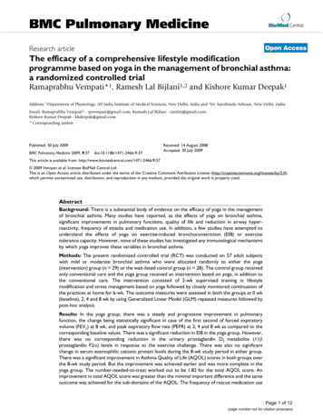
BMC Physiology 2003, 3http://www.biomedcentral.com/1472-6793/3/5Table 1: Effects of fasting on body weight, adipose weight and glucose in intact and adrenalectomized mice.GroupnBody weight (g)Adipose weight (g)Blood glucose (mg/dl)Intact/Ad lib fedIntact/FastedADX/Ad lib fedADX/Fasted6610922.8 0.8a16.1 0.4b23.6 0.4a19.5 0.7b0.14 0.04a0.02 0.002b0.22 0.02a0.04 0.006b108.8 15.1a60.3 4.3b124.1 6.6a52.6 4.8bData are expressed as mean SEM. Groups with different letters are statistically different (p 0.05), reflecting ANOVA followed by Tukey-Kramerpost hoc tests comparing every group to every other group. Thus groups with the same letter do not differ from each other at a p 0.05 level.diabetic mice [17] (Table 2), and adrenalectomy alsoameliorated this response to insulin deficiency (Table 2).Serum corticosterone was elevated in the Sham-STZ groupand was decreased in both ADX-STZ and ADX-STZ-Fastgroups (Fig. 3A). Adrenalectomy did not result in complete elimination of corticosterone but instead resulted inlow but detectable levels of serum corticosterone as previously noted [9] and described above. Serum insulin wasreduced to the same extent in Sham-STZ, ADX-STZ, and inADX-STZ-Fast groups, compared to control mice, aswould be expected with streptozotocin injection (Fig. 3B).Serum leptin was also reduced to the same levels in allexperimental groups compared to control mice (Fig. 3C).Hypothalamic AGRP mRNA was significantly elevated insham-operated diabetic (vs. sham-operated euglycemic)mice as expected [5,17] (Fig. 4A). However, adrenalectomy completely blocked the elevation of AGRP mRNA indiabetic mice whether or not they were fasted (Fig. 4A).Hypothalamic POMC mRNA was reduced in diabeticmice as expected [5,17] (Fig. 4B). Adrenalectomy blockedthis reduction in POMC mRNA in diabetic mice whetheror not they were fasted (Fig. 4B). As with AGRP mRNA,NPY mRNA was elevated in diabetic mice, and adrenalectomy blocked this induction of NPY mRNA in diabeticmice whether or not they were fasted (Fig. 4C). In summary, hypothalamic AGRP, POMC and NPY mRNA allexhibited the expected changes in association with streptozotocin-induced diabetes [5,17] (Fig. 4A,4B,4C), andadrenalectomy largely or completely blocked all theseresponses to insulin deficiency whether or not the micewere fasted (Fig 4A,4B,4C).Study 3: Effects of corticosterone implants onhypothalamic gene expressionImplantation of corticosterone implants for 48 hours significantly increased serum corticosterone, compared toplacebo-implanted mice, as expected (Fig. 5A). Interestingly, the corticosterone implants also significantlyincreased serum insulin and leptin (Fig. 5B and 5C). Incontrast, the corticosterone implants did not significantlyinfluence body weight or food intake compared to pla-cebo-implanted mice (body weight of 24.5 0.6 g for theplacebo group compared to 24.4 0.6 g for the corticosterone implant group; food intake of 4.3 0.1 g/day forthe placebo group compared to 4.1 0.5 g/day for the corticosterone implant group), although epididymal adiposetissue depot was slightly smaller in the corticosteroneimplant group (0.19 0.01 g for the placebo group compared to 0.15 0.01 g for the corticosterone implantgroup; p 0.05). Corticosterone implants also had noinfluence on blood glucose (134.8 10.9 mg/dl for theplacebo group compared to 128.8 4.3 mg/dl for the corticosterone implant group).Implantation of corticosterone implants for 48 hours significantly elevated hypothalamic AGRP expression compared to the placebo group (Fig. 6A). HypothalamicPOMC mRNA was also significantly elevated in the corticosterone implant group (Fig. 6B). Hypothalamic NPYmRNA exhibited a trend toward an increase although theeffect was not significant (p 0.14; Fig. 6C). Thereforehypothalamic AGRP and POMC mRNAs were bothinduced by the glucocorticoid implant, whereas the NPYmRNA was not significantly influenced.ConclusionsIn the present study, adrenalectomy and corticosteroneimplants were used to address the role of elevated corticosterone in mediating the effects of fasting and insulindeficiency on hypothalamic gene expression, resulting inseveral novel observations. First, we have demonstratedthat adrenalectomy can block the elevation of AGRPmRNA and the reduction in POMC mRNA with fasting,consistent with previous findings by Ponsalle et al. concerning NPY [14], and suggesting that increases in ocortin tone. Second, we have also shown thathypothalamic responses to insulin deficiency require elevation in corticosterone. Third, we have demonstratedusing corticosterone implants, that elevated corticosterone is sufficient to enhance hypothalamic AGRP expression, but not to reduce hypothalamic POMC mRNA.Together these results suggest that an elevation of corticosterone is necessary for reduction in melanocortin tonePage 4 of 13(page number not for citation purposes)
BMC Physiology 2003, 3http://www.biomedcentral.com/1472-6793/3/5A.*AGRP mRNA(% Control)5004003002001000B.Fed FastIntactFed FastADX140POMC mRNA(% Control)120100806040*200C.300Fed FastIntactFed FastADX*NPY mRNA(% Control)250200150100500Fed FastIntactFed FastADXFigureof2 fasting on hypothalamic gene expression in intact and fasted miceEffectsEffects of fasting on hypothalamic gene expression in intact and fasted mice. (A) Hypothalamic AGRP is induced byfasting in intact mice however the fasting associated induction is blocked by adrenalectomy. (B) Fasting reduces hypothalamicPOMC mRNA in intact mice but not in adrenalectomized mice. (C) Similar to AGRP, hypothalamic NPY mRNA is induced inintact mice but not in adrenalectomized mice. Data are expressed as mean SEM. Groups with different letters are statisticallydifferent (p 0.05), reflecting ANOVA followed by Tukey-Kramer post hoc tests comparing every group to every other group.Thus groups with the same letter do not differ from each other at a p 0.05 level.Page 5 of 13(page number not for citation purposes)
BMC Physiology 2003, 3http://www.biomedcentral.com/1472-6793/3/5Table 2: Effects of adrenalectomy, diabetes and fasting on body weight, adipose weight, food intake and glucose.GroupnBody weight (g)Adipose weight (g)Food intake (g/day)Glucose (mg/dl)ControlSham/STZADX/STZADX/STZ/Fast9811524.3 0.5a17.8 0.8b21.5 0.9c19.0 0.3b0.20 0.01a0.02 0.01b0.07 0.02c0.03 0.01c4.3 0.1a6.1 0.6b4.8 0.1cN/A138.1 6.5a578.5 17.1b368.8 20.4c44.8 13.3dData are expressed as mean SEM. Groups with different letters are statistically different (p 0.05), reflecting ANOVA followed by Tukey-Kramerpost hoc tests comparing every group to every other group. Thus groups with the same letter do not differ from each other at a p 0.05 level.during fasting and with diabetes and is sufficient toinduce hypothalamic AGRP mRNA but not to reducePOMC expression.Although a reduction in plasma leptin plays an importantrole in mediating neuroendocrine responses to fasting [6]and diabetes [17], we have previously demonstrated thatfasting can regulate hypothalamic melanocortin toneindependently of both leptin and insulin [5]. The presentstudies suggest that corticosterone may play a role inmediating these leptin-independent effects. Fasting wasassociated with elevated corticosterone (Fig. 1A) andreduced insulin and leptin (Fig. 1B and 1C) as well asmelanocortin tone (Fig. 2A and 2B) as expected. However,preventing the elevation of corticosterone completelyblocked the effect of fasting on hypothalamic AGRP andPOMC mRNA (Fig. 2A and 2B), without preventing effectsof fasting on plasma leptin or insulin. Adrenalectomy alsoblocked the effects of fasting on hypothalamic NPYmRNA (Fig. 2C), confirming the findings of Ponsalle et al.[14]. These data suggest that leptin's effect to reverseeffects of fasting on melanocortin tone may require andbe mediated by a reduction in glucocorticoids.Glucocorticoids were also elevated, as expected, in insulin-deficient diabetes (Fig. 3A), and serum insulin, leptinand melanocortin tone were also reduced in this condition (Fig. 3B and 3C, 4A and 4B). As with fasting, thereduction in plasma leptin could largely be a reflection ofthe reduction in adipose weight, as well as plasma insulin.In turn, as with fasting, the reduction of plasma leptincould be the cause of the elevation of hypothalamic AGRPand NPY mRNA, as well as the reduction of hypothalamicPOMC mRNA. On the other hand, blocking the rise incorticosterone by adrenalectomy prevented the reductionin hypothalamic POMC mRNA and the induction ofAGRP mRNA, without reversing either the reduced leptinor the reduced adiposity, characteristic of insulin-deficientdiabetic mice (Fig. 4A and 4B). These data suggest thatreversal of effects of insulin deficiency by leptin [7] mayrequire and be mediated by a reduction in glucocorticoids. We had previously shown that fasting could regulate hypothalamic gene expression in insulin-deficientmice even though in these mice fasting did not influenceplasma leptin or insulin [5]. Since adrenalectomy blockedthe fasting-induced reduction in melanocortin tone indiabetic mice (as it had in euglycemic mice in the firststudy), these results suggest that effects of fasting onmelanocortin tone in diabetic mice may be mediated atleast in part by a rise in plasma corticosterone (itself possibly secondary to the reduction in plasma leptin).Implanting corticosterone implants for 48 hours led to anelevation in both hypothalamic AGRP and POMC mRNA(Fig. 6B). Since adrenalectomy blocked the elevation ofAGRP mRNA by fasting and insulin deficiency, elevationof corticosterone would be expected to increase hypothalamic AGRP mRNA, as was indeed observed. However, thecorticosterone implants produced the unexpected effect ofelevating hypothalamic POMC mRNA, a rare if notunique circumstance in which hypothalamic AGRP andPOMC mRNA are regulated in the same direction. Thebasis of this unexpected phenomenon is not yet clear.However, since leptin and insulin both stimulate hypothalamic POMC [18], the elevation of insulin and leptin inthe corticosterone-implanted mice (Fig. 5B and 5C), theformer possibly mediated by glucocorticoid-inducedinsulin resistance and the latter possibly mediated by theresulting hyperinsulinemia, might mediate the observedeffect of corticosterone implants on hypothalamic POMCin these implanted mice. On the other hand, leptin inhibits hypothalamic AGRP mRNA [4], yet AGRP mRNA wasnevertheless elevated in the face of elevated insulin andleptin. These data suggest that in fasting and insulin-deficient diabetes, as in leptin-deficient ob/ob mice [9,19], glucocorticoids dominate leptin and insulin levels indetermining AGRP expression, whereas insulin and leptindominate glucocorticoids in determining hypothalamicPOMC expression. These data are also consistent with theobservation that in streptozotocin-induced diabetic rats,replacement of insulin is not sufficient to normalize elevated serum corticosterone or the elevated hypothalamicAGRP mRNA while it does partially normalize plasmaleptin and hypothalamic POMC mRNA [17].Page 6 of 13(page number not for citation purposes)
BMC Physiology 2003, ticosterone(ng/ml)b300200a100acc0B.STZADXFast- - - 3.0aInsulin(ng/ml)2.52.01.51.0b0.50C.bSTZADXFast - - a1.6Leptin peptide(ng/ml)-b1.2b0.80.4bb0STZADXFast- - - Figureof3 adrenalectomy, streptozotocin injection and fasting on levels of blood hormonesEffectsEffects of adrenalectomy, streptozotocin injection and fasting on levels of blood hormones. (A) Streptozotocininjection elevates levels of serum corticosterone however adrenalectomy prevents the induction associated with streptozotocin. Both serum insulin (B), and serum leptin (C) are reduced to the same extent in all experimental conditions. Data areexpressed as mean SEM. Groups with different letters are statistically different (p 0.05), reflecting ANOVA followed byTukey-Kramer post hoc tests comparing every group to every other group. Thus groups with the same letter do not differfrom each other at a p 0.05 level.Page 7 of 13(page number not for citation purposes)
BMC Physiology 2003, RP mRNA(%)A8006004002000BaSTZADXFast- -POMC mRNA(%)160cac - aa120a80b400CSTZADXFast-1400 - - b1200NPY mRNA(%)1000800600bc400ca2000STZADXFast- - - Figureof4 adrenalectomy, streptozotocin injection and fasting on levels of hypothalamic gene expressionEffectsEffects of adrenalectomy, streptozotocin injection and fasting on levels of hypothalamic gene expression. (A)Hypothalamic AGRP mRNA is induced by streptozotocin injection, however the induction is partially blocked by adrenalectomy. (B) Hypothalamic POMC mRNA is reduced by streptozotocin injection, but adrenalectomy completely blocks thereduction associated with streptozotocin. (C) Similar to AGRP, NPY mRNA is induced by streptozotocin injection and partiallyblocked by adrenalectomy. Data are expressed as mean percentage SEM of the control group. Data are expressed as mean SEM. Groups with different letters are statistically different (p 0.05), reflecting ANOVA followed by Tukey-Kramer post hoctests comparing every group to every other group. Thus groups with the same letter do not differ from each other at a p 0.05 level.Page 8 of 13(page number not for citation purposes)
BMC Physiology 2003, orticosterone(ng/ml)12008004000Control ImplantB40*Insulin(ng/ml)3020100Control ImplantCLeptin peptide(ng/ml)16*12840Control ImplantFigureof5 corticosterone implants on levels of blood hormonesEffectsEffects of corticosterone implants on levels of blood hormones. Corticosterone implants elevates levels of serum corticosterone (A), insulin (B), and leptin (C). Statistical significance was determined by unpaired student's t-test (p 0.05) and isdenoted by *.Page 9 of 13(page number not for citation purposes)
BMC Physiology 2003, P mRNA(%)300*250200150100500Control ImplantB350POMC mRNA(%)300*250200150100500Control ImplantCNPY mRNA(%)2001000Control ImplantFigureof6 corticosterone implants on hypothalamic gene expressionEffectsEffects of corticosterone implants on hypothalamic gene expression. Corticosterone implants induces both hypothalamic AGRP (A) and POMC (B) mRNA. (C) NPY mRNA is also elevated by corticosterone implants but the effect did notreach statistical significance. Data are expressed as a mean percentage SEM of placebo implant group. Statistical significancewas determined by unpaired student's t-test (p 0.05) and is denoted by *.Page 10 of 13(page number not for citation purposes)
BMC Physiology 2003, 3After the present paper was submitted for review, webecame aware of a recent study by Savontaus et al. examining the role of glucocorticoids in the regulation ofhypothalamic gene expression in rats [20], using aroughly similar experimental design as in the presentstudy. In several respects the conclusions from this studyare similar to the present study (for example, corticoisterone implants increased hypothalamic AGRP and POMCmRNA, and adrenalectomy blocked the effect of fastingon hypothalamic POMC mRNA). However, in one keyrespect the results reported by Savontaus et al. differedfrom those in the present paper: adrenalectomy failed toblock the effects of fasting on hypothalamic AGRP andNPY, even though corticosterone implants were observedto induce AGRP mRNA. Since we observed that adrenalectomy also blocks the effects of diabetes on hypothalamicgene expression in the present study, we suspect the difference between the present study and that of Savontaus etal. is probably due to different regulatory requirements inmice and rats. For example, it is clear that the regulationof AGRP differs in rats and mice, since in mice hypothalamic AGRP mRNA is elevated by a deficiency in leptinreceptor function [4] but in rats this is not true [21]. However, hypothalamic POMC is regulated similarly in ratsand mice, and this is probably the more important element regulating metabolic phenotype.Taken together, these data suggest that elevated glucocorticoids are sufficient to drive the increase in hypothalamicAGRP mRNA during fasting and diabetes but not thereduction in POMC mRNA. Interestingly, at least withrespect to food intake and body weight over this 48-hourperiod, the elevation of POMC appears to compensate forthe elevation in AGRP to produce no net functional effecton body weight homeostasis in the presence ofsignificantly elevated plasma corticosterone. After morechronic exposure, this delicate balance could plausiblybecome disrupted, leading to glucocorticoid-mediateddevelopment of the metabolic syndrome [10]. Thus interactions between insulin, leptin, and glucocorticoids, andthe neuropeptide responses they generate within themelanocortin system, allow complex metabolic responsesto nutritional status but may also allow the developmentof metabolic pathologies.MethodsAnimalsThe appropriate Institutional Animal Review Board hadapproved all studies.To study the effect of adrenalectomy on fasting-inducedchanges in hypothalamic gene expression (Study 1), twomonth-old male C57Bl/6J mice were obtained from TheJackson Laboratory (Bar Harbor, ME). Mice were individually housed with free access to food and water 12 h light-dark cycle (lights on at 07:00 h). Of the 31mice, 19 mice were adrenalectomized and the rest underwent a sham operation. Adrenalectomy was performed, asdescribed previously, by bilateral flank incision underanesthesia (2.5% 2-2-2-tribromoethanol, 0.015–0.017ml/g body weight, i.p.) [9]. Sham adrenalectomy entailedthe same procedure as adrenalectomy, except that theadrenal glands were grasped but not removed. After thesurgery all mice were given one injection of dexamethasone (45 mg/kg b.w. i.p.) to facilitate recovery from surgery. The drinking water was replaced with normal saline(0.9% NaCl) to compensate for the loss of mineralocorticoids. Body weight was monitored for one week followingthe operation at which time it was verified that the animals had recovered initial body weight as expected. Allmice were placed in fresh cages with new bedding, andfood was removed from half of the adrenalectomizedmice and half of the sham operated mice for 48 hours.This resulted in four groups; sham ad lib fed (n 6), sham48 hour fast (n 6), adrenalectomized ad lib fed (n 10)and adrenalectomized 48 hour fast (n 9).To study the effects of adrenalectomy, insulin deficiencyand fasting (Study 2) 33 male C57Bl/6J mice wereobtained at 2 months of age from The Jackson Laboratory(Bar Harbor, ME) and were individually housed with freeaccess to feed and water under 12:12 h light-dark cycle asdescribed above. Mice were treated with daily injections ofdexamethasone (45 mg/kg body weight i.p.) for threeweeks during which time they were eitheradrenalectomized or sham operated as described above.Half the sham-operated and all the adrenalectomizedmice were injected with streptozotocin 10 days after thesurgery (160 mg/kg body weight; freshly purchased fromSigma, St. Louis, MO and dissolved in 25 mM Citric AcidBuffer pH 4.5) while the rest received buffer alone by tailvein injection 10 days after surgery. Dexamethasonetreatment continued until four days after the streptozotocin injection. Mice were then monitored for two weekswithout the dexamethasone injection at which point halfthe adrenalectomized-STZ treated group were fasted whilethe rest of the mice were ad lib fed. This result
BioMed Central Page 1 of 13 (page number not for citation purposes) BMC Physiology Research article Open Access Role of glucocorticoids in mediating effects of fasting and diabetes on hypothalamic gene expression Hideo Makimura 1,2, Tooru M Mizuno , Fumiko Isoda1,2, Joe Beasley2,3, Jeffrey H Silverstein2,3 and Charles V Mobbs*1,2