Transcription
(2022) 22:234Nerdal et al. BMC 5Open AccessRESEARCHThe relationship of acute deliriumwith cognitive and psychiatric symptomsafter stroke: a longitudinal studyVilde Nerdal1†, Elise Gjestad1,2†, Ingvild Saltvedt3,4, Ragnhild Munthe‑Kaas5,6, Hege Ihle‑Hansen7,8, Truls Ryum1,Stian Lydersen9 and Ramune Grambaite1,2,10*AbstractObjective: Delirium, a common complication after stroke, is often overlooked, and long-term consequences arepoorly understood. This study aims to explore whether delirium in the acute phase of stroke predicts cognitive andpsychiatric symptoms three, 18 and 36 months later.Method: As part of the Norwegian Cognitive Impairment After Stroke Study (Nor-COAST), 139 hospitalized strokepatients (49% women, mean (SD) age: 71.4 (13.4) years; mean (SD) National Institutes of Health Stroke Scale (NIHSS)3.0 (4.0)) were screened for delirium with the Confusion Assessment Method (CAM). Global cognition was measuredwith the Montreal Cognitive Assessment (MoCA), while psychiatric symptoms were measured using the HospitalAnxiety and Depression Scale (HADS) and the Neuropsychiatric Inventory-Questionnaire (NPI-Q). Data was analyzedusing mixed-model linear regression, adjusting for age, gender, education, NIHSS score at baseline and premorbiddementia.Results: Thirteen patients met the criteria for delirium. Patients with delirium had lower MoCA scores compared tonon-delirious patients, with the largest between-group difference found at 18 months (Mean (SE): 20.8 (1.4) versus(25.1 (0.4)). Delirium was associated with higher NPI-Q scores at 3 months (Mean (SE): 2.4 (0.6) versus 0.8 (0.1)), andhigher HADS anxiety scores at 18 and 36 months, with the largest difference found at 36 months (Mean (SE): 6.2 (1.3)versus 2.2 (0.3)).Conclusions: Suffering a delirium in the acute phase of stroke predicted more cognitive and psychiatric symptomsat follow-up, compared to non-delirious patients. Preventing and treating delirium may be important for decreasingthe burden of post-stroke disability.Keywords: Mental status and dementia tests, Cognitive dysfunction, Anxiety, Depression, Cerebrovascular event,Confusion†Vilde Nerdal and Elise Gjestad are joint first authorship.*Correspondence: ramune.grambaite@ntnu.no10Health Services Research Unit (HØKH), Akershus University Hospital,Lørenskog, NorwayFull list of author information is available at the end of the articleStroke is ranked the third largest contributor to deathand disability adjusted life years worldwide [1]. Cognitiveimpairment and psychiatric symptoms are prevalent inboth the acute and chronic phases of stroke [2, 3]. Thesesymptoms can interfere with restoration of daily functionand independent living [4], placing emotional and economical burdens on patients, their families, and society[5, 6]. Identifying risk factors for post-stroke cognitive The Author(s) 2022. Open Access This article is licensed under a Creative Commons Attribution 4.0 International License, whichpermits use, sharing, adaptation, distribution and reproduction in any medium or format, as long as you give appropriate credit to theoriginal author(s) and the source, provide a link to the Creative Commons licence, and indicate if changes were made. The images orother third party material in this article are included in the article’s Creative Commons licence, unless indicated otherwise in a credit lineto the material. If material is not included in the article’s Creative Commons licence and your intended use is not permitted by statutoryregulation or exceeds the permitted use, you will need to obtain permission directly from the copyright holder. To view a copy of thislicence, visit http:// creat iveco mmons. org/ licen ses/ by/4. 0/. The Creative Commons Public Domain Dedication waiver (http:// creat iveco mmons. org/ publi cdoma in/ zero/1. 0/) applies to the data made available in this article, unless otherwise stated in a credit line to the data.
Nerdal et al. BMC Neurology(2022) 22:234impairment and psychiatric symptoms is crucial for correct preventive measures and treatment.The first 7 days of stroke, often referred to the acutephase [7], has key implications for long-term outcomes [8, 9], and complications during this phase hasbeen shown to increase the risk of post-stroke sequelae [10]. Delirium is a common complication in acutestroke and has been suggested as a potential risk factorfor later dependency and dementia [2, 11]. The Diagnostic and Statistical Manual of Mental Disorders, 5thedition (DSM-5), defines delirium as an acute and fluctuating disturbance of attention, cognition and/or consciousness, which occurs due to medical conditions andcannot be better explained by a pre-existing neurocognitive disorder [12]. Studies have found a prevalence ofdelirium in the acute phase of stroke ranging from 8 to48% [13]. A Norwegian stroke unit found that 10% ofstroke patients had delirium [14].Delirium often occurs as a response to brain injury[15], such as stroke [16], but the etiology is poorlyunderstood. The lack of knowledge of causal mechanisms of delirium, combined with its fluctuating andheterogeneous nature, can challenge detection of thecondition. The gold standard for diagnosing delirium isa clinical evaluation by professionals using the DSM-5criteria [17]. However, briefer screening tools are usually more feasible in acute settings. The ConfusionAssessment Method (CAM) [18] is a screening toolfound noninferior to diagnosis made by trained neurologists using the DSM-5 criteria, with sensitivity of 76%and specificity of 98% [19].Experiencing delirium can be stressful and emotionally challenging for the patient [20]. Stroke patients withdelirium have higher mortality rates and longer hospitalstays [21]. Furthermore, the condition increases the riskof falls and hospital acquired infections [22]. It predisposes for worse functional outcomes [23, 24] and lessfunctional independence by the time of discharge [21].Studies have found delirium to be associated withpost-stroke cognitive impairment for up to 2 years afterstroke [25]. However, the literature is inconsistent, andother studies have found the negative effect of deliriumdiminishing after 3 months [26] and after 12 months[27]. As neither of the previous studies adjusted for prestroke dementia or stroke severity, the effect of deliriumon cognition and psychiatric symptoms is left somewhatunclear.Fleischmann et al. [17] theorizes that delirium may hinder the standard course of stroke rehabilitation, not justby delaying physical training, but also by interfering withcognitive interventions. This further highlights the relevance of examining whether the condition is associatedwith poorer long-term cognitive outcomes.Page 2 of 12Several of the clinical consequences of delirium, such aslonger hospitalization and poorer functional outcomes,are also associated with increased risk of developing psychiatric symptoms, such as depression [28, 29]. However,recent studies have mainly addressed post-stroke depression in general stroke populations, leaving the impactsof delirium largely unexplored. Further, there is an evengreater absence of research on how delirium is linked toa broader range of psychiatric symptoms, such as anxiety, which often overlaps with depressive symptoms [30].This study aims to explore whether delirium in the acutephase of stroke predicts global cognitive function, as wellas symptoms of depression, anxiety, and general psychiatric distress over the course of 3 years.MethodParticipantsThe present sub-study is a part of the Norwegian Cognitive Impairment After Stroke Study (Nor-COAST). NorCOAST is a longitudinal multicenter, prospective cohortstudy that recruited 815 participants from five Norwegian hospitals from May 2015 to March 2017 [31]. Theinclusion criteria were diagnosis of acute stroke, admittance to hospital within 7 days of symptom onset, ageover 18 years, and fluency in a Scandinavian language.Stroke was diagnosed according to the World HealthOrganization criteria or with findings of acute intra-cerebral hemorrhage or infarction on Magnetic ResonanceImaging (MRI). The only exclusion criterion was a lifeexpectancy of less than 3 months.Our sub-study included only patients from BærumHospital, Vestre Viken Health Trust, excluding patientsfrom the other four hospitals. This hospital implementeda consistent regime for delirium screening, ensuring thatall patients were screened regularly during the first 2 daysafter admittance.Aphasia was not an exclusion criterion in the NorCOAST study, nor in this particular study. Patients withaphasia were however automatically excluded fromMoCA assessments by research nurses, but not forHADS or NPI-Q assessments.Premorbid function and health historyHealth history was collected from medical records, andinterviews with either the patient or the caregiver wereused to register premorbid function, including TheGlobal Deterioration Scale (GDS) [32] and the CharlsonComorbidity Index [33]. The Global Deterioration Scale(GDS) [32] was used to assess cognitive function beforethe stroke, and after three, 18 and 36 months. Pre-strokeGDS was assessed by the local principal investigator atinclusion based on interview of the patients, the proxyand medical records. GDS has seven stages, where 4–7
Nerdal et al. BMC Neurology(2022) 22:234are considered dementia stages and 1–3 are consideredpre-dementia stages, with stage 3 being equivalent tomild cognitive impairment (MCI) [34]. The CharlsonComorbidity Index [33] was used to classify the extentof comorbid diseases as mild (0–2), moderate [3–5] orsevere ( 5), and was registered upon admission.Stroke characteristics and complicationsStroke severity was measured using the National Institutes of Health Stroke Scale (NIHSS) [35] at day one ofhospitalization. NIHSS has 15 items measuring specificsymptoms on a 3- or 4-point ordinal scale (0 no impairment). The highest score possible for non-comatosepatients is 42. The NIHSS item measuring language difficulties was also used to identify patients with moderate and severe aphasia (i.e., causing interference withconversation). Further, complications during stroke weredefined as the number of infections, falls, progressionin stroke symptoms and seizures during hospitalization,which were then pooled into a sum score and recordedupon discharge.DeliriumDelirium was diagnosed using CAM [18]. Screeningswere performed by nurses specialized in stroke care onceduring every shift during the first 48 hours of hospitalization, resulting in a total of six screenings per patient.The majority of patients were screened at fixed intervals (7:00, 15:00 and 21:00). Delirium is defined basedon four features in CAM: 1) the acute onset of fluctuations or changes in the mental status of the patient, 2)inattention, 3) disorganized thinking, and 4) an alteredlevel of consciousness. To increase the sensitivity of theinattention item, a task was added, asking the patients toname the days of the week backward [36–38]. The diagnosis of delirium required the presence of feature 1 and2, plus feature 3 or 4. Feature 2 was considered presenteither if indicated by the inattention item in CAM, or ifthe patient named less than 7 days backwards and aphasiawas not indicated. Screening with CAM was integratedin the clinical observations of the patients. The patient’sfamilies were not involved in the CAM screening. Prestroke GDS-score and study notes on pre-stroke history provided a reference point for the patient’s normalbehavior.Cognitive outcomeGlobal cognitive function was measured using the Montreal Cognitive Assessment (MoCA) [39]. MoCA assessesseveral cognitive domains including executive function,memory, language, visuospatial abilities, attention andworking memory. The maximum score is 30, with higherscores indicating better cognition. Scores of 26 and abovePage 3 of 12are considered normal cognitive function [39]. Patientswere tested at baseline and after three, 18 and 36 months.Baseline assessment of cognitive function was performedeither at discharge, or at day seven for patients withlonger stays. Follow-up assessments were performed byresearch nurses and physicians specializing in stroke atan outpatient clinic. If the patients were unable to attendthe clinic, follow-up was completed by telephone.Psychiatric outcomesThe Hospital Anxiety and Depression Scale (HADS) [40]was used to assess symptoms of anxiety and depression.HADS consists of 14 items assessing two subscales (anxiety: HADS-A and depression: HADS-D). The items arerated on a 4-point scale from 0 (not at all) to 3 (most ofthe time), and a score of 8 or more on either subscalemay indicate clinically relevant symptoms [41]. The Neuropsychiatric Inventory-Questionnaire (NPI-Q) [42] wasused to assess symptoms according to 12 neuropsychiatric domains, such as delusions, hallucinations, agitation,motor disturbance and aggression. The score for eachdomain ranged from 0 (no symptoms) to 4 (severe symptoms), with the total score reflecting the sum of the individual domains. Both NPI-Q and HADS were assessed atthree, 18 and 36 months post-stroke.StatisticsA statistical analysis plan was completed prior to conducting the analyses. MoCA sum scores were used toanalyze cognitive symptoms, while sum scores fromHADS-A, HADS-D and NPI-Q were used to analyzepsychiatric symptoms. Imputation of the mean value ofmissing items was performed if 50% of the items haddata [43] for MoCA, HADS and NPI-Q. The number ofmissing data on single items was low. Details are shownin Supplementary Table S1.Normality of residuals was checked by visual inspection of Q-Q plots. Differences between groups (deliriumvs. non-delirious) were analyzed using t-tests for continuous variables, and chi-square test for categorical variables. This was conducted to explore whether the resultsfrom the Bærum sample would be representative for thetotal Nor-COAST sample. In cases with expected countunder 5, we used the unconditional z-pooled test as recommended by Lydersen, Langaas and Bakke [44].Mixed-model linear regression was applied withMoCA, HADS and NPI-Q one at a time, as dependentvariables. Independent variables were delirium, time as acategorical covariate and their interaction. The analyseswere adjusted for age, gender, education, NIHSS scoreat baseline and premorbid dementia. Premorbid dementia was defined as being diagnosed with dementia, premorbid GDS score over 3 and/or premorbid usage of
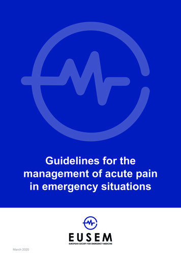
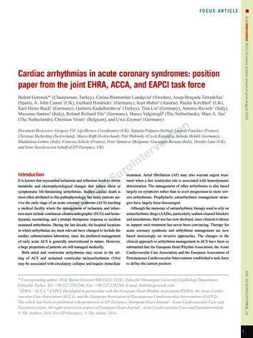
Nerdal et al. BMC Neurology(2022) 22:234anti-dementia medication and/or other previous treatment for dementia.Linear mixed-models includes and analyzes participants with available data on at least one time point, andgive unbiased estimated if data are missing at random[45]. Sensitivity analyses (excluding patients deceasedat 18 and/or 36 months and patients with premorbiddementia) were conducted, as well as analyses adjustedfor comorbidity and complications. Finally, analysesexcluding outliers and analyses excluding patients withmoderate and severe aphasia were conducted for HADSand NPI-Q.In this paper, the term “significant” refers to two tailedp-values less than .05, and 95% confidence intervals (CIs)are reported where relevant. SPSS 27 was used for theanalyses.ResultsParticipantsAs shown in Fig. 1, 139 of the 141 patients included in theNor-COAST study from Bærum Hospital, were screenedPage 4 of 12for delirium. Of these patients, 133 had the data necessary to be included in the mixed-model linear regressionanalyses.At 3 months, 132 patients attended the follow-up. At18 months, 111 patients participated, one of which didnot participate at the previous follow-up. At 36 months,92 patients participated in the follow-up. Compared tothe four other hospitals participating in the Nor-COASTstudy (St. Olavs Hospital, Haukeland Hospital, UllevålHospital and Ålesund Hospital), patients at Bærum Hospital were significantly younger, had more years of education, higher MoCA scores at baseline, less pre-strokedementia, less comorbidity and lower pre-stroke GDSscore (Table 1).Of the 139 screened with CAM, 13 (9.4%) were diagnosed with delirium. Demographics and clinical characteristics of patients with delirium and non-deliriouspatients are shown in Table 2. Patients with deliriumwere older and had lower MoCA score at baseline. Further, a higher percentage of patients with delirium hadGDS scores 3 (in the dementia range) at three, 18 andFig. 1 Overview of the study sample. CAM (Inouye, 1994) was used to screen for delirium. Each patient was screened six times during thefirst 48 hours. Delirium was considered present if the patients had acute onset of fluctuations in mental status and inattention, in addition todisorganized thinking and/or altered level of consciousness at one or more of the screenings. Patients were included in mixed models if they hadbeen screened at baseline and returned for at least one additional follow-up
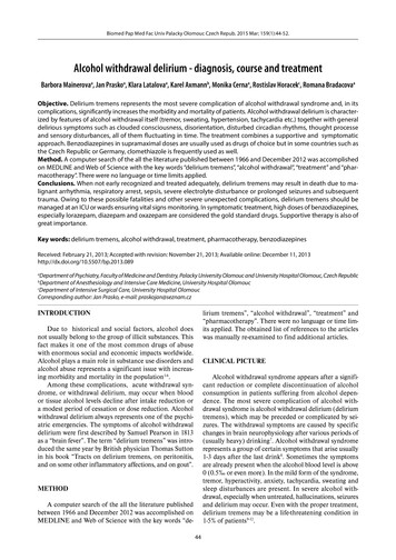
Nerdal et al. BMC Neurology(2022) 22:234Page 5 of 12Table 1 Group differences in demographics and clinical characteristics for Bærum Hospital and all other hospitalsPatients at Bærum Hospital(n 141)Patients from all otherhospitals (n 674)t/ x2pAge, M (SD)71.4 (13.4)74.0 (11.3)2.21.02*Years of education, M (SD)13.9 (3.5)11.5 (3.6) 6.9.000**Gender, n female (%)68 (49%)302 (36%)NIHSS at b aselinea, M (SD)3.0 (4.0)3.67 (5.04)MoCA at baselineb, M (SD)24.7 (4.6)22.6 (5.1)Premorbid dementia, n (%)2 (1.4%)63 (9.5%) .03.78 4.610.1.75.45.000**.001**Complicationsc 0, n (%)34 (24.3%)160 (25%).04.85Charlson Comorbidity Index (CCI), M (SD)3.6 (1.8)4.2 (2.0)3.4.001**Pre-stroke Global Deterioration Scale 3d, n (%)138 (98.6%)554 (80%)27.8.000**Intracerebral hemorrhage, n (%)16 (11.5%)62 (9.2%)0.6.42Arterial ischemic stroke, n (%)120 (86.1%)543 (81.6%)2.1.14aHigher values indicating more severe stroke symptoms. NIHSS at baseline assessed at day 1 of admittance to hospitalbLower values indicating poorer global cognitive function. MoCA assessment at baseline was performed either at discharge or 7 days after admittance for patientswith longer hospital staycInfections, seizures, neurological progression and falls registered during hospitalizationdValues 3 indication no to very mild cognitive decline**indicating p-level .01*indicating p-level .0536 months (Table 2). Patients were hospitalized for mean0.84 days, and 88% were admitted within day one ofsymptom debut.Delirium as a predictor for cognitive outcomesCognitive assessments for patients with and withoutdelirium are described in Table 3 and displayed in Fig. 2.Delirium at baseline predicted significantly lower MoCAscore at all timepoints. The largest difference in meanscores between the two groups was 4.2 points (95% CI:1.4 to 7.1) at 18 months. The results were substantiallythe same in sensitivity analyses excluding patients withpremorbid dementia, patients that were deceased at 18and/or 36 months, and when adjusting complications,and comorbid diseases (results not shown). The variancecomponents in the mixed models are shown in Table 4.Delirium as a predictor for psychiatric outcomesDelirium predicted significantly higher NPI-Q scores,compared to non-delirious patients after 3 months(Table 3). Anxiety symptoms measured by HADSA increased continuously for patients with delirium,resulting in significantly higher anxiety scores at 18 and36 months, compared to non-delirious patients. The largest difference in mean scores for anxiety between the twogroups was found at 36 months (2.8 points (95% CI: 5.5to 0.3)). Mean scores for NPI-Q, HADS-A and HADSD are displayed in Figs. 3, 4 and 5 respectively.DiscussionThis study examined whether delirium in the acute phaseof stroke predicts cognitive and psychiatric symptomsover the course of 3 years. We demonstrated that patientswith delirium had significantly poorer global cognition(MoCA scores) than non-delirious patients at three, 18and 36 months. Delirium also predicted significantlyhigher levels of neuropsychiatric symptoms (NPI-Q)after 3 months, as well as higher anxiety levels (HADS-A)at 18 and 36 months.MoCA is estimated to have a sensitivity of 90% fordetecting MCI using a cut-off score of 26 [39]. This is inline with a previous publication from the Nor-COASTstudy by Munthe-Kaas et al. [46], who found that aMoCA cut-off of 26 had high specificity and sensitivity for identifying patients with cognitive impairment(according to the DSM-5 criteria) 3 months post-stroke.Further, Nasreddine et al. [39] suggest that MoCA scoresin the range of 11.4–21.0 indicate potential dementia.These estimations suggest that the average MoCA scoreof stroke patients with delirium in this study (21.5 at36 months) could indicate more severe clinical outcomes,compared to the MoCA scores of non-delirious patients(25.1 at 36 months).Several studies have reported delirium as a risk factorfor cognitive impairment in hospitalized patients [36,47–50], and in patients with stroke [25–27]. However,the independent effect of delirium on post-stroke cognitive impairment remains somewhat unexplored, as fewstudies have adjusted for other risk factors such as stroke
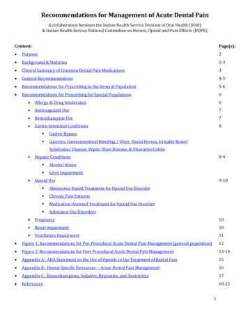
Nerdal et al. BMC Neurology(2022) 22:234Page 6 of 12Table 2 Group differences in demographics and clinicalcharacteristics for patients with and without deliriumDelirium (n 13) Nondelirious(n 126)t/ x2p.000**Age, M (SD)79.5 (6.0)70.6 (13.7)4.34Years of education,M (SD)12.5 (3.5)13.9 (3.4) 1.43 .15Gender, n female (%)6 (46%)62 (49%).04.84NIHSS at baslinea, M(SD)4.5 (4.6)2.8 (3.8)1.35.17MoCA at b aselineb,M (SD)20.0 (2.2)25.1 (4.7) 3.10 .002**Premorbid dementia,n (%)02.19Complicationsc 0,n (%)7 (54.6)27 (21.4) 2.15 .053.6 (1.9) 1.20 .24Charlson Comorbidity 4.1 (1.3)Index (CCI), M (SD).91Global Deterioration Scale (GDS) 3dPre-stroke, n (%)12 (92%)124 (98%).19.663 months, n (%)4 (30%)5 (4%)14.0.003**18 months, n (%)4 (31%)11 (9%)5.9.024*36 months, n (%)3 (23%)10 (8%)3.2.079Moderate to severe aphasiaeBaseline, n (%)3 (23%)19 (15%).570.573 months, n (%)04 (3%).420.6818 months, n (%)03 (2%).320.8136 months, n (%)0001.0aHigher values indicating more severe stroke symptoms. NIHSS at baseline doneat day 1 of admittance to hospitalbLower values indicating poorer global cognitive function. MoCA assessmentat baseline was done either at discharge or 7 days after admittance for patientswith longer hospital staycInfections, seizures, neurological progression and falls registered duringhospitalizationdValues 3 indication no to very mild cognitive decline. Values 3 indicatingpotential dementiaeAmount of patients with a level of aphasia causing interference withconversation, indicated by the value 2 (moderate) or 3 (severe) in the NIHSS itemmeasuring aphasia**indicating p-level .01*indicating p-level .05severity, pre-stroke dementia, complications, and comorbid diseases. In this study, the patients with deliriumhad fewer years of education, more complications, morecomorbid diseases, a higher mean NIHSS score, and weresignificantly older, than the non-delirious patients. Itshould be stressed that the association between deliriumand poorer MoCA scores remained significant even whenadjusting for these covariates in the sensitivity analyses,suggesting an independent effect of delirium on globalcognition. Further, these results were found despite theBærum sample having several more protective factors[51], such as lower age, longer education, and milderstrokes, compared to the total Nor-COAST sample(Table 1). This raises a question as to whether deliriumwould predict more severe cognitive impairment in oldersamples with larger strokes.The results for HADS and NPI-Q remained significant when adjusting for aphasia. This could be due tothe majority of patients having mild strokes and the ratesof aphasia being low. Additionally, we only adjusted formoderate and severe aphasia (i.e., interfering with conversation). In future studies it would be beneficial toadjust for mild aphasia and explore the effect of aphasiaand other complications in samples with more severestrokes.Although patients with delirium had higher HADS-Ascores than non-delirious patients in this study, the average score was not above the commonly used cut-off scoreof 8 [41]. However, adjusting HADS cut-off scores to thespecific clinical sample can provide a higher specificityand sensitivity [52, 53]. Sagen et al. [53] found a cut-offscore of 4 to be optimal in Norwegian stroke populationsfor detecting clinical symptoms. This would imply thatthe anxiety levels in patients with delirium are of clinical importance at 18 and 36 months (Mean (SD): 5.6 (1.2)and 6.2 (1.3) respectively). Though Kowalska et al. [54]recently found delirium to be a risk factor for anxiety3 months after stroke, our findings suggest that the subsequent anxiety symptoms can be present or even increaseover a longer timeframe.In the present study, both HADS-A and HADS-Dscores were higher in patients with delirium compared tonon-delirious patients. However, group differences wereonly significant for HADS-A. This corresponds to severalother Norwegian stroke studies, finding anxiety symptoms to be more prevalent than depressive symptomsusing HADS [53, 55, 56]. However, a meta-analysis from2017 found higher prevalence of post-stroke depressivedisorders than post-stroke anxiety (33.5% versus 9.8%),using DSM or World Health Organization criteria (ICD10) [57]. A potential explanation for this discrepancy isthat anxiety is more commonly overlooked in older samples, as symptoms like withdrawal and avoidant copingstrategies can be falsely attributed to aging [58]. Thiscould lead to an underestimation of anxiety symptoms instroke populations.MacLullich, Beaglehole, Hall & Meagher [59] emphasizes stress as a potential mediator between deliriumand anxiety symptoms. Systems related to the physiological stress response, such as the limbic hypothalamicpituitary-adrenal axis (HPA axis) and glucocorticoids,have been found to precipitate and/or sustain deliriumboth in acute disease [60, 61] and in stroke specifically[62]. Intense or prolonged stress responses can initiate
Nerdal et al. BMC Neurology(2022) 22:234Page 7 of 12Table 3 Assessments for patients at Bærum Hospital with and without delirium during first 48 hoursDelirium nMean (SE)Non-delirious nMean (SE)Difference Estimate (95% CI)pMoCA1021.7 (1.4)11524.7 (0.4)2.9 (0.1 to 5.7).04*NPIQ102.4 (0.6)1170.8 (0.1).005**HADS Depression83.7 (1.3)1003.7 (0.3) 1.6 ( 2.7 to 0.47)HADS Anxiety84.0 (1.1)1003.1 (0.3)MoCA920.8 (1.4)100NPIQ90.6 (0.6)100HADS Depression76.2 (1.4)HADS Anxiety7MoCANPIQ3 months0.04 ( 2.6 to 2.7).97- 0.9 ( 3.4 to 1.4).4425.0 (0.4)4.2 (1.4 to 7.1).004*1.0 (0.2)0.44 ( 0.8 to 1.7)47933.7 (0.3)- 2.44 ( 5.2 to 0.3).085.6 (1.2)933.1 (0.3)- 2.6 ( 5.0 to 0.1).04*521.5 (1.5)8325.1 (0.4)3.6 (0.5 to 6.7).02*51.7 (0.7)840.7 (0.2).17HADS Depression56.3 (1.4)754.1 (0.4) 1.0 ( 2.5 to 0.5)- 2.3 ( 5.1 to 0.64).12HADS Anxiety56.2 (1.3)753.3 (0.3)- 2.8 ( 5.5 to - 0.3).03*18 months36 monthsMean (SE) are descriptive data. The difference with CI and p-value are from linear mixed models with covariates delirium, time and their interaction as categoricalcovariates, adjusted for age, gender, NIHSS-score at baseline and premorbid dementia.* Indicating p .05 and ** indicating p .01Fig. 2 MoCA total scores for patients with and without delirium at three, 18 and 36 months. Descriptive mean total-scores for MoCA after three, 18and 36 months for patients from Bærum with and without deliriumincreased vigilance and symptoms of anxiety [63, 64],and several studies address dysregulations of the HPAaxis and glucocorticoid production as central for thisassociation [64–66]. Further, older age is associated withincreased dysregulations of the HPA axis, causing cortisol levels to be sustained for a longer period of time aftermajor stressors [67, 68]. The Bærum sample had a lowermean age than the total Nor-COAST sample and thegeneral stroke population in Norway [69]. Consideringthe age-related risk of HPA dysregulation, the associationbetween delirium and anxiety may be even stronger in alarger and more representative population.A limitation of this study was using CAM, rather thanclinical evaluation, to assess for delirium. CAM has notbeen validated for stroke patients. The CAM-ICU [70]has however been evaluated as a valid instrument fordiagnosing delirium in patients with stroke [71]. TheCAM-ICU consist of the same four features as CAM
Nerdal et al. BMC Neurology(2022) 22:234Page 8 of 12Table 4 Variance components in linear mixed model reported inTable 3Variance ComponentDependent variableWithin participantsBetweenparticipantsMoCA13.38 (1.7)3.7 (.38)NPI-Q1.8 (.18)1.0 (.24)HADS Depression4.16 (.46)8.4 (1.4)HADS Anxiety3.76 (.41)6.1 (1.0)Variance components are from linear mixed models with delirium, time andtheir interaction as categorical covariates, adjusted for age, gender, NIHSS-scoreat baseline and premorbid dementia(acute onset of changes or fluctuations in mental status;inattention; disorganized thinking and/or altered level ofconsciousness) [70, 72]. CAM-ICU is however adaptedto mechanically ventilated patients with more severestrokes, as the questions are nonverbal. Our sample hadmild strokes and low rates of aphasia, making the originalCAM more relevant.As delirium has a fluctuating and heterogenous nature,it can be easy to overlook or mislabel delirium on othercerebral dysfunction with similar expression. Detecting individual deviations in attention can be particularlychallenging without any point of reference [38]. TheFig. 3 NPI-Q total scores for patients with and without delirium at three, 18 and 36 months. Descriptive mean total-scores for NPI-Q after three, 18and 36 months for patients from Bærum with and without deliriumFig. 4 HADS-D scores for patients with and without delirium at three, 18 and 36 months. Descriptive mean for HADS-A after three, 18 and36 months for patients
Nerdal et al. BMC Neurology Page 10 of 12 Conclusion Patients suering from delirium in the acute phase of stroke had poorer global cognition and more psychiatric symptoms over the course of 3 years, compared to non-delirious patients. e results suggest that stroke patients with delirium may benet from long-term follow-up of