Transcription
Theranostics 2018, Vol. 8, Issue 113007IvyspringTheranosticsInternational PublisherResearch Paper2018; 8(11): 3007-3021. doi: 10.7150/thno.21966Flow-dependent epigenetic regulation of IGFBP5expression by H3K27me3 contributes to endothelialanti-inflammatory effectsSuowen Xu1, Yanni Xu1, 2, Meimei Yin1, Shuya Zhang1, 3, Peng Liu1, 2, Marina Koroleva1, Shuyi Si2, Peter J.Little4, 5, Jaroslav Pelisek6, Zheng Gen Jin1, 1.2.3.4.5.6.Aab Cardiovascular Research Institute, Department of Medicine, University of Rochester School of Medicine and Dentistry, Rochester, New York, USAInstitute of Medicinal Biotechnology Peking Union Medical College and Chinese Academy of Medical Sciences, Beijing, ChinaKey Laboratory of Fertility Preservation and Maintenance of Ministry of Education, Department of Biochemistry and Molecular Biology, Ningxia MedicalUniversity, Yinchuan, ChinaSchool of Pharmacy, The University of Queensland, Pharmacy Australia Centre of Excellence (PACE), Woolloongabba QLD 4102, AustraliaXinhua College of Sun Yat-sen University, Guangzhou, ChinaDepartment of Vascular and Endovascular Surgery, Klinikum rechts der Isar der Technischen Universitaet Muenchen, Germany Corresponding author: Zheng Gen Jin, Ph.D.; Aab Cardiovascular Research Institute (CVRI), Department of Medicine, University of Rochester School ofMedicine and Dentistry; 601 Elmwood Avenue, Box CVRI, Rochester, NY 14642; Phone: 585-276-7691; Fax: 585-276-9829; E-mailZheng-gen Jin@urmc.rochester.edu Ivyspring International Publisher. This is an open access article distributed under the terms of the Creative Commons Attribution (CC BY-NC) /4.0/). See http://ivyspring.com/terms for full terms and conditions.Received: 2017.07.17; Accepted: 2018.02.17; Published: 2018.04.30AbstractRationale: Atherosclerosis is a chronic inflammatory and epigenetic disease that is influenced by differentpatterns of blood flow. However, the epigenetic mechanism whereby atheroprotective flow controlsendothelial gene programming remains elusive. Here, we investigated the possibility that flow alters endothelialgene expression through epigenetic mechanisms.Methods: En face staining and western blot were used to detect protein expression. Real-time PCR was usedto determine relative gene expression. RNA-sequencing of human umbilical vein endothelial cells treated withsiRNA of enhancer of zeste homolog 2 (EZH2) or laminar flow was used for transcriptional profiling.Results: We found that trimethylation of histone 3 lysine 27 (H3K27me3), a repressive epigenetic mark thatorchestrates gene repression, was reduced in laminar flow areas of mouse aorta and flow-treated humanendothelial cells. The decrease of H3K27me3 paralleled a reduction in the epigenetic “writer”-EZH2, thecatalytic subunit of the polycomb repressive complex 2 (PRC2). Moreover, laminar flow decreased expressionof EZH2 via mechanosensitive miR101. Genome-wide transcriptome profiling studies in endothelial cellstreated with EZH2 siRNA and flow revealed the upregulation of novel mechanosensitive gene IGFBP5(insulin-like growth factor-binding protein 5), which is epigenetically silenced by H3K27me3. Functionally,inhibition of H3K27me3 by EZH2 siRNA or GSK126 (a specific EZH2 inhibitor) reduced H3K27me3 levels andmonocyte adhesion to endothelial cells. Adenoviral overexpression of IGFBP5 also recapitulated theanti-inflammatory effects of H3K27me3 inhibition. More importantly, we observed EZH2 upregulation, andIGFBP5 downregulation, in advanced atherosclerotic plaques from human patients.Conclusion: Taken together, our findings reveal that atheroprotective flow reduces H3K27me3 as achromatin-based mechanism to augment the expression of genes that confer an anti-inflammatory response inthe endothelium. Our study exemplifies flow-dependent epigenetic regulation of endothelial gene expression,and also suggests that targeting the EZH2/H3K27me3/IGFBP5 pathway may offer novel therapeutics forinflammatory disorders such as atherosclerosis.Key words: atherosclerosis, endothelial cells, epigenetic, EZH2, H3K27me3, IGFBP5IntroductionAtherosclerosis is a devastating global disease ic plaques has a preferential distributionin aortic regions of turbulent/disturbed flow, buthttp://www.thno.org
Theranostics 2018, Vol. 8, Issue 11regions with steady laminar flow are protectedagainst atherosclerosis development [8-12]. Theunderlying mechanisms of this geographicaldistribution of atherosclerotic lesions remain largelyunclear, but are likely related to hemodynamicforces-induced signal transduction and geneexpression. In particular, laminar flow increases theexpression of many anti-atherosclerotic genes, such askruppel-like factor 2 (KLF2), endothelial nitric oxidesynthase (eNOS), and nuclear factor erythroid2–related factor 2 (Nrf2), which critically regulateendothelial homeostasis, but decreases those“pro-atherogenic” genes, such as monocytechemoattractant protein-1 (MCP1), and endothelin-1(ET1) [8-14]. In contrast, disturbed flow skewsendothelial cells towards the direction of endothelialdysfunction [8-12]. An understanding of mechanismsthat mediate the differential gene expression patternbetween laminar flow and disturbed flow regionsoffers us therapeutic opportunities to intervene inendothelial dysfunction and atherosclerosis.Atherosclerosis can be regulated by severalepigenetic mechanisms, such as DNA methylation/demethylation [15], histone methylation/demethylation and acetylation/deacetylation [16, 17],microRNAs [18] and long non-coding RNAs [19, 20].Research in the past decade has indicated that theseepigenetic mechanisms modulate multiple aspects ofendothelial function [21], lineage commitment [22],and atherogenesis [4]. In the context of epigeneticmodifications mediated by flow, recent evidence hasshown that flow shear stress regulates endothelialgene expression by DNA methylation [23-25], histoneacetylation [26], miRNAs [27-29] and long non-codingRNAs [30] . However, it remains elusive whether flowregulates histone methylation and the expression ofnovel mechanosensitive genes that regulateendothelial function. Among the epigenetic modifiers,EZH2 in the polycomb repressive complex 2 (PRC2)has gained considerable interest for its regulatory rolein maintaining pluripotency, cell differentiation,organ development and cancer [31]. PRC2, whichdirectly trimethylates histone 3 lysine 27 (H3K27me3)and causes chromatin compaction and transcriptionalsilencing, is composed of EED, SUZ12, RbAp46/48,and EZH2 [31]. In contrast, PRC2-mediatedH3K27me3 formation can be reversed by two histonedemethylases, named UTX (ubiquitously transcribedX-chromosome tetratricopeptide repeat protein, alsoknown as KDM6A) and JMJD3 (jumonji domaincontaining-3, also known as KDM6B) [31]. Severallines of evidence have implicated EZH2 in thedevelopment and progression of atherosclerosis. Forexample, recent studies observed increased H3K27dimethylation in endothelial cells from human early3008and advanced atherosclerotic plaques [16] despite aglobal decrease in H3K27 methylation level [32, 33].EZH2 is also responsive to fluid shear stress andinvolved in laminar flow-mediated cell quiescence[34]. EZH2 regulates transendothelial migration ofleukocytes to microvessels [35] as well as tumorangiogenesis [36]. In addition, EZH2 has recently beenshown to restrict myocardin-dependent smoothmuscle differentiation from the mesothelium [37] andpromote the formation of foam cell andatherosclerosis development in hyperlipidemicApoE-/- mice [38]. Moreover, EZH2 was alsoupregulated in hyperhomocysteinemic ApoE-/- miceand contributed to foam cell formation [39]. Theseevidences suggest that EZH2 could play apro-atherogenic role in atherosclerosis development.Since EZH2 is a histone methyltransferase thatresponds to laminar flow [34], it remains unknownwhether atheroprotective flow controls EZH2mediated histone methylation patterns, which in turnalter endothelial gene expression, thus regulatingendothelial function. Here, we show thatEZH2/H3K27me3 is reduced by laminar flow inendothelial cells both in vivo and in vitro. Throughgenome-scale RNA sequencing (RNA-seq)-basedcomparison, chromatin-immunoprecipitation (ChIP)and luciferase assays, we demonstrated thatatheroprotective flow upregulates novel mechanosensitive genes, such as IGFBP5 (insulin-like growthfactor-binding protein 5) through an H3K27me3dependent inhibitory mechanism to repressesendothelial inflammation. Finally, we observed thatEZH2 is upregulated, while IGFBP5 is downregulatedin advanced atherosclerotic plaques from patientsundergoing carotid endarterectomy, indicating sis.Materials and methodsStudy populationHuman atherosclerotic plaques were obtainedfrom patients undergoing carotid endarterectomydeposited in the Biobank of Department of Vascularand Endovascular Surgery (Klinikum rechts der Isarder Technischen Universität Muenchen, Germany)[16, 33]. Atherosclerotic plaques were histologicallycharacterized according to the American HeartAssociation (AHA) Stary classification [40, 41] anddivided into early (types I–III) or advanced (typesV–VII) atherosclerosis stages. In this study, carotidarteries (n 10) with advanced stage of atherosclerosis(types VI) were used. Plaque-free healthy carotidarteries (n 10) were obtained from the Department ofTrauma Surgery as previously described [16, 33].http://www.thno.org
Theranostics 2018, Vol. 8, Issue 11Details of inclusion and exclusion criteria areprovidedinSupplementaryMaterial.Thedemographic data of atherosclerotic patients andhealthy donors are summarized in Table S1-2. Thestudy was approved by the ethics committee ofTechnischen Universität Muenchen with patients’written informed consent. The study was performedaccording to the guidelines of the Declaration ofHelsinki.RNA isolation and quantitative real-time PCRRNA was extracted from formalin-fixed,paraffin-embedded (FFPE) carotid artery samplesusing the High Pure RNA Paraffin Kit (#3270289001,Roche, Mannheim, Germany) according to themanufacturer’s instructions [16, 33]. Two 10 μmsections of each sample were pooled for RNAisolation. Total RNA from cultured cells and micetissues were isolated with RNeasy Minikit and TRIzolReagent, respectively. Total RNA was converted intocomplementaryDNA(cDNA)usingtheHigh-Capacity cDNA Reverse Transcription Kit(#4374966, Applied Biosystems, Foster City, CA)following the manufacturer’s instructions. Then,quantitative real-time PCR (qRT-PCR) was performedusing iQ SYBR Green Supermix (#1708886, Bio-Rad)as previously described in detail [42]. Thecomparative cycle threshold (Ct) method (2 ΔΔCt) wasused to determine the relative gene expression afternormalization to housekeeping gene GAPDH. The Ctvalues of EZH2 in one control sample and oneatherosclerotic patient sample were below thedetection limit of qRT-PCR and thus not included foranalysis. The sequences of primers are listed in TableS3.En face immunofluorescent stainingEn face staining was performed as previouslydescribed in detail [43], with minor modifications [42,44]. Mouse aortas were collected after perfusion withsaline and neutrally buffered 4% PFA. Aorticsegments were then permeabilized with 0.1% TritonX-100 in PBS for 10 min and blocked with 10% normalgoat serum (Invitrogen) in Tris-buffered saline (TBS)containing 2.5% Tween-20 for 30 min at roomtemperature. Next, aorta segments were incubatedwith rat anti-VE-Cadherin (1:100; #555289, BDBioscicence), rabbit anti-EZH2 (#6263, ProSci) orrabbit anti-H3K27me3 (#39155, Active Motif)antibody overnight at 4 C. After rinsing withwashing buffer 3 times, aortic segments wereincubated with Alexa Fluor 488-conjugated goatanti-rat and Alexa Fluor 546-conjugated goatanti-rabbit secondary antibodies (1:1,000; ThermoFisher Scientific) for 1 h in darkness at room3009temperature. Finally, aortic segments were carefullywhole mounted (with lumen side up) in the ProLongGold-antifade Mounting Media (Invitrogen) forconfocal microscopy (IX81, Olympus) with 60x or 40xoil lens. All animal procedures conformed to theGuideline for the Care and Use of Laboratory Animalspublished by the U.S. National Institutes of Healthand were approved by the Institutional Animal Careand Use Committee of the University of RochesterMedical Center.Primary isolation of mouse endothelial cells,smooth muscle cells and macrophagesMouse endothelial cells were isolated from adultC57BL/6J mouse lungs using collagenase I digestion(3 mg/mL in serum-free DMEM) at 37 C in anincubator for 45 min, followed by CD31- andICAM2-dynabeads-based double positive selection[45, 46]. The isolated mouse lung endothelial in staining. More than 99% of cultured cellswere VE-cadherin positive. The isolation of vascularsmooth muscle cells (VSMCs) was performed usingthe explant method as previously described [47], withminor modifications. VSMCs migrated from thoracicaorta explants at around 8 days, then, sproutedVSMCs were digested with 0.05% Trypsin-EDTA andreseeded for confocal microscopy or RNA isolation.More than 85% of cultured cells were smooth musclealpha-actin (α-SMA) positive. Mouse peritonealmacrophages were isolated from the peritoneumcavity by injecting 5 mL ice-cold PBS/0.1% BSA [48].Cells were cultured in complete media for 4 h, then,non-adherent cells were removed by washing.Macrophages were stained with CD68, and more than99% of attached cells were CD68 positive.Cell cultureHuman umbilical vein endothelial cells(HUVECs) were isolated from fresh human umbilicalcords and cultured in Medium 200 containing 5% FBSand 1X low serum growth supplement (LSGS) [42, 44].HUVECs were collected in accordance with theUniversity of Rochester human subjects review boardprocedures that prescribe to the Declaration ofHelsinki. Different donors of human coronary arteryendothelial cells (HCAECs) were used in this study,which were from Lifeline Cell Technologies(Frederick, MD, #FC-0032, Lot No. 01181, 01354) andCell Applications Inc. (#300K-05A, Lot No. 1858, SanDiego, CA). HCAECs were cultured in MesoEndoCell Growth Media supplemented with GrowthSupplement (#212K-500, Cell Applications Inc.) plus5% FBS [42]. For cell authentication, HUVECs andHCAECs were identified by cobblestone cellhttp://www.thno.org
Theranostics 2018, Vol. 8, Issue 11morphology, staining of endothelial cell-markerVE-Cadherin (BD Bioscicence, #555661) andfunctional uptake of DiI-oxidized LDL (10 µg/mL, 4h; Alfa Aesar, #J64164). HCAECs tested negative formycoplasma, bacteria, yeast, fungi, Hepatitis B and C,as stated on the Vendors’ certificates of analysis onindividual lots of cells. HUVECs (confluence 80-90%)and HCAECs (confluence 60-70%) were subculturedon the day before experiments. For flow experiments,ECs cultured in fresh complete media were exposedto laminar flow [42] or disturbed flow [49] in a 60 mmor 100 mm cone and plate viscometer. Due to the factthat prolonged culture of ECs renders ECs lessresponsive to flow [50], HUVECs and HCAECs werepassaged on the day before flow treatment. HUVECsand HCAECs at passage 2 to 5 were used for all theexperiments. THP-1 cells (gifted by Y. Cai), apro-monocytic cell line, were cultured as previouslydescribed [42]. THP-1 cells were authenticated by theability to differentiate into macrophages after PMA(100 nM, 48 h) stimulation and form foam cells afterloading with oxidized LDL for 24 h. For cell culturestudies, all compounds were prepared as stocksolution at 10 mM in dimethylsulfoxide (DMSO) andworking solutions were freshly prepared before use.Monocyte adhesion assayTNFα is a commonly used stimulus in triggeringmonocyte adhesion/rolling/transmigration [51, 52],by enhancing the production of MCP1 [51, 53] andother pro-inflammatory and pro-migratory cytokines(such as IL6) [54]. Therefore, TNFα was used in theTHP-1 monocyte adhesion assay, which wasperformed as previously described in detail [55].Quantification of adherent THP-1 monocytes wasperformed by manual counting of monocytes afterwashing with pre-warmed HUVEC complete mediathree times.siRNA and miRNA transfectionTo deplete EZH2, HUVECs at 75-80% confluencewere used for transfection. In brief, Lipofectamine2000 or RNAi Max (6 μL; Thermo Fisher Scientific)[42] was mixed with Opti-MEM (250 μL; ThermoFisher Scientific), and then two independent siRNAoligos against human EZH2 (20 nM; #SR301494-A, -B,Origene, Rockville, MD) or non-target control siRNA(25 nM; #SR30004, Origene) diluted in 250 μLOpti-MEM was added to the solution, mixed gently,and incubated at room temperature for 20 min. After3-4 h of transfection, the medium was changed toM200 complete medium and cells were treated 48 hafter transfection. Additional Silencer Select EZH2siRNA (#s4916, Thermo Fisher Scientific) or Silencer Select Negative Control #1 siRNA (#S25503, Thermo3010Fisher Scientific) were used for RNA sequencing. Thesame method was used to transfect HUVECs withAnti-miRTM negative control (100 nM; #AM17010,Ambion) or Anti-miRTM hsa-miR-101-3p miRNAinhibitor (100 nM; #AM17000, Ambion) using RNAiMax as the transfection reagent.Adenoviral EZH2 overexpressionEZH2 adenovirus was purchased from VectorBiolab (#1371, Malvern, PA). HUVECs were infectedwith EZH2 adenovirus as indicated.Extraction of purified histones from culturedcellsCore histones were purified using a commercialHistone Purification Kit (Active Motif, #40025,Carlsbad, CA, USA) following the manufacturer'sinstructions, with minor modifications. Briefly,HUVECs were treated with a potent and specificEZH2 inhibitor GSK126 (#A1275, Active Biochem,Hongkong) for the indicated time without changingthe media. After washing twice with pre-warmed (37 C) serum-free M200 media, 0.4 mL ice-coldExtraction Buffer was added to a 60 mm dish. Then, aplastic scraper was used to collect the cell proteinextracts. The cells were rotated in Extraction Bufferovernight on a VWR rotating platform at 4 C torelease histones. The following day, cell extracts weretransferred to fresh tubes and centrifuged in amicrocentrifuge at maximum RCF for 5 min at 4 C.Crude histones were neutralized with 0.1 mL 5XNeutralization Buffer and purified using columnsprovided. For Western blots, boiled histone lysateswere separated by 12-15% SDS-PAGE. The samemembrane was stripped and reprobed withanti-Histone 3 (H3) for equal loading.Western blot analysisCells were harvested in freshly-prepared lysisbuffer (20 mM Tris-HCl pH 7.5, 150 mM NaCl, 1%Triton X-100, 1 mM EDTA, 1 mM EGTA, 2.5 mMsodium pyrophosphate, 1 mM β-Glycerolphosphate,50 mM NaF, 1 mM Na3VO4, and 1X protease inhibitorcocktail) by cell lifters. After clarification at 4 C, 12,000 g for 10 min, total cell lysate was collected forSDS-PAGE gel analysis as previously described indetail [56]. After transfer, membranes were blocked inPBS-diluted LI-COR blocking buffer at roomtemperature for 1 h. Then, the blots were incubatedwith 3% BSA-diluted primary antibodies (listed inTable S4) at 4 C overnight or room temperature for 1h, followed by incubation with LI-COR IRDye 680RD goat anti-mouse IgG (H L) or IRDye 800CWgoat anti-rabbit IgG (H L) or IRDye 680RD donkeyanti-goat IgG (H L) (dilution at 1:10,000) at roomtemperature for 30 min. Images were visualized usinghttp://www.thno.org
Theranostics 2018, Vol. 8, Issue 11an Odyssey Infrared Imaging System (LI-COR).Densitometric analysis of blots was performed usingNIH Image J software (http://imagej.nih.gov/ij/).Chromatin immunoprecipitation coupled withquantitative real-time PCR (ChIP-qPCR)ChIP assays were performed using EZ-MagnaCHIPTM A/G ChIP kits (#17-10086, EMD Millipore),with slight modifications [57]. Equal amounts ofBioruptor-sheared chromatin were incubated withcontrol rabbit IgG, or rabbit H3K27me3 antibodies at 4 C overnight. Chromatin-associated proteins werewashed and then digested with 1 µL Proteinase K (10mg/mL) for 2 h at 62 C with gentle shaking andincubated at 95 C for 10 min for reverse crosslinkingand inactivation of Proteinase K. Chromatin DNAwere then purified with spin columns provided withthe kit. Additional ChIP DNA purification kits werefrom Active Motif (#58002, Carlsbad, CA). qRT-PCRwas performed using ChIP primers encompassing theproximal IGFBP5 gene promoter (Table S3). For eachPCR reaction, 1 µL ChIP’ed DNA template was addedfor each 10 µL reaction. 1% of starting chromatin isused as input. Data are presented as % input using the2 ΔΔCt method.Luciferase reporter assayThe predicted IGFBP5 promoter region (-1000 bpto 45 bp relative to transcription start site) wassynthesized by GeneScript and subcloned intopGL3-basic plasmid (Promega). Luciferase activityassay was performed as previously described [55]. Inbrief, 100 ng Luciferase plasmid was transfected intoHUVECs for 24 h using Lipofectamine 2000 asdescribed above. COS7 cells were co-transfected with10 ng pRL-TK plasmid (Promega) for normalization oftransfection efficiency. The ratio of luciferase units topRL-TK units was calculated and normalized. Dataare presented as fold over control after normalization.Genome-wide RNA sequencing studyRNA sequencing (RNA-seq) is a useful approachfor probing the pathological mechanisms of vasculardiseases [58]. RNA-seq was performed at theUniversity of Rochester Genomics Research Center(URGRC) as previously described [57]. For flowRNA-seq,HUVECsweresubjectedtoatheroprotective laminar flow for 48 h using cellsunder static conditions as the control (n 2). ForRNA-seq in HUVECs depleted of EZH2, cells weretransfected with control siRNA (20 nM) or EZH2siRNA (20 nM) for 48 h before RNA was prepared forRNA-seq (n 3). DESeq2-1.10.1 was used to performdata normalization and differential expressionanalysis with an adjusted p-value threshold of 0.05.3011The significance test used in DeSeq2 analysis was theWald test, and a log2 fold change between groups ispresented. Adjusted p-values were derived using theBenjamini and Hochberg multiple test correction.Accession numberThe RNA-seq data reported in this paper .nih.gov/geo/)withtheaccession number GEO: GSE87534. In Figure S4,published datasets were mined from the GEOdatabase with the accession number of GEO: GDS5083[59].StatisticsData are expressed as mean SEM. Statisticalanalysis was performed using GraphPad Prismsoftware 5.0 (GraphPad, La Jolla, CA). Student’s t testand one-way analysis of variance (ANOVA) with posthoc Bonferroni tests were used for comparisonsbetween two groups and multiple comparisons,respectively. A p value less that 0.05 was consideredstatistically significant.ResultsLaminar flow specifically reduces repressiveepigenetic mark H3K27me3 in endothelialcells in vitro and in vivo.PRC2isanevolutionarilyconservedmulti-protein complex that leads to gene silencingthrough H3K27me3 [31]. To examine the potentialinvolvement of PRC2/H3K27me3 in ogramming, we first performed a RNA-seq studyon human endothelial cells exposed to long-termatheroprotective laminar flow (12 dyne/cm2, 48 h),with an aim to assess the effect of long-term flow onthe endothelial transcriptome. We focused on thegenes in the PRC2 (Figure 1A), including SUZ12,EED, RbAp46 (RBBP7), RbAp48 (RBBP4), and EZH2.Our data showed that treatment with laminar flowdecreased gene expression of EZH2 and RpAp46,without influencing the other three components ofPRC2 (Table S5). Since histone methylation isdynamically controlled by the equilibrium of histonemethylation and demethylation [60], we also lookedat the effect of laminar flow on the expression of theH3K27me3 demethylases including KDM6A (alsoknown as UTX) and KDM6B (also known as JMJD3)[60]. Our RNA-seq data suggest that laminar flowdoes not significantly alter KDM6A, or KDM6B geneexpression (Table S5), implicating that laminar flowcould potentially influence net PRC2-dependentH3K27me3 modification. We also observed that,similar to the effect of EZH2 enzymatic inhibitorhttp://www.thno.org
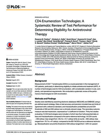
Theranostics 2018, Vol. 8, Issue 11GSK126 [61], laminar flow treatment significantlydownregulated the global level of H3K27me3 inendothelial cells (Figure 1B), indicating that ation. However, laminar flow does notsignificantly affect global protein expression ofhistone 3 lysine 9 trimethylation (H3K9me3) oracetylation (H3K9ac) (Figure 1C), suggesting thespecific inhibitory effects of laminar flow onH3K27me3. To address whether our in vitro3012observations can be translated into in vivo conditions,we compared the differential expression ofH3K27me3 in two different regions of mouse aorta:one is the inner curvature of the aortic arch exposed todisturbed flow, and the other is thoracic aortagenerally exposed to laminar flow. We observed amarked decrease in H3K27me3 staining in the laminarflow area of mouse aorta (Figure 1D). We alsoobserved that the expression pattern of H3K27me3 invascular endothelium is heterogeneous.Figure 1. Laminar flow specifically reduces repressive epigenetic mark H3K27me3 in endothelial cells in vitro and in vivo. (A) Diagram of PRC2. PRC2 iscomposed of EED, SUZ12, RbAp46/48, and EZH2, which imposes the H3K27me3 epigenetic mark to the target gene promoter, leading to gene silencing. (B) Laminar flowdecreases global H3K27me3 levels. HUVECs were exposed to laminar flow for 48 h, and then total histones were purified as described in the methods section. GSK126, a specificinhibitor of EZH2 activity was used as positive control (n 4-5, **P 0.01, ***P 0.001 vs. static control or DMSO). (C) Laminar flow does not affect the level of H3K9me3 orH3K9ac. HUVECs were exposed to laminar flow. Cell alignment in response to flow was observed to ensure successful induction of laminar flow. Then, epigenetic marksH3K9me3 and H3K9ac were determined by Western blot. The same membranes were stripped and incubated with histone 3 (H3) as loading controls (n 3). (D) En faceimmunofluorescent staining of H3K27me3 in mouse aorta. The aortic arch and thoracic aorta were collected from 3-month-old ApoE-/- mice fed a normal chow diet for en facestaining. Red, H3K27me3; green, VE-cadherin; blue, DAPI; scale bar 20 µm (n 5).http://www.thno.org
Theranostics 2018, Vol. 8, Issue 113013Figure 2. Laminar flow decreases the expression of chromatin modifier EZH2 in vitro and in vivo. (A) HUVECs were exposed to laminar flow for the indicated timepoints, and then EZH2 protein expression was determined by Western blot (*P 0.05, ***P 0.001 vs. static control, n 6). (B) HCAECs were exposed to laminar flow for 24 h,and then EZH2 protein expression was determined by Western blot (**P 0.01, n 3). (C) Illustration of atheroprone (inner curvature of aortic arch, #1) and atheroprotective(thoracic aorta, #2) regions of mouse aorta. The picture was manually drawn by powerpoint. (D) EZH2 mRNA expression in aortic endothelium from atheroprone andatheroprotective regions of mouse aorta. Intima-enriched RNA was isolated from the aortic arch and thoracic aorta of ApoE-/- mice fed a normal chow diet for 3 months. RNAwas reverse transcribed and cDNA was used for real-time PCR quantification of EZH2 using GAPDH as internal control (**P 0.01 vs. aortic arch, n 8). (E) En faceimmunofluorescent staining of EZH2 in mouse aorta. The aortic arch and thoracic aorta were collected from 3-month-old ApoE-/- mice fed a normal chow diet for en face staining.Red, EZH2; green, VE-cadherin; blue, DAPI; scale bar 20 µm (n 5).Laminar flow decreases the expression ofchromatin modifier EZH2 in vitro and in vivo.Since EZH2 is the histone methyltransferase thatimposes H3K27me3 epigenetic mark, we next askedwhether the decrease of H3K27me3 is due to thedecrease of EZH2 expression. We first determined thetissue and cell distribution of EZH2. Consistent g/home/gene/EZH2),we observed that EZH2 was mostly expressed in thespleen, consolidating the key role of EZH2 inimmunity [62, 63]. Other organs enriched in EZH2include lung, brain and aorta, which are endothelialcells-enriched organs (Figure S1A). We nextcompared EZH2 expression in vascular cells, usingprimary isolated mouse lung endothelial cells, aorticvascular smooth muscle cells and peritonealmacrophages. We observed that EZH2 wasprominently expressed in endothelial cells (FigureS1B). This expression pattern suggests EZH2 mayhave an important role in regulating immunity andendothelial functions. We then evaluated the timecourseoflaminarflow-inducedEZH2downregulation. We observed that laminar flowsignificantly decreased EZH2 protein expressionstarting from 6 h, with maximal reduction at 24 h(Figure 2A). The flow-dependent reduction of EZH2was recapitulated in HCAECs exposed to laminarflow (Figure 2B). In agreement with the RNA-seqdata, we observed that laminar flow does notsignificantly affect the protein expression of PRC2components EED and SUZ12 (Figure S2). To examinewhether disturbed flow increases EZH2 expression,we compared the protein expression of EZH2 underdisturbed flow and laminar flow conditions, usingcells under static conditions as a control. We observedthat, compared with static conditions, disturbed flowhas no significant effect on EZH2 mRNA and proteinexpression (Figure S3). To corroborate our in vitrohttp://www.thno.org
Theranostics 2018, Vol. 8, Issue 11study, we analyzed mRNA and protein expression ofEZH2 in mouse aorta regions with disturbed flow(inner curvature of aortic arch) and laminar flow(thoracic aorta) (Figure 2C). Interestingly, weobserved a significant reduction of EZH2 mRNA andprotein expression in arterial regions with laminarflow compared with that of regions with disturbedflow (Figure 2D-E).miR101 is involved in laminar flow-inducedEZH2 downregulation.To dissect the mechanism whereby laminar flowreduces EZ
Medical Center. Primary isolation of mouse endothelial cells, smooth muscle cells and macrophages Mouse endothelial cells were isolated from adult C57BL/6J mouse lungs using collagenase I digestion (3 mg/mL in serum-free DMEM) at 37 C in an incubator for 45 - and min, followed by CD31 ICAM2-dynabeads-based double positive selection [45, 46].