Transcription
ArticleReprogramming to recover youthfulepigenetic information and restore eived: 31 July 2019Accepted: 22 October 2020Published online: 2 December 2020Check for updatesYuancheng Lu1, Benedikt Brommer2,3,11, Xiao Tian1,11, Anitha Krishnan3,4,11, Margarita Meer5,6,11,Chen Wang2,3, Daniel L. Vera1, Qiurui Zeng1, Doudou Yu1, Michael S. Bonkowski1,Jae-Hyun Yang1, Songlin Zhou2,3, Emma M. Hoffmann3,4, Margarete M. Karg3,4, Michael B. Schultz1,Alice E. Kane1, Noah Davidsohn7, Ekaterina Korobkina3,4, Karolina Chwalek1, Luis A. Rajman1,George M. Church7, Konrad Hochedlinger8, Vadim N. Gladyshev5, Steve Horvath9,Morgan E. Levine6, Meredith S. Gregory-Ksander3,4,12, Bruce R. Ksander3,4,12, Zhigang He2,3,12 &David A. Sinclair1,10,12 Ageing is a degenerative process that leads to tissue dysfunction and death.A proposed cause of ageing is the accumulation of epigenetic noise that disrupts geneexpression patterns, leading to decreases in tissue function and regenerativecapacity1–3. Changes to DNA methylation patterns over time form the basis of ageingclocks4, but whether older individuals retain the information needed to restore thesepatterns—and, if so, whether this could improve tissue function—is not known. Overtime, the central nervous system (CNS) loses function and regenerative capacity5–7.Using the eye as a model CNS tissue, here we show that ectopic expression of Oct4(also known as Pou5f1), Sox2 and Klf4 genes (OSK) in mouse retinal ganglion cellsrestores youthful DNA methylation patterns and transcriptomes, promotes axonregeneration after injury, and reverses vision loss in a mouse model of glaucoma andin aged mice. The beneficial effects of OSK-induced reprogramming in axonregeneration and vision require the DNA demethylases TET1 and TET2. These dataindicate that mammalian tissues retain a record of youthful epigenetic information—encoded in part by DNA methylation—that can be accessed to improve tissue functionand promote regeneration in vivo.The metaphor of the epigenetic landscape, which was first invoked toexplain embryonic development8, is increasingly being seen as relevantto the later stages of life9. Evidence from yeast and mammals supportsan information theory of ageing10,11, in which the loss of epigeneticinformation disrupts youthful gene expression patterns1–3, leading tocellular dysfunction and senescence12,13.DNA methylation patterns are laid down during embryonic development to establish cell type and function. During ageing, for reasonsthat are currently unclear, these patterns change in ways that can beused to calculate DNA methylation age—a representation of biological age that can predict future health and lifespan4. In cell culture, theectopic expression of the four Yamanaka transcription factors OCT4,SOX2, KLF4 and MYC (OSKM) can reprogram cultured somatic cells tobecome pluripotent stem cells14—a process that erases cellular identityand resets DNA methylation age4,15. In a premature-ageing mouse modelof Hutchinson–Gilford progeria syndrome, cyclic transgene-mediatedexpression of the four genes encoding these transcription factors1alleviates symptoms and extends lifespan, raising the possibility thatOSKM might also counteract normal ageing16. The continuous expression of all four factors in mice, however, often induces teratomas17–19 oris fatal within days16, ostensibly due to tissue dysplasia18.Ageing is generally considered to be a unidirectional process, akin toan increase in entropy. However, living systems are open, not closed,and in some cases biological age can be fully reset, for example in‘immortal’ jellyfish and through the cloning of animals by nucleartransfer. Having previously found evidence for epigenetic noise as anunderlying cause of ageing3,13, we wondered whether mammalian cellsmight retain a faithful copy of epigenetic information from earlier inlife that could serve as instructions to reverse ageing10.Reset of ageing signatures rather than identityOur first goal was to find a way to reset the epigenome without erasingcell identity. Among the genes expressing the Yamanaka factors, MycDepartment of Genetics, Blavatnik Institute, Paul F. Glenn Center for Biology of Aging Research, Harvard Medical School, Boston, MA, USA. 2Department of Neurology, Boston Children’sHospital, Harvard Medical School, Boston, MA, USA. 3Department of Ophthalmology, Harvard Medical School, Boston, MA, USA. 4Schepens Eye Research Institute of Mass Eye and Ear, HarvardMedical School, Boston, MA, USA. 5Division of Genetics, Department of Medicine, Brigham and Women’s Hospital, Harvard Medical School, Boston, MA, USA. 6Department of Pathology, YaleSchool of Medicine, New Haven, CT, USA. 7Department of Genetics, Wyss Institute for Biologically Inspired Engineering, Harvard University, Boston, MA, USA. 8Department of Molecular Biology,Cancer Center and Center for Regenerative Medicine, Massachusetts General Hospital, Boston, MA, USA. 9Department of Human Genetics, David Geffen School of Medicine, University ofCalifornia Los Angeles, Los Angeles, CA, USA. 10Laboratory for Ageing Research, Department of Pharmacology, School of Medical Sciences, The University of New South Wales, Sydney, NewSouth Wales, Australia. 11These authors contributed equally: Benedikt Brommer, Xiao Tian, Anitha Krishnan, Margarita Meer. 12These authors jointly supervised this work: Meredith S. Gregory-Ksander,Bruce R. Ksander, Zhigang He, David A. Sinclair. e-mail: david sinclair@hms.harvard.edu124 Nature Vol 588 3 December 2020
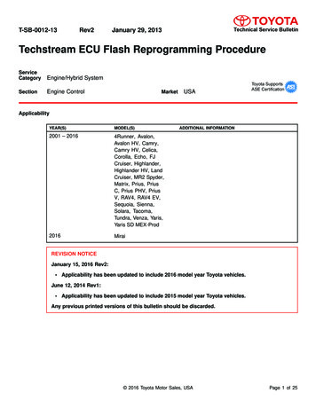
OSK stimulates axon regenerationAlmost all species experience a decline in regenerative potential during ageing. In mammals, one of the first systems to lose regenerativepotential is the CNS5–7. Retinal ganglion cells (RGCs) of the CNS projectaxons away from the retina to form the optic nerve. If damaged, RGCsof embryos and neonates can regenerate axons, but this capacity islost within days after birth, probably due to a molecular program thatis intrinsic to the RGCs6,22. Attempts to reverse optic-nerve damagehave been only moderately successful, and there are currently notreatments that can restore eyesight either in late-stage glaucomaor in old age23.To test whether it is possible to provide adult RGCs with the regenerative capacity of younger RGCs, we induced OSK in an optic-nervecrush-injury model. To deliver genes into the retina, Tet-Off AAV2scarrying an OSK polycistron were injected into the vitreous body,resulting in DOX-responsive OSK gene expression in around 40% ofRGCs, as estimated by immunostaining (Fig. 1b, Extended Data Fig. 2a,b). Even after 15 months of continuous induction, OSK expression didnot induce any tumours or structural changes in the retina (ExtendedData Fig. 2c–i, Supplementary Videos 1–3).In a separate cohort, RGCs were infected with Tet-Off AAV2 andoptic-nerve crush was performed two weeks later (Fig. 1c). Afteranother two weeks, Alexa Fluor 555-conjugated cholera toxin subunitB (CTB)—an anterograde axonal tracer—was delivered by intravitrealinjection to quantify axon regeneration. Independent of RGC proliferation (Extended Data Fig. 3a, b), the greatest extent of axon regenerationand RGC survival occurred when all three genes were delivered andexpressed as a polycistron within the same AAV particle (Fig. 1d, e).Notably, when polycistronic OSK was induced for 12–16 weeks, regenerating and sprouting RGC axon fibres extended over 5 mm into thechiasm, where the optic nerve connects to the brain (Extended DataFig. 3c, d). Suppression of polycistronic OSK by DOX treatment prevented both regenerative and survival effects (Fig. 1d, e, ExtendedData Fig. tTADOXTRETet-Off AAV2sTREOSKbOSKRPERodsConesGene ofinterestRBPMSKLF4Cross-section50002005001,000 1,500Distance from injury (μm)******1,000Estimated number ofregenerating axonsg12-month-old; 5 wpcTet-Offd2EGFPOct4Sox2Klf4Oct4 Sox2Oct4 Sox2 Klf4Oct4 Sox2 Klf4Oct4 Sox2 Klf4 offeRBPMSKLF4fd2EGFPPerfusion 2 weeks 2 daysP 1 10–4OSK offRetina flat-mountGanglion cellsOptic nerveAAV2***CTBP 1 10–4OSKBipolar cellsAmacrine cells 2 weeks1,000Horizontal cells***CrushrtTA4-week-old; 2 wpcTet-OnEstimated number ofregenerating axonsaOSKis an oncogene that reduces the lifespan of mice20 and is not requiredfor the initiation of cellular reprogramming21. We therefore excludedMYC from our experiments and expressed only the transcription factorsOCT4, SOX2 and KLF4 (OSK) in fibroblasts from old mice, after whichwe monitored the effect on genes that are known to be altered with age,including Lmnb1, Chaf1b, and those from the H2a and H2b families13.Even without MYC, the expression of OSK for five days promoted ayouthful mRNA profile, with no apparent loss of cellular identity orinduction of NANOG—a transcription factor that is indicative of pluripotency and oncogenicity (Extended Data Fig. 1a–c).Next we tested the long-term safety of ectopically expressing OSKin vivo. To deliver and control OSK expression in mice, we engineereda dual adeno-associated virus (AAV) system under the tight controlof a tetracycline response element (TRE) promoter (Fig. 1a). The TREpromoter can be activated either by reverse tetracycline-controlledtransactivator (rtTA) in the presence of the tetracycline derivative doxycycline (DOX) (‘Tet-On’) or by tetracycline-controlled transactivator(tTA) in the absence of DOX (‘Tet-Off’). Extraneous AAV sequences wereremoved from the vector to accommodate OSK as a polycistron. To testif AAV-mediated OSK expression was toxic in vivo, we co-infected young(5-month-old) and aged (20-month-old) mice with AAV9-rtTA;TRE-OSKvia intravenous delivery. OSK expression was then induced by providingDOX in the drinking water, our rationale being that the weak tropismof AAV for the digestive system might avoid the dysplasia and weightloss that is seen in transgenic mouse models18 (Extended Data Fig. 1d–i).After 10–18 months of continuous OSK induction, no increase in tumourincidence or negative effects on overall health were observed, indicating that cellular identity was maintained in OSK-expressing cells(Extended Data Fig. 1j–n).P 1 10–4800600400d2EGFP Ganglion cellsOSKd2EGFP Amacrine cellsOSKP 0.00120002005001,000 1,500Distance from injury (μm)Fig. 1 AAV2-delivered polycistronic OSK promotes axon regeneration andRGC survival after optic nerve injury. a, Schematic of the Tet-On and Tet-Offdual AAV vectors. OSK denotes Oct4-P2A-Sox2-T2A-Klf4. b, Schematic (top) andrepresentative retinal wholemount (bottom left) and cross-sections (bottomright) (n 10), two weeks after intravitreal injection of AAV2-tTA;TRE-OSK,showing Klf4 (green) expression in the RGCs (blue, visualized by RBPMS asmarker). Scale bars, 1 mm (left); 100 μm (right). c, Experimental outline of theoptic-nerve crush-injury study using the Tet-Off system. 555-CTB was used foranterograde axonal tracing. d, The number of axons 16 days after crush injury,at distances distal to the lesion, in mice treated with AAV2 encoding:destabilized enhanced green fluorescent protein (d2EGFP); Oct4; Sox2; Klf4;Oct4 and Sox2; all three OSK genes on three monocistronic AAV2s (indicated asseparated by signs); or all three OSK genes on a single polycistronic AAV2(n 5, 4, 4, 4, 4, 4 and 7 eyes, respectively). In d, e, suppression of polycistronicOSK expression by DOX (n 5 eyes) is shown as OSK off. e, Representativeimages of longitudinal sections through the optic nerves of 4-week-old (young)mice after receiving intravitreal injections of AAV2-tTA;TRE-OSK in thepresence (n 5) or absence (n 7) of DOX. CTB-labelled axons are shown 16 dayspost-crush. Asterisks indicate the optic-nerve crush site. f, Representativeconfocal images of longitudinal sections through the optic nerve showingCTB-labelled axons in 12-month-old (old) mice, 5 weeks post-crush, withAAV2-mediated expression of either GFP or OSK. Representative results fromn 5 eyes. Scale bars (e, f), 200 μm. g, The number of axons 16 days post-crushat multiple distances distal to the lesion site in transgenic Cre lines thatselectively express OSK either in RGCs (Vglut2-Cre) or in amacrine cells(Vgat-Cre) after intravitreal injection of AAV2-FLEx-tTA;TRE-OSK (n 4 eyes foreach condition). Further details are given in Extended Data Fig. 4e–h. One-wayANOVA with Bonferroni correction (d, g), with comparisons to d2EGFP shownin f. All data are presented as mean s.e.m.When Oct4, Sox2 and Klf4 were co-delivered by separate monocistronic AAVs, no effect on axon regeneration was observed; this wasostensibly due to the lower frequency of co-infection or inconsistent stoichiometry (Fig. 1d, Extended Data Fig. 3f–i). When deliveredsingly, Oct4 or Sox2 alone increased RGC survival slightly (ExtendedData Fig. 3e), but none of the single factors alone had any effect onregenerative capacity (Fig. 1d). Because Klf4 can repress rather thanpromote axonal growth24, we tested a dual-cistron of only Oct4 andSox2 and observed no regenerative effect.Nature Vol 588 3 December 2020 125
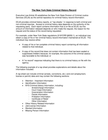
ArticleOSK counters injury-induced DNA methylation ageingCurrently, there is no known treatment that can induce robust RGC axonregeneration when the treatment is started after crush injury. Usingour Tet-On AAV system, which rapidly induces OSK expression (Fig. 2a,Extended Data Figs. 2b, 5d, e), we observed significant improvementin axon regeneration and RGC survival, even when OSK was inducedafter injury. The longer the duration of the OSK induction the greaterthe distance the axons extended, with no sign of RGC proliferation(Fig. 2b, c, Extended Data Fig. 5f).Given the effectiveness of OSK induction after injury and the abilityof Yamanaka factors to reverse DNA methylation age in vitro4,15,31,32, wespeculated that OSK might act to regenerate axons by counteracting theeffect of injury on DNA methylation. The DNA methylation age of RGCswas calculated on the basis of a ribosomal DNA methylation clock33,which provided the best site coverage (67 out of 72 CpG sites) and washighly correlated with chronological age (Extended Data Fig. 5g). At day4 after injury, RGCs experienced an acceleration of DNA methylationage, whereas OSK expression counteracted this effect (Fig. 2d). Patterns of global DNA methylation were also altered by injury in a mannersimilar to that of ageing, without affecting average DNA methylationlevels (Extended Data Fig. 5h, i). Notably, OSK reversed the global DNAmethylation changes caused by injury, and this reversal was enrichedat genes that are associated with light detection and synaptic transmission (Fig. 2e, Extended Data Fig. 5j, k).4-week- 2 weeks 2–4 weeks 2 daysoldPre-inj. DOXPost-inj. DOXde0.6P 0.867P 0.007P 0.9520.40.2Methylated (%)Water10001,500 P 0.8251,000P 0.00750002 wpccOffOffPre-inj. onPost-inj. onP 0.0012,000P 0.016Post-inj. OnCTB PerfusionIntact Injured InjuredGFPGFPOSKf1,5001,000GFP GFP OSKIntact***InjuredP 1 10–4Tet2 WTTet2 WT (OSK)Tet2 cKOTet2 cKO (OSK)P 3 10–450000–***4 wpcEstimated number ofregenerating axonsbCrushNumber of RGCs per mm2artTA/TRE-OSKDNA methylation ageMany regenerative approaches that work well in young individualsoften fail in older ones25,26. In 12-month-old mice, OSK treatment doubled RGC survival, similar to the results observed in 1- and 3-month-oldmice (Extended Data Fig. 4a). Although axon regeneration was slightlyweaker in the older mice (Extended Data Fig. 4b), when treatment wasextended for 3 more weeks (thus measuring regeneration 5 weeks afterthe optic-nerve crush), robust axon regeneration still occurred, withno increase in RGC number (Fig. 1f, Extended Data Fig. 4c, d). Thesedata indicate that ageing does not greatly diminish the ability of OSKtranscription factors to induce axon regeneration.To test whether OSK was acting directly on RGCs or through amacrinecells27—the other type of retinal cells that AAV2 can infect after intravitreal injection—we introduced tTA in a double-floxed inverse orientation (AAV2-FLEx-tTA) with AAV2-TRE-OSK into two Cre transgenicmouse lines, so that OSK was selectively expressed either in RGCs(Vglut2-Cre) or amacrine cells (Vgat-Cre) (Extended Data Fig. 4e, f).OSK expression in RGCs alone promoted axon regeneration, whereasexpression in amacrine cells did not (Fig. 1g, Extended Data Fig. 4g, h).The survival frequency of OSK-positive RGCs was roughly three timesthat of nearby OSK-negative cells or that of RGCs in GFP-expressingretinas, resulting in a greater relative abundance of OSK-positive RGCsafter injury (Extended Data Fig. 4i–k). Therefore, the ability of OSK toincrease survival seems to be mediated within the RGC and is largelycell-autonomous. The mRNA levels of Stat3, a pro-regeneration gene28,robustly increased in response to OSK and were maintained post-injury(Extended Data Fig. 5a, Supplementary Table 1). Stat3 upregulation,however, does not seem to be mediated by the PTEN–mTOR–S6K orthe SOCS3 pathways, two canonical Stat3 regulatory pathways thataffect RGC axon regeneration29,30 (Extended Data Fig. 5b, c, Supplementary Table 1).200 500 1,000 1,500Distance from injury (μm)Fig. 2 DNA demethylation is required for OSK-induced axon regenerationafter injury. a, Strategies for induction of OSK pre- and post-injury. b, Survivalof RGCs (number of RGCs per mm2) at 2 and 4 weeks post-crush (wpc) inresponse to OSK induction either pre- or post-injury (inj.) (n 4 eyes, exceptfor the post-injury ‘on’ group at 2 weeks post-crush, for which n 7 eyes).c, Representative longitudinal sections through the optic nerve showingCTB-labelled axons at 4 weeks post-crush, without (top) or with (bottom) theinduction of OSK after injury. Asterisks indicate the optic-nerve crush site.Results are representative from n 4 eyes. Scale bars, 200 μm. d, RibosomalDNA methylation age (in months) of 6-week-old RGCs isolated from axon-intactretinas infected with or without GFP-expressing AAV2 (intact), or fromaxon-injured retinas infected with GFP- or OSK-expressing AAV2 at 4 dayspost-crush (injured) (n 6, 4, 8 and 8 eyes, respectively). DNA methylation ageestimates of neurons tend to be lower than their chronological ages but remaincorrelated (see Extended Data Fig. 5g and Methods). e, Hierarchical clusteredheat map of DNA methylation levels of CpGs that significantly changed in RGCsafter crush injury (intact GFP vs injured GFP, q 0.05) and the effect of OSKat 4 days post-crush (injured OSK). f, Axon regeneration in response to OSKexpression (AAV2-tTA; TRE-OSK), 16 days post-crush in Tet2flox/flox mice infectedwith saline (Tet2 wild type (WT); n 3 for each condition) or AAV2-Cre(Tet2 cKO; n 4 for each condition). One-way ANOVA with Bonferroni’s multiplecomparison test (b, d, f). For all bar graphs, data are mean s.e.m.upregulated by OSK35,36 (Extended Data Fig. 6a), we tested the effect ofknocking down their encoding genes Tet1 or Tet2 (using short hairpin(sh)RNA delivered by AAV37) in the context of damaged, OSK-treatedRGCs (Extended Data Fig. 6b–d).The knockdown of either Tet1 or Tet2 blocked the ability of OSK toincrease Stat3 mRNA levels and to promote RGC survival and axonregeneration (Extended Data Fig. 6e–g). Knocking out Tet2 in RGCsby delivering AAV2-Cre30 intravitreally to Tet2flox/flox mice also abrogated the increase in regeneration and survival (Fig. 2f, Extended DataFig. 6h–j). OSK-expressing RGCs showed no evidence of DNA replicationin a 5-bromo-2′-deoxyuridine (BrdU) assay (Extended Data Fig. 3a, b),and knockdown of Tdg38 completely abolished the beneficial effectsof OSK (Extended Data Fig. 7a–d); however, a global reduction of DNAmethylation mediated by overexpression of the TET1 catalytic domain39had no protective or regenerative effect (Extended Data Fig. 7e–h).These data indicate that active DNA demethylation is necessary—but isnot sufficient by itself—in order for OSK to protect RGCs and to regenerate axons (Extended Data Fig. 7i).Axon regeneration in human neuronsRegeneration requires DNA demethylationNext we asked whether the DNA methylation changes induced by OSKare necessary for RGC survival and axon regrowth. DNA demethylationis catalysed by ten-eleven translocation methylcytosine dioxygenases(TET1–TET3), either passively through DNA replication or actively—even in non-replicating cells—by reversion to cytosine via thymineDNA glycosylase (TDG)34. Because TET1 and TET2—but not TET3—were126 Nature Vol 588 3 December 2020Paralleling the mouse RGC data, the expression of OSK in differentiatedhuman neurons effectively counteracted axonal loss and the advancement of DNA methylation age induced by vincristine—a chemotherapeuticdrug that induces axon injury—in the absence of cell proliferation or globalDNA demethylation (Extended Data Fig. 8a–g). Nine days after damage,the neurite area was 15-fold greater in the rejuvenated OSK-transducedcells compared with control cells (Extended Data Fig. 8h, i).
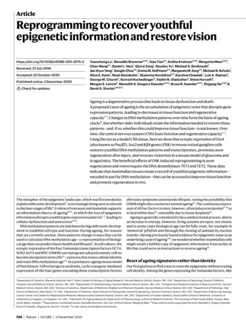
estoration of vision in old miceThe finding that OSK induction could effectively restore the DNA methylation age of RGCs after injury indicated that vision loss caused bynatural ageing might be reversible too. To test this, we treated 3- and11-month-old mice with OSK AAVs and induced OSK expression for4 weeks (Fig. 4a). Optomotor response and PERG measurements werethen carried out. In the absence of OSK induction, there was a significantreduction in visual acuity and in RGC electrical activity by 12 months ofage—a loss that was restored by OSK induction (Fig. 4b, Extended DataFig. 9e). Restoration of visual acuity was not seen in 18-month-old mice(Extended Data Fig. 9e, f), probably because assessment was hinderedby an age-dependent increase in corneal opacity43.Because there was no obvious increase in the number of RGCs orin axon density in the 12-month-old mice treated with OSK AAV(Extended Data Fig. 9g, h), we hypothesized that the visual improvements were due to transcriptomic changes. RGCs from 12-month-oldmice, either untreated or treated with AAVs for 4 weeks, were purifiedby fluorescence-activated cell sorting and analysed by RNA sequencing. Compared with RGCs from young (5-month-old) mice, the mRNAlevels of 464 genes were altered during ageing. Around 90% of thesewere restored to youthful levels by OSK treatment (Fig. 4c, d, ExtendedData Fig. 9i, Supplementary Table 2). Of the 268 age-downregulatedgenes, 44 are linked to sensory perception and axon targeting44, raising the possibility that they contribute to the decline in vision causedby ageing (Fig. 4d, Extended Data Figs. 9j, 10a). Another 196 genesthat were upregulated during ageing are involved in ion transport orneuronal projection development, including Efemp1 (Extended Data3020100ce07142128Days post-injectionStriped pattern rotationLeft eye12BaselineTreated (–OSK)96P13P10N1N1–3N2 N2–612Amplitude (μV)IOP (mmHg)Recovery of vision in mice with glaucomaAAV expression 4 weeks 4 weeksSalineMicrobeadsP 1 10–4Right eye0.1 cycles per degree6P13N10N1–3N2 N2–6fBaselineTreated ( OSK)P190Acuity90 180 270 360Time (ms)SalineBeads (PBS)0.4 cyclesper degreeP 0.025dOptomotor acuity(cycles per degree)We then tested whether OSK induction could restore the function ofRGCs in a disease setting. Glaucoma, a leading cause of age-relatedblindness worldwide, is characterized by a progressive loss of RGCs andtheir axons, and often coincides with increased intraocular pressure.We induced increased intraocular pressure unilaterally for 21 days byinjecting microbeads into the anterior chamber of the eye40 (Fig. 3a, b),using saline injections as non-glaucomatous controls. At the 4-weektime point, after a significant decrease in axonal density and the number of RGCs (baseline, Extended Data Fig. 9a, b), AAVs were injectedintravitreally and OSK expression was induced for another 4 weeks(Fig. 3a). Compared with glaucomatous eyes that received either PBSor AAVs with no OSK induction ( OSK), the OSK-treated glaucomatouseyes ( OSK) presented with a restored axon density equivalent to thatin the non-glaucomatous eyes, with no evidence of RGC proliferation(Extended Data Fig. 9c, d).A significant increase in visual acuity relative to the pre-treatmentfunction baseline was observed for eyes treated with OSK: the optomotor response assay indicated that half of the visual acuity lost fromincreased intraocular pressure was restored (Fig. 3c, d). A similarimprovement associated with OSK expression was also seen in pattern electroretinogram (PERG) analysis (N1 P1), a readout of electricalwaves generated specifically by RGCs (Fig. 3e, f). To our knowledge, thisis the first example of vision-loss reversal after glaucomatous injuryhas occurred; previous attempts have focused on neuroprotectiondelivered at an early stage to prevent further disease progression41,42(Supplementary Information).HistologyIncreased IOPbFunctionpost-treatmentAmplitude (μV)a0.45P 0.999P 0.999P 0.0030.300.150Beads (–OSK)Beads ( OSK)P 0.010P 0.00415Amplitude (μV)OSK-mediated recovery of the neurite area, as well as both the number and the length of axons, were blocked by Tet2 knockdown but notby inhibition of the mTOR–S6K pathway (Extended Data Fig. 8j–s).Again, overexpression of the TET1 catalytic domain alone failed topromote axon regeneration (Extended Data Fig. 8t). The ability of OSKto reprogram neurons and to promote axon growth in a Tet2-dependentmanner therefore seems to be conserved across mice and humans.P 0.999P 0.999P 0.002P 1 10–41050BaselineTreatedBaselineTreatedFig. 3 Four weeks of OSK expression reverses vision loss afterglaucomatous damage has already occurred. a, Experimental outline of theinduced glaucoma studies using 8-week-old mice. b, Intraocular pressure (IOP)measured weekly by rebound tonometry for the first 4 weeks after the injectionof microbeads (n 29 mice) or saline (n 10 mice). c, High-contrast visualstimulation to measure optomotor response. Reflexive head movements weretracked in response to the rotation of a moving striped pattern that increasedin spatial frequency. d, Optomotor response of each mouse before treatment(baseline) and 4 weeks after intravitreal injection of AAVs (treated; see f for key)(n 10, 7, 12 and 12 mice for each condition; similar results from 3 independentexperiments combined). e, Representative PERG waveforms in response to areversing contrast checkerboard pattern, recorded from the same eye both atthe pre-injection baseline and at 4 weeks after OSK (top) or OSK (bottom)AAV injection. f, Mean PERG amplitudes of recordings from each mouse in d atthe baseline before treatment and at 4 weeks after intravitreal AAV injection(n 10, 7, 12 and 12 mice for each condition; similar results from 3 independentexperiments combined). OSK (not induced), AAV2-rtTA;TRE-OSK; OSK(induced), AAV2-tTA;TRE-OSK. For b, a two-way ANOVA with Bonferronicorrection between groups was used; for d, f, a two-way mixed ANOVA withBonferroni correction between groups was used for the overall effect of timeand treatment, and before and after treatments were analysed using a pairedtwo-tailed Student’s t-test. Data in b, d and f are presented as mean s.e.m.Fig. 10b–d), a TET1- and TET2-regulated gene that is associated withglaucoma45 and age-related macular degeneration46.To gain insights into how DNA methylation changes might be involvedin the effects of OSK, we used machine learning to identify a methylationageing signature based on 1,226 sites from genome-wide DNA methylation datasets, derived from the first principal component (PC1) inmulti-age training samples (Extended Data Fig. 10e, SupplementaryTable 3). In an independent set of test samples, OSK reversed the DNAmethylation ageing signature in the context of both injury and ageing(Fig. 4e, Extended Data Fig. 10f). Notably, the signature CpG sites wereassociated with genes that are involved in synaptic and neuronal processes and are enriched for the binding of polycomb repressive complexNature Vol 588 3 December 2020 127
ArticleVision?aOptomotor acuity(cycles per degree) 4 weeks Transcriptome?DNA methylation?Old miceYoungP 0.228P 2 10–4YoungOld0.30.20.10OldOld(–OSK) Old( OSK)d81.561.0log2( OSK/–OSK)log2(1 CPM)P 0.0340.4AAV2cP 0.814–OSK OSKb0.50420 2Significant in ageingSignificant in bothSignificant in both (sensory) Up by OSK Down in ageing 4 6 402 2log2(Old/young)46P 0.999ef0.6P 0.999P 0.028P 0.034Optomotor acuity(cycles per degree)DNA methylationageing signatureP 0.0110.40.20–0.2–OSK OSK0.4model system, we present evidence that the ectopic expression ofOSK transcription factors safely induces in vivo epigenetic restoration of aged CNS neurons, without causing a loss of cell identity orpluripotency. Instead, OSK promotes a youthful epigenetic signatureand gene-expression pattern that causes the neurons to function asthough they were young again. The requirement for active demethylation in this process supports the idea that changes in DNA methylationpatterns are involved in the ageing process and its functional reversal(Extended Data Fig. 10n). However, we do not wish to imply that DNAmethylation is the only epigenetic mark involved in this process. It islikely to involve other transcription factors and epigenetic modifications, such as the PRC2 complex and H3K27me3, both of which areinvolved in maintaining stem cell plasticity and are associated withDNA methylation clock sites4,49,50.Perhaps the most interesting question raised by these data is howcells encode and store youthful epigenetic information. Possibiliti
epigenetic information and restore vision Yuancheng Lu 1, Benedikt Brommer 2,3 ,11, Xiao Tian 1,11, Anitha Krishnan 3,4 ,11, . regeneration and vision require the DNA demethylases TET1 and TET2. These data . Paul F. Glenn Center for Biology of Aging Research, Harvard Medical School, Boston, MA, USA. 2Department of Neurology, Boston Children's