Transcription
Journal of Pathology and Translational Medicine 2016; 50: 270-277http://dx.doi.org/10.4132/jptm.2016.03.19 ORIGINAL ARTICLE Stromal Expression of MicroRNA-21 in Advanced ColorectalCancer Patients with Distant MetastasesKyu Sang Lee1 · Soo Kyung Nam1Jiwon Koh2 · Duck-Woo Kim3Sung-Bum Kang3 · Gheeyoung Choe1,2Woo Ho Kim2 · Hye Seung Lee1Department of Pathology, Seoul NationalUniversity Bundang Hospital, Seongnam;2Department of Pathology, Seoul NationalUniversity College of Medicine, Seoul;3Department of Surgery, Seoul NationalUniversity Bundang Hospital, Seongnam, Korea1Received: February 12, 2016Revised: March 15, 2016Accepted: March 18, 2016Corresponding AuthorHye Seung Lee, PhDDepartment of Pathology, Seoul National UniversityBundang Hospital, 82 Gumi-ro 173beon-gil,Bundang-gu, Seongnam 13620, KoreaTel: 82-31-787-7714Fax: 82-31-787-4012E-mail: hye2@snu.ac.krBackground: The aim of this study was to determine the regional heterogeneity and clinicopathological significance of microRNA-21 (miR-21) in advanced colorectal cancer (CRC) patients withdistant metastasis. Methods: miR-21 expression was investigated by using locked nucleic acid–fluorescence in situ hybridization in the center and periphery of the primary cancer and in distantmetastasis from 170 patients with advanced CRC. In addition, α-smooth muscle actin and desmin were evaluated to identify cancer-associated fibroblasts (CAFs) by using immunohistochemistry. Results: The miR-21 signal was observed in the cancer stroma. The expression of miR-21 (ascore of 1–4) in the center and periphery of the primary cancer and in distant metastasis was observed in specimens from 133 (78.2%), 105 (61.8%), and 91 (53.5%) patients, respectively. miR-21expression was heterogeneous in advanced CRC. Discordance between miR-21 expression inthe center of the primary cancer and either the periphery of the primary cancer or distant metastasis was 31.7% or 44.7%, respectively. miR-21 stromal expression in the periphery of the primarycancer was significantly associated with a better prognosis (p .004). miR-21 expression wassignificantly associated with CAFs in the center of the primary cancer (p .001) and distant metastases (p .041). Conclusions: miR-21 expression is observed in cancer stroma related to theCAF quantity and frequently presents regional heterogeneity in CRC. Our findings indicate thatthe role of miR-21 in predicting prognosis may be controversial but provide a new perspective ofmiR-21 level measurement in cancer specimens.Key Words: Colorectal neoplasms; MicroRNA-21; Neoplasm metastasis; Genetic heterogeneityMicroRNAs (miRNAs) are small (18–25 nucleotides), endogenous, non-coding RNAs that act as post-transcriptional modulators of all cellular processes, including proliferation, differentiation, and apoptosis.1 Alterations in miRNA expression areassociated with the deregulation of oncogenes and tumor suppressor genes.2 The discovery that miRNA expression is deregulated in cancer suggests the potential of miRNA as biomarkersfor cancer.3 Therefore, targeting miRNAs is regarded as a potential therapeutic strategy and may be a promising diagnostic tool.MiRNA-21 (miR-21) is consistently upregulated in variouscancers, including the colon, stomach, lung, and breast cancer.4-7Previous studies proved that miR-21 expression correlates withcarcinogenesis.8-11 Some studies indicated that miR-21 expressionis closely associated with poor prognosis in various cancers.4,12,13In colorectal cancer (CRC), miR-21 functions as an onco-miRNAdue to its key roles in proliferation, invasion, and metastasis.10,14Although the mechanisms underlying the regulation of miR-21in CRC remain to be defined, miR-21 can promote tumorigenesis in CRC. Some studies reported that plasma miR-21 is a potential noninvasive biomarker for early detection and prognosisof CRC.15-17 Additionally, miR-21 expression is greatly increasedin chemotherapy-resistant CRC cells.18,19 Recent reports suggestthat increased expression of miR-21 predicts poor prognosis inpatients with CRC.20-22 However, these results were restrictedto patients with stage II CRC.21-24 Thus, the prognostic value ofmiR-21 alterations in patients with advanced CRC presentingmetastases is controversial.We previously evaluated cancer-associated fibroblasts (CAFs)in advanced CRC with synchronous or metachronous distant metastases.25 CAFs facilitate the communication between tumorcells and the tumor microenvironment. In addition, they regulatetumor invasion and metastasis.26,27 Interestingly, most studies reported that miR-21 was predominantly observed in CAFs, notin cancer cells.21,22,24 These findings point to a dynamic malignant 2016 The Korean Society of Pathologists/The Korean Society for Cytopathology270This is an Open Access article distributed under the terms of the Creative Commons Attribution Non-Commercial License (http://creativecommons.org/licenses/by-nc/3.0) which permits unrestricted non-commercial use, distribution, and reproduction in any medium, provided the original work is properly cited.pISSN 2383-7837eISSN 2383-7845
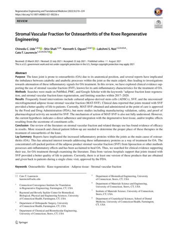
miR-21 in Advanced Colorectal Cancer 271role of CAFs through miR-21 expression in CRC. Nevertheless,the correlation between miR-21 and CAF status is yet to be demonstrated in CRC.Recently, systemic chemotherapy and targeted therapy havebeen used in patients with advanced CRC, increasing patient survival.28 Despite advances in medicine, some patients with CRCrespond poorly.29 Although the reasons for drug resistance are notfully understood, they may be related to the presence of tumorheterogeneity.30-32 Previous reports indicated that regional heterogeneity of miRNA expression is observed in various cancertypes.33-35 Therefore, variation of miR-21 expression betweenthe primary tumor and metastatic sites needs to be elucidated inpatients with advanced CRC.The aim of this study was to evaluate the clinical significanceof miR-21 and analyze its heterogeneity in patients with advanced CRC presenting metastases. Additionally, we analyzed thecorrelation between CAFs and miR-21 status.MATERIALS AND METHODSPatients and samplesTo evaluate the clinicopathological significance and heterogeneity of miR-21 status, 170 patients with advanced CRC presenting synchronous or metachronous metastases who had undergone surgical resection at Seoul National University BundangHospital between May 2003 and December 2009 were enrolled.The clinicopathological characteristics were obtained from thepatients’ medical records and pathology reports. Follow-up information, including patient’s outcome and the interval betweenthe date of surgical resection and death was collected. Data frompatients lost to follow-up or those who had died from causesother than CRC were censored.Ethical statementAll samples were obtained from surgically resected tumorsexamined pathologically at the Department of Pathology, SeoulNational University Bundang Hospital. All samples and medical record data were anonymized before use in this study and theparticipants did not provide written informed consent. The useof medical record data and tissue samples for this study was approved by the Institutional Review Board of Seoul National University Bundang Hospital (reference No. B-1109/136-302).Tissue array methodtive areas of the donor blocks were obtained and rearranged intonew recipient blocks (Superbiochips Laboratories, Seoul, Korea).A single core was 2 mm in diameter and those containing 20%tumor cells were considered valid cores.miRNA in situ hybridizationTissue microarray slides were processed by using locked nucleic acid (LNA)–fluorescence in situ hybridization (FISH) oligonucleotide probes for miR-21 and U6, both labeled with fluorescein at the 5'-end, according to the protocol described in thepreparation protocol. After deparaffinization, the slides were incubated with proteinase-K and endogenous peroxidase wasblocked with 3% H2O2. Next, the slides were incubated withhybridization mix containing 1 nM LNA U6 snRNA probe(Exiqon, Vedbaek, Denmark) and 20 nM doubled-DIG LNAmiR-21 probe (Exiqon) in ThermoBrite (Abbott Laboratories,Abbott Park, IL, USA) for 1 hour at 50 C. Next, the slides wereincubated in blocking solution and antifluorescein–horseradishperoxidase antibody (1:125, PerkinElmer, Shelton, CT, USA)for 1 hour. The signals were then amplified using TyramideSignal Amplification (TSA-plus FITC, 1:50, PerkinElmer) for10 minutes at room temperature. After incubation, the slideswere mounted directly with SlowFade Gold antifade reagentwith DAPI (Invitrogen, Carlsbad, CA, USA). All steps beginning with hybridization were performed in the dark.The experimental data were interpreted according to the instructions in the RNAscope FFPE Assay Kit (Advanced CellDiagnostics, Hayward, CA, USA): no staining observed at 40 magnification (score of 0); difficult to see at 40 magnification(score of 1); difficult to see at 20 magnification, but detectablestaining at 40 magnification (score of 2); difficult to see at 10 magnification, but detectable staining at 20 magnification (scoreof 3); and detectable staining at 10 magnification (score of 4).A score of 1–4 indicates miR-21 overexpression (Fig. 1).ImmunohistochemistryArray slides were labeled by immunohistochemistry usingantibodies for smooth muscle actin (SMA; 1:1,000, Neomarkers,Fremont, CA, USA) and desmin (1:300, Dako, Glostrup, Denmark) after a microwave antigen retrieval procedure, except forSMA. The staining procedures were carried out using the ultraView Universal DAB Kit (Ventana Medical Systems, Tucson,AZ, USA) and an automated stainer (BenchMark XT, VentanaMedical Systems), according to the manufacturer’s instructions.Surgically resected primary CRC specimens were formalinfixed and paraffin-embedded (FFPE). For each case, 9http://jpatholtm.org/
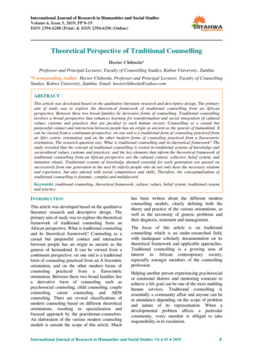
272 Lee KS, et al.ABFig. 1. MicroRNA-21 (miR-21) staining using locked nucleic acid–based in situ hybridization in colorectal cancer. (A) miR-21 expression isobserved in cancer stromal tissue. (B) No miR-21 expression is observed in normal colonic mucosa.Table 1. Heterogeneity of miR-21 expression with respect to tumor location in advanced CRCmiR-21 expressionPeripheryDistant gativePositive24 (14.1)13 (7.6)20 (11.8)17 (10.0)41 (24.1)92 (54.1)59 (34.7)74 (43.5)Total170 (100)170 (100)Values are presented as number (%). p-values are calculated by using chi-square test or Fisher exact test.miR-21, microRNA-21; CRC, colorectal cancer.Calculation of CAFs using digital pathologyStatistical analysesCRC cells were considered as internal negative controls. Intestinal muscular layer or medium- to large-sized vessels were considered as internal positive controls for desmin and SMA. Samples showing inappropriate staining in internal negative or positivecontrols were considered non-informative and were excludedfrom the analysis. Slides were scanned using an Aperio ScanScopeH CS instrument (Aperio Technologies Inc., Vista, CA, USA)at 20 magnification. Because desmin-positive muscularis mucosa and propria are positive for SMA staining, the area of CAFs(mm2) was calculated by subtracting the areas of desmin staining from that of SMA staining (SMA-desmin).The association between the clinicopathological features andmiR-21 status was analyzed by using the chi-square or Fisherexact test, as appropriate. The correlation between miR-21 expression and CAFs was examined by using the Mann-Whitneytest. The patients’ survival was analyzed by using the KaplanMeier method and the log-rank test was used to determine ifthere were any significant differences between the survival curves.A p-value .05 was considered statistically significant. All statistical analyses were performed using the SPSS statistics ver. 21software (IBM Corp., Armonk, NY, USA).RESULTSMicrosatellite instabilityMicrosatellite instability (MSI) was assessed in 160 CRC caseswith available tissue. MSI results were generated by comparingthe allelic profiles of five microsatellite markers (BAT-26, BAT25, D5S346, D17S250, and D2S123) in the tumors and corresponding normal samples. Polymerase chain reaction productsfrom the FFPE tissues were analyzed using an automated DNAsequencer (ABI 3731 Genetic Analyzer, Applied Biosystems, Foster City, CA, USA) according to the protocol.http://jpatholtm.org/miR-21 stromal expression and regional heterogeneity inadvanced CRC patientsThe miR-21 signal was predominantly observed in the stromal compartment of the cancer and the normal mucosa was negative for miR-21 (Fig. 1). The snRNA U6 signal was observedin the nucleus of all cell types. To evaluate the regional heterogeneity of miR-21 expression, we examined tissues from threesites, including the center and periphery of primary cancer aswell as distant metastases for each patient with advanced CRC.In the center of primary tumors, a miR-21 FISH score of 0 washttp://dx.doi.org/10.4132/jptm.2016.03.19
miR-21 in Advanced Colorectal Cancer 273observed in 37 (21.8%), a score of 1 in 16 (9.4%), a score of 2 in56 (32.9%), a score of 3 in 46 (27.1%), and a score of 4 in 15(8.8%) patients with CRC. In the periphery of primary tumors,a miR-21 FISH score of 0 was observed in 65 (38.2%), a scoreof 1 in 14 (8.2%), a score of 2 in 46 (27.1%), a score of 3 in 44(25.9%), and a score of 4 in one (0.6%) patient with CRC. Additionally, in distant metastatic tumors, a miR-21 FISH scoreof 0 was observed in 79 (46.5%), a score of 1 in 13 (7.6%), ascore of 2 in 43 (25.3%), a score of 3 in 32 (18.8%), and a scoreof 4 in three (1.8%) patients with CRC. miR-21 stromal expression (a score of 1–4) in the center and periphery of primary tumor as well as in distant metastasis was observed in 133 (78.2%),105 (61.8%), and 91 (53.5%) patients with CRC, respectively.The heterogeneity of miR-21 status according to tumor location is shown in Table 1. Of the 170 cases, discordance betweenmiR-21 expression in the center and periphery was noted in 54Table 2. The association between clinicopathological parameters and expression of miR-21 in 170 advanced CRC patients with metastasisCenterVariableAge (yr)MeanSexMaleFemalepT stage0-23-4DifferentiationLGHGLocation of primary tumorRight colonLeft colonRectumLN metastasisAbsentPresentLymphatic invasionAbsentPresentPerineural invasionAbsentPresentVenous invasionAbsentPresentTumor borderExpandingInfiltrativeDistant metastasisSynchronousMetachronouspTNM stage at initial diagnosisI, IIIII, IVMSI lueNegativePositive60.759.7908021 (12.4)16 66860.459.669 (40.6)64 (37.6).59931 (18.2)34 (20.0)23 (13.5)14 (8.2)80 (47.1)53 (31.2).8251482233 (19.4)4 (2.4)115 (67.6)18 (10.6).6624169608 (4.7)12 (7.1)17 (10.0)33 (19.4)57 (33.5)43 (25.3)311397 (4.1)30 (17.6)24 (14.1)109 (64.1)551159 (5.3)28 9 (34.7)46 (27.1).28136 (21.2)43 (25.3)54 (31.8)37 (21.8).07336 (21.2)29 (17.1)67 (39.4)38 (22.4).27545 (26.5)34 (20.0)58 (34.1)33 (19.4).36750 (29.4)15 (8.8)98 (57.6)7 (4.1).00270 (41.2)9 (5.3)78 (49.5)13 (7.6).57520 (11.8)23 (13.5)22 (12.9)21 (12.4)46 (27.1)38 (22.4)20 (11.8)31 (18.2)28 (16.5)21 (12.4)38 (22.4)32 (18.8).9036 (3.5)59 (34.7)25 (14.7)80 (47.1).01715 (8.8)64 (37.6)16 (9.4)75 (44.1).81346 (27.1)87 (51.2).23815 (8.8)50 (29.4)40 (23.5)65 (38.2).04228 (16.5)51 (30.0)27 (15.9)64 (37.6).42219 (11.2)18 (10.6)64 (37.6)69 (40.6).72829 (17.1)36 (21.2)54 (31.8)51 (30.0).38841 (24.1)38 (22.4)42 (24.7)49 (28.8).4551185224 (14.1)13 (7.6)94 (55.3)39 (22.9).49742 (24.7)23 (13.5)76 (44.7)29 (17.1).28658 (34.1)21 (12.4)60 (35.3)31 (18.2).291121584 (2.4)33 (19.4)8 (4.7)125 (73.5).3144 (2.4)61 (35.9)8 (4.7)97 (57.1).7176 (3.5)73 (42.9)6 (3.5)85 (50.0).7991106023 (13.5)14 (8.2 )87 (51.2)46 (21.7).71445 (26.5)20 (11.8)65 (38.2)40 (23.5).33152 (30.6)27 (15.9)58 (34.1)33 (19.4).776201505 (2.9)32 (18.8)15 (8.8)118 (69.4).7094 (2.4)61 (35.9)16 (9.4)89 (52.4).0749 (5.3)70 (41.2)11 (6.5)80 (47.1).888157335 (21.9)1 (0.6)122 (76.3)2 (1.3).65059 (36.9)2 (1.3)98 (61.3)1 (0.6).30474 (46.3)3 (1.9)83 (51.9)0.06960.0.299.260.925Values are presented as number (%). p-values are calculated by using chi-square test or Fisher exact test.miR-21, microRNA-21; CRC, colorectal cancer; T, tumor; LG, low grade; HG, high grade; LN, lymph node; MSI, microsatellite instability; MSS, microsatellitestable; MSI-L, microsatellite instability–low; MSI-H, microsatellite 2016.03.19http://jpatholtm.org/
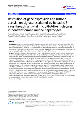
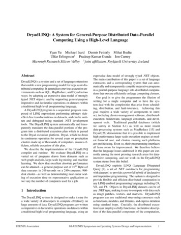
274 Lee KS, et al.Prognostic significance of miR-21 stromal expression inadvanced CRC patientscases (31.7%). Discordance between the center and distant metastasis was detected in 76 cases (44.7%). Thus, regional heterogeneity of miR-21 stromal expression was very common in patients with advanced CRC.All 170 patients with advanced CRC were successfully followedup for inclusion in the survival analysis (Fig. 2). The mean follow-up time was 42 months (range, 1 to 105 months) and 73patients (42.9%) died from cancer during the follow-up period.Kaplan-Meier analysis showed that miR-21 stromal expressionin the periphery of primary tumors was significantly associatedwith a better prognosis (p .004). There was no significant correlation between the patients’ prognosis and miR-21 expressionin the center of primary tumors and distant metastases (p .925and p .863, respectively).Clinical implications of miR-21 stromal expression inadvanced CRC patientsTable 2 shows the relationships between miR-21 status andthe clinicopathological parameters of patients with advancedCRC. In the periphery of primary tumor, miR-21 expression wascorrelated with less aggressive features, including histologic lowgrade differentiation (p .002). In addition, lymphatic invasionand lymph node metastasis was frequently observed in patientswith CRC negative for miR-21 (p .042 and p .017, respectively). There was no statistically significant correlation between theclinicopathological factors and miR-21 expression in the centerof the primary tumors and distant metastatic tumors (p .05).The expression of miR-21 in distant metastasis was different according to metastatic site (Table 3). miR-21 stromal expressionwas more frequently observed in lung metastasis and peritonealseeding (p .046).Correlation between miR-21 stromal expression and CAFvalueThe regional heterogeneous values for CAF in CRC are shownin our previous study.25 The area occupied by CAFs was the lowest in distant metastases (median, 0.91; interquartile range [IQR],0.68 to 1.18) than any other sites (median, 1.12; IQR, 0.88 to1.41 in the center of primary tumors and median, 1.22; IQR,0.96 to 1.54 in the periphery of primary tumors). Mann-Whitney test showed that miR-21 expression was significantly associated with CAFs in the center of primary tumors (p .001)and distant metastases (p .041). In the periphery of primary tumors, miR-21 overexpression was not correlated with CAFs (p .102) (Fig. 3).Table 3. Expression of miR-21 in distant metastasis according tometastatic site in 170 advanced CRC patientsSite of metastasisTotalLiverLungSeedingDistant nodesOvaryTotal763737218170miR-21 in metastasisNegativePositive42 (24.7)13 (7.6)13 (7.6)011 (6.5)7934 (20.0)24 (14.1)24 (14.1)2 (1.2)7 (4.1)91p-value.046DISCUSSIONMany predictive and prognostic molecular markers have beensuggested for CRC. However, they are not reliably accepted dueto a lack of reproducibility and validation. Previous studies pro-Values are presented as number (%).miR-21, microRNA-21; CRC, colorectal cancer.0.60.40.2p .9250.00204060 80 100 120Time (mo)A1.0NegativePositive0.8Overall survival0.8Overall survival1.0NegativePositive0.60.40.2p .0040.00204060 80 100 120Time (mo)BNegativePositive0.8Overall survival1.00.60.40.2p .8630.00204060 80 100 120Time (mo)CFig. 2. Kaplan-Meier curves illustrating the overall survival of patients with advanced colorectal cancer in relation to microRNA-21 expression.(A) Center. (B) Periphery. (C) Distant 10.4132/jptm.2016.03.19
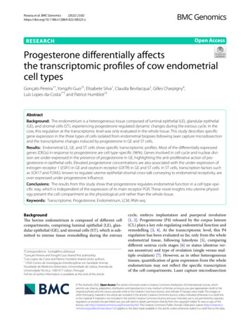
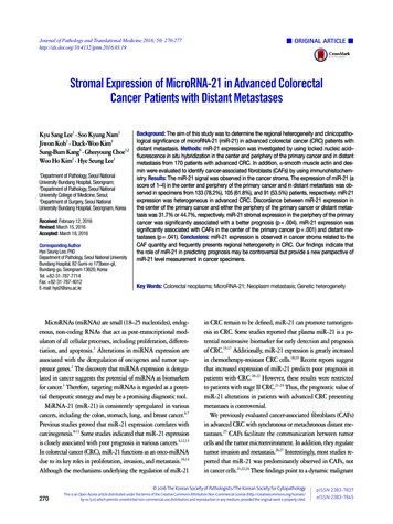
miR-21 in Advanced Colorectal Cancer 2751.00.50NegativePositivemiR-21 (center)1.00.50A1.5p .102CAF (median, 95% CI)1.5p .001CAF (median, 95% CI)CAF (median, 95% CI)1.5NegativePositivemiR-21 (periphery)1.00.50Bp .041NegativePositivemiR-21 (metastasis)CFig. 3. Correlation between microRNA-21 (miR-21) expression and cancer associated fibroblasts (CAFs) by Mann-Whitney analysis. (A) Center. (B) Periphery. (C) Distant metastasis. CI, confidence interval.posed that high expression of miR-21 might predict poor survival in patients with CRC.20-24 However, elevated miR-21 leveland poor prognosis correlated only in a subgroup of patientswith stage II CRC.21-24 Interestingly, our results contradict thoseof previous miR-21 expression studies. In our study, miR-21 expression in the periphery of primary tumors was significantlyassociated with a better prognosis in patients with advancedstage CRC (p .004) (Fig. 2). Otherwise, there was no significantcorrelation between the prognosis and miR-21 expression inthe center of primary tumors and in distant metastases (p .925and p .863, respectively) (Fig. 2). This discrepancy may be explained by the fact that our cohort was largely comprised of patients with stage IV CRC (98 cases, 57.6%). They received various personalized treatments and these might reflect the statisticaldifference.Bullock et al.36 recently demonstrated that stromal miR-21 expression induced a pro-metastatic mechanism of CRC via activation of matrix metalloproteinase-2. These data highlight theimportance of miR-21 deregulation in CRC metastasis. Becauseall patients in our study presented with advanced CRC with metastasis, miR-21 may be expressed at higher levels than thatobserved in previous reports. In our study, miR-21 expression inthe center and periphery of primary tumors and in distant metastasis was observed in 78.2%, 61.8%, and 53.5% of the patients, respectively, whereas it was observed in 27.4% of the patients in another study.22 Our data suggest that the lack of miR21 expression in patients presenting with CRC with metastasisis associated with a rather poor prognosis and more frequentlymphatic invasion and lymph node metastasis (Table 2, Fig. 1),presumably because, in these patients, the metastatic mechahttp://dx.doi.org/10.4132/jptm.2016.03.19nism is controlled by other regulatory pathways. Further studiesare necessary to prove that other mechanisms induce CRC metastasis, independently of miR-21. The evaluation of the prognostic value of miR-21 expression in patients with advanced CRCpresenting distant metastasis might be of little importance.Previous studies in various cancers indicated that miR-21 localizes mainly in the cancer stroma and more particularly in thestromal fibroblast-like cells.21,22,24 This localization may be dueto molecules secreted by cancer cells, which influence the microenvironment.37-39 Meanwhile, the mechanisms of regulationof miR-21 in CAFs remain unknown. Only few studies examined the correlation between the value of CAFs and miR-21 expression.40 Our study revealed that miR-21 expression was associated with CAF value in the center of primary tumors anddistant metastases (p .001 and p .041, respectively) (Fig. 3).As expected, the value of CAF was greater in stroma of CRCspecimens presenting miR-21 expression. Therefore, we shouldbe careful when evaluating stromal miR-21 expression. In mostprevious studies, miR-21 expression was evaluated quantitatively by image analysis.21,24,36 This method may mislead us whendistinguishing miR-21 high expression from a simple increasein the number of CAFs. Thus, accurate observation should beperformed to distinguish miR-21 expression from CAF increase.To clarify miR-21 expression in CRC, being aware of the heterogeneity in miR-21 expression level is crucial because regional heterogeneity can lead to sampling bias. In recent studies, intra-tumor heterogeneity of various miRNAs was detected incolorectal, pancreatic, and breast cancer.34,35,41 Our study indicatesmiR-21 regional heterogeneity, which constitutes approximately 40% of the total cohort (Table 1). Thus, a reliable assessmenthttp://jpatholtm.org/
276 Lee KS, et al.of CRC miR-21 expression may include sampling of the primarytumor in several locations and metastasis.In conclusion, we demonstrated that miR-21 is expressed inadvanced CRC and that this upregulation is mainly confined tothe cancer stroma. Our FISH data indicated that miR-21 expression is related to CAF value in the center of primary tumors anddistant metastasis. We determined that miR-21 expression frequently presents regional heterogeneity in CRC. Such data couldlead to the new perspective of miR-21 level measurement. Theevaluation of miR-21 expression for the prediction of survivalin patient with advanced CRC is a matter of debate.sis by targeting multiple genes in renal cell carcinoma. Urology2011; 78: 474.9. Hiyoshi Y, Kamohara H, Karashima R, et al. MicroRNA-21 regulates the proliferation and invasion in esophageal squamous cellcarcinoma. Clin Cancer Res 2009; 15: 1915-22.10. Asangani IA, Rasheed SA, Nikolova DA, et al. MicroRNA-21 (miR21) post-transcriptionally downregulates tumor suppressor Pdcd4and stimulates invasion, intravasation and metastasis in colorectalcancer. Oncogene 2008; 27: 2128-36.11. Lu Z, Liu M, Stribinskis V, et al. MicroRNA-21 promotes cell transformation by targeting the programmed cell death 4 gene. Oncogene 2008; 27: 4373-9.12. Mathé EA, Nguyen GH, Bowman ED, et al. MicroRNA expressionConflicts of Interestin squamous cell carcinoma and adenocarcinoma of the esopha-No potential conflict of interest relevant to this article wasreported.gus: associations with survival. Clin Cancer Res 2009; 15: 6192-200.13. Yan LX, Huang XF, Shao Q, et al. MicroRNA miR-21 overexpression in human breast cancer is associated with advanced clinicalAcknowledgmentsstage, lymph node metastasis and patient poor prognosis. RNAThis research was supported by a grant of the Korea HealthTechnology R&D Project through the Korea Health IndustryDevelopment Institute (KHIDI), funded by the Ministry ofHealth & Welfare, Republic of Korea (grant number: HI14C1813,https://www.khidi.or.kr/eps).2008; 14: 2348-60.14. Yu Y, Nangia-Makker P, Farhana L, Rajendra SG, Levi E, Majumdar AP. miR-21 and miR-145 cooperation in regulation of coloncancer stem cells. Mol Cancer 2015; 14: 98.15. Kanaan Z, Rai SN, Eichenberger MR, et al. Plasma miR-21: a potential diagnostic marker of colorectal cancer. Ann Surg 2012; 256: 544-51.REFERENCES16. Ng EK, Chong WW, Jin H, et al. Differential expression of microRNAs in plasma of patients with colorectal cancer: a potential mark-1. Carrington JC, Ambros V. Role of microRNAs in plant and animaldevelopment. Science 2003; 301: 336-8.2. Zhang B, Pan X, Cobb GP, Anderson TA. microRNAs as oncogenesand tumor suppressors. Dev Biol 2007; 302: 1-12.3. Kwak PB, Iwasaki S, Tomari Y. The microRNA pathway and cancer. Cancer Sci 2010; 101: 2309-15.4. Chan SH, Wu CW, Li AF, Chi CW, Lin WC. miR-21 microRNA expression in human gastric carcinomas and its clinical association.Anticancer Res 2008; 28: 907-11.5. Gabriely G, Wurdinger T, Kesari S, et al. MicroRNA 21 promotesglioma invasion by targeting matrix metalloproteinase regulators.Mol Cell Biol 2008; 28: 5369-80.er for colorectal cancer screening. Gut 2009; 58: 1375-81.17. Huang Z, Huang D, Ni S, Peng Z, Sheng W, Du X. Plasma microRNAs are promising novel biomarkers for early detection of colorectal cancer. Int J Cancer 2010; 127: 118-26.18. Yu Y, Sarkar FH, Majumdar AP. Down-regulation of miR-21 induces differentiation of chemoresistant colon cancer cells and enhancessusceptibility to therapeutic regimens. Transl Oncol 2013; 6: 180-6.19. Valeri N, Gasparini P, Braconi C, et al. MicroRNA-21 induces resistance to 5-fluorouracil by down-regulating human DNA MutS homolog 2 (hMSH2). Proc Natl Acad Sci U S A 2010; 107: 21098-103.20. Xia X, Yang B, Zhai X, et al. Prognostic role of microRNA-21 incolorectal cancer: a meta-analysis. PLoS One 2013; 8: e80426.6. Qian B, Katsaros D, Lu L, et al. High miR-21 expression in breast21. Kjaer-Frifeldt S, Hansen TF, Nielsen BS, et al. The prognostic im-cancer associated with poor disease-free survival in early stage dis-portance of miR-21 in stage II colon cancer: a population-basedease and high TGF-beta1. Breast Cancer Res Treat 2009; 117: 131-40.study. Br J Cancer 2012; 107: 1169-74.7. Markou A, Tsaroucha EG, Kaklamanis L, Fotinou M, Georgoulias22. Kang WK, Lee JK, Oh ST, Lee SH, Jung CK. Stromal expression ofV, Lianidou ES. Prognostic value of mature microRNA-21 and mi-miR-21 in T3-4a colorectal cancer is an independent predictor ofcroRNA-205 overexpression in non-small cell lung cancer by quan-early tumor relapse. BMC Gastroenterol 2015; 15: 2.titative real-time RT-PCR. Clin Chem 2008; 54: 1696-704.8. Zhang A, Liu Y, Shen Y, Xu Y, Li X. miR-21 modulates cell apopto-http://jpatholtm.org/23. Zhang JX, Song W, Chen ZH, et al. Prognostic and predictive valueof a microRNA signature in stage II colon cancer: a microRNA exhttp://dx.doi.org/10.4132/jptm.2016.03.19
miR-21 in Advanced Colorectal Cancer 277pression analysis. Lancet Oncol 2013; 14: 1295-306.24. Nielsen BS, Jørgensen S, Fog JU, et al. High levels of microRNA-21in the stroma of colorectal cancers predict short disease-free survivalin stage II colon cancer patients. Clin Exp Metastasis 2011; 28: 27-38.25. Kwak Y, Lee HE, Kim WH, Kim DW, Kang SB, Lee HS. The clinical3CA mutations in primary colorectal adenocarcinomas and theircorresponding metastases. Clin Cancer Res 2010; 16: 790-9.33. Villegas-Ruiz V, Juárez-Méndez S, Pérez-González OA, et al. Heterogeneity of microRNAs expression in cervical cancer cells: overexpression of
University College of Medicine, Seoul; 3Department of Surgery, Seoul National University Bundang Hospital, Seongnam, Korea Background: The aim of this study was to determine the regional heterogeneity and clinicopatho-logical significance of microRNA-21 (miR-21) in advanced colorectal cancer (CRC) patients with distant metastasis.