Transcription
RESEARCH ARTICLETranslational regulation of protrusionlocalized RNAs involves silencing andclustering after transportKonstadinos Moissoglu1†, Kyota Yasuda1,2,3†, Tianhong Wang1†,George Chrisafis1, Stavroula Mili1*1Laboratory of Cellular and Molecular Biology,Center for Cancer Research, NationalCancer Institute, National Institutes of Health, Bethesda, United States; 2Program ofMathematical and Life Sciences, Graduate School of Integrated Science for Life,Hiroshima University, Higashi-Hiroshima, Japan; 3Laboratory for ComprehensiveBioimaging, RIKEN Center for Biosystems Dynamics Research, Suita, JapanAbstract Localization of RNAs to various subcellular destinations is a widely used mechanism*For correspondence:voula.mili@nih.govthat regulates a large proportion of transcripts in polarized cells. In many cases, such localizedtranscripts mediate spatial control of gene expression by being translationally silent while in transitand locally activated at their destination. Here, we investigate the translation of RNAs localized atdynamic cellular protrusions of human and mouse, migrating, mesenchymal cells. In contrast to themodel described above, we find that protrusion-localized RNAs are not locally activated solely atprotrusions, but can be translated with similar efficiency in both internal and peripheral locations.Interestingly, protrusion-localized RNAs are translated at extending protrusions, they becometranslationally silenced in retracting protrusions and this silencing is accompanied by coalescence ofsingle RNAs into larger heterogeneous RNA clusters. This work describes a distinct mode oftranslational regulation of localized RNAs, which we propose is used to regulate protein activitiesduring dynamic cellular responses.†DOI: https://doi.org/10.7554/eLife.44752.001Competing interests: Theauthors declare that nocompeting interests exist.IntroductionThese authors contributedequally to this workFunding: See page 28Received: 27 December 2018Accepted: 08 July 2019Published: 10 July 2019Reviewing editor: Robert HSinger, Albert Einstein Collegeof Medicine, United StatesThis is an open-access article,free of all copyright, and may befreely reproduced, distributed,transmitted, modified, builtupon, or otherwise used byanyone for any lawful purpose.The work is made available underthe Creative Commons CC0public domain dedication.Targeting of RNA molecules to distinct subcellular destinations has emerged as a widely-used mechanism that controls the cytoplasmic distribution of a large proportion of expressed transcripts in arange of organisms (Buxbaum et al., 2015; Medioni et al., 2012; Meignin and Davis, 2010). SuchRNA localization events are functionally important during development, cell-fate decisions, or physiologic responses of somatic cells, including synaptic plasticity and cell migration (Condeelis andSinger, 2005; Holt and Bullock, 2009; Holt and Schuman, 2013; Yasuda and Mili, 2016). LocalizedRNAs are thought to mediate these functional outcomes by directing and restricting protein production in specific subcellular compartments, thus contributing to the molecular compartmentalizationof polarized cells. To achieve spatially-controlled protein production, translation of localized RNAs isthought to be suppressed during transport and activated at their final destination (Besse and Ephrussi, 2008; Buxbaum et al., 2015; Jung et al., 2014).This model is supported by several lines of evidence. Localized RNAs are associated during transport with RNA-binding proteins (RBPs) that function as translational repressors, which block the initiation or elongation steps of translation. For example, the RBPs Khd1 and Puf6 repress thetranslation of the yeast ASH1 mRNA until it reaches the tip of the emerging yeast bud. Puf6 inhibitstranslation by binding to the initiation factor eIF5B, thus preventing ribosome subunit binding to themRNA (Deng et al., 2008; Gu et al., 2004; Paquin et al., 2007). Supporting the coordinateMoissoglu et al. eLife 2019;8:e44752. DOI: https://doi.org/10.7554/eLife.447521 of 31
Research articleCell Biology Chromosomes and Gene Expressionregulation of RNA transport and translation, certain RBPs perform multiple functions and can operate to both repress translation and support RNA trafficking (Abaza and Gebauer, 2008; Besse andEphrussi, 2008). Such multifunctional RBPs include FMRP, a known translational repressor, whichcan also link its target RNAs to the kinesin motor-based transport machinery (Davidovic et al.,2007; Dictenberg et al., 2008; Richter et al., 2015). Additionally, IMP-1/ZBP-1 is required both fortransport as well as for translational repression of the beta-actin mRNA, by blocking recruitment ofthe 60S ribosomal subunit and initiation of translation (Condeelis and Singer, 2005;Hüttelmaier et al., 2005).Once the RNA reaches its final destination, spatial control of gene expression is achieved by localde-repression of translation. Exemplifying this notion, at the yeast bud tip, the Puf6 and Kdh1 RBPsare phosphorylated by the Yck1 kinase, leading to release of eIF5B and translation activation(Deng et al., 2008). Similarly, phosphorylation of ZBP1 by the Src kinase disrupts ZBP1-RNA bindingleading to translation of beta-actin RNA (Hüttelmaier et al., 2005).Various other mechanisms maintain localized RNAs in a silenced state, and are relieved in a spatial manner. The Drosophila nanos mRNA is deadenylated and translationally repressed in the bulkcytoplasm of Drosophila embryos through the action of the RBP Smaug and the CCR4/NOT deadenylase. At the posterior pole, the Oskar protein relieves this inhibition and leads to de-repression ofnanos translation (Jeske et al., 2011; Zaessinger et al., 2006). In neuronal dendrites, translation ofRNAs can be suppressed by miRNAs (Schratt et al., 2006), and degradation of components of theRISC complex controls synaptic protein synthesis (Ashraf et al., 2006).Transported RNAs can also be maintained in a translationally-repressed state through oligomerization or multiplexing into higher-order RNP particles or granules (Carson et al., 2008;Chekulaeva et al., 2006; De Graeve and Besse, 2018). These particles (also referred to, in the caseof neurons, as neuronal transport granules) share protein components as well as liquid-droplet properties with other phase-separated RNA granules, such as P-bodies and stress granules (De Graeveand Besse, 2018; Gopal et al., 2017). Containment within such granules is thought to retain RNAsin a repressed state, inaccessible to the translation machinery. Local signals can release such‘masked’ RNAs and allow their translation (Buxbaum et al., 2014; Kotani et al., 2013).We have been investigating a group of RNAs that are localized at protrusions of migrating cells.We refer to these RNAs as ‘APC-dependent’ because their localization requires the tumor-suppressor protein APC (Mili et al., 2008; Wang et al., 2017). Localization of APC-dependent RNAs at protrusions requires a particular subset of modified microtubules, namely detyrosinated microtubules,and is mechanically regulated in response to the stiffness of the extracellular environment(Wang et al., 2017; Yasuda et al., 2017). Specifically, increased actomyosin contractility on stiff substrates, through activation of a signaling pathway involving the RhoA GTPase and its effector forminmDia, leads to formation of a detyrosinated microtubule network, which in turn supports RNA localization at protrusions. Localization of APC-dependent RNAs at protrusions is important for efficientcell migration (Wang et al., 2017). We hypothesize that the positive effect of APC-dependent RNAson cell migration is mediated through local RNA translation at protrusions.Here, we use polysome association, single-molecule translation imaging reporters, and in situimaging of endogenous nascent proteins to determine whether APC-dependent RNAs are translatedat protrusions and whether their translation is affected by their location in the cytoplasm. We findthat indeed, localized RNAs are translated at protrusions, but interestingly they are also translatedwith similar efficiency regardless of their location within the cell. Intriguingly, we observe that continuous transport to the periphery leads to coalescence of single RNAs into larger clusters that aretranslationally silenced. We further show that such silencing and clustering occurs at retracting protrusions. Therefore, in contrast to the model described above, APC-dependent RNAs are not locallyactivated solely at protrusions. Instead, after transport to the periphery, and upon protrusion retraction, they become translationally silent and segregate into multimeric RNA granules. We proposethat this mechanism is used to regulate protein activities during dynamic cellular responses.Moissoglu et al. eLife 2019;8:e44752. DOI: https://doi.org/10.7554/eLife.447522 of 31
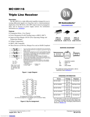
Research articleCell Biology Chromosomes and Gene ExpressionResultsDisrupting the localization of APC-dependent RNAs at protrusions doesnot affect their translationAs a first step towards assessing whether localization of APC-dependent RNAs at protrusions is coupled to their translation status, we disrupted RNA localization at protrusions and determinedwhether that affected the efficiency of their translation. To measure translation efficiency, we fractionated cell extracts on sucrose gradients to resolve RNAs according to the number of bound ribosomes (Figure 1A). To facilitate a larger scale analysis, we divided each gradient into four fractionsbased on UV absorbance traces. Fraction one includes free RNPs and the 40S and 60S ribosomalsubunits, fraction 2 includes 80S monosomes, and fractions 3 and 4 include light and heavy polysomes, respectively. mRNAs in fractions 1 and 2 largely correspond to non-translated mRNAs,Figure 1. Disrupting localization of APC-dependent RNAs, through competition, does not alter their translation. (A) Outline of experimental procedure.Sucrose gradients are divided into four fractions based on UV absorbance, an equal amount of spike RNA is added to each, and RNA presence isquantitatively assessed with nanoString analysis. (B, C) Representative absorbance profiles of polysome gradients of control, puromycin treated (B) orPkp4-cUTR-expressing cells (C). Inset in (B) shows an enlargement of the polysome region. (D) Heat maps showing RNA presence in polysome gradientfractions, based on nanoString analysis, under the indicated conditions. Gene names are shown on the left. Values indicate averages of 3 independentexperiments. Statistically significant differences compared to the corresponding control fractions are indicated by asterisks (2-way ANOVA withDunnett’s multiple comparisons test).DOI: https://doi.org/10.7554/eLife.44752.002The following source data is available for figure 1:Source data 1. File containing values used for generation of the heatmaps and statistics of Figure 1D.DOI: https://doi.org/10.7554/eLife.44752.003Moissoglu et al. eLife 2019;8:e44752. DOI: https://doi.org/10.7554/eLife.447523 of 31
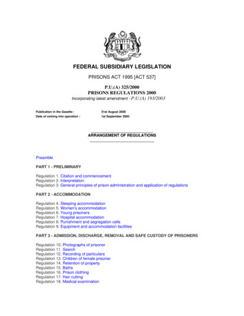
Research articleCell Biology Chromosomes and Gene Expressionwhereas mRNAs in fractions 3 and 4 are actively translated. To correct for variations introduced bysample manipulation during RNA purification, an equal amount of in vitro transcribed spike RNAwas added to each fraction. The recovered RNA from each fraction was then used to simultaneouslydetect the levels of multiple RNA species along the four fractions (Figure 1A). For detection, weused nanostring analysis which allows for direct RNA counting and thus avoids biases introduced byreverse transcription and amplification. We designed probes to detect 20 protrusion-enriched RNAs,that we have previously defined as APC-dependent, 6 RNAs encoding ribosomal proteins or ribosome biogenesis factors, which we have previously shown are also enriched at protrusions but in anAPC-independent manner, and eight control RNAs which based on our prior analysis are notenriched at protrusions (Wang et al., 2017) (Figure 1D).To first validate whether our experimental approach can detect changes in translation, we isolated polysome gradient fractions from control cells or cells treated with puromycin, an antibioticthat terminates translation and dissociates polysomes. Indeed, puromycin treatment led to anincrease in monosomes and ribosomal subunits and a decrease in heavy polysomes (Figure 1B).(Note that NIH/3T3 cells used in these experiments have a reduced baseline amount of ribosomesengaged in translation (i.e. in light and heavy polysomes) compared to HEK293 cells shown inFigure 1A). Nanostring analysis of recovered RNA revealed that under control conditions a higherproportion of most RNAs exists in fractions 3 and 4, while treatment with puromycin significantlyshifted the distribution of RNAs, in all three groups examined, towards fractions 1 and 2 (Figure 1Dand Figure 1—source data 1). These results therefore indicate that indeed mRNAs present in fractions 3 and 4 correspond to actively translated transcripts, and furthermore, that our nanostringbased methodology can quantitatively detect changes in translation efficiency.To disrupt the localization of APC-dependent RNAs at protrusions, we employed two differentmethodologies. One is a competition-based method that relies on overexpression of an exogenousconstruct carrying the 3’UTR of the APC-dependent RNA, Pkp4. Our prior characterization showedthat inducible expression of such competitive-UTR (cUTR) constructs leads to a preferential mislocalization of APC-dependent RNAs from protrusions (see Wang et al., 2017 for a detailed characterization of the stable cell lines used). A second method relies on the requirement for detyrosinatedmicrotubules for localization of APC-dependent RNAs at protrusions (Wang et al., 2017;Yasuda et al., 2017). Brief treatment with the chemical compound parthenolide disrupts detyrosinated microtubules (Yasuda et al., 2017) and significantly reduces RNA localization at protrusions,measured through the degree of RNA enrichment in isolated protrusion samples (Figure 2A).To test whether mislocalization from protrusions is accompanied by changes in translation, polysome gradient fractions were isolated from control and cUTR- or parthenolide-treated cells and analyzed for the presence of various RNA species. In both cases, cUTR overexpression or parthenolidetreatment did not affect the overall gradient profile, suggesting that no overt changes in translationresulted from either treatment (Figures 1C and 2B). Furthermore, both approaches did not result insignificant changes in the translation state of the control RNAs, the APC-independent RNAs or themislocalized APC-dependent RNAs analyzed (Figures 1D and 2B and Figure 1—source data 1, Figure 2—source data 1). Therefore, disrupting the peripheral localization of APC-dependent RNAsdoes not affect their translational efficiency, suggesting a lack of coupling between RNA transportand translational control.We note that both cUTR overexpression and parthenolide treatment resulted in a small apparentreduction in the amount of APC-dependent RNAs sedimenting in the heavy polysome fraction (fraction 4) and an apparently corresponding increase of RNA amounts in polysome fraction 3. Thesechanges are small, they are not statistically significant and do not alter the conclusion that APCdependent RNAs remain translationally active. Nevertheless, we make a note of this observationbecause it is manifested quite consistently by APC-dependent RNAs under conditions that disrupttheir peripheral localization. In light of data presented below, we believe that this might reflect areal change in the organization of a small fraction of these RNPs (see discussion).Single-molecule translation reporters of protrusion-localized RNAsWhile the above results suggest that transport and translation of APC-dependent RNAs are notcoordinated, they also raise the possibility that assessing the translation status in a whole-cell extractderived from heterogeneous cell populations might not offer the required sensitivity to detect localchanges occurring on a single-cell level. To address this, we took advantage of the recentlyMoissoglu et al. eLife 2019;8:e44752. DOI: https://doi.org/10.7554/eLife.447524 of 31
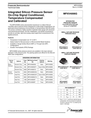
Research articleCell Biology Chromosomes and Gene ExpressionFigure 2. Disrupting localization of APC-dependent RNAs, through perturbation of detyrosinated microtubules, does not alter their translation.(A) Schematic on the left indicates experimental procedure used for isolation of protrusions. Migration of cells through microporous filters was inducedby addition of LPA and protrusion (Ps) and cell body (CB) samples were isolated from control or parthenolide (PTL) treated cells. The indicated RNAswere detected through nanoString analysis to calculate Ps/CB enrichment ratios (n 3; error bars: standard error). *: p-value 0.04 by two way ANOVAwith Bonferroni’s multiple comparisons test against the corresponding control. Parthenolide treatment specifically reduces the enrichment of APCdependent RNAs at protrusions. (B) Representative absorbance profiles of polysome gradients of control and PTL-treated cells, and heat maps showingRNA presence in polysome gradient fractions, based on nanoString analysis. Gene names are shown on the left. Values indicate averages of 3independent experiments. No statistically significant differences were detected by 2-way ANOVA with Dunnett’s multiple comparisons test against thecorresponding control fractions.DOI: https://doi.org/10.7554/eLife.44752.004The following source data is available for figure 2:Source data 1. File containing values used for generation of the heatmaps and statistics of Figure 2B.DOI: https://doi.org/10.7554/eLife.44752.005developed SunTag-based reporters that allow imaging of translation of single RNA molecules in livecells (Morisaki et al., 2016; Wang et al., 2016; Wu et al., 2016; Yan et al., 2016) (Figure 3A).These reporters carry a coding sequence which includes, at the 5’ end, a series of SunTag peptideepitopes, which are recognized by a single chain antibody fragment fused to superfolder-GFP (scFvGFP). The concentration of GFP-fused antibodies on a series of nascent peptides generated duringMoissoglu et al. eLife 2019;8:e44752. DOI: https://doi.org/10.7554/eLife.447525 of 31
Research articleCell Biology Chromosomes and Gene ExpressionFigure 3. Validation of single-molecule translation reporter assay. (A) Schematic of translation reporter constructs for labeling of RNA and nascentprotein chains. (B) Live cell imaging snapshot of a cell expressing the control translation reporter. The mCherry channel detects the 3x-mCherry-PCPprotein. Bright spots correspond to RNA molecules. Diffuse signal results from free 3x-mCherry-PCP. The GFP channel detects the scFv-GFP antibody.Bright spots overlap with RNA spots (merge image) and correspond to nascent protein at translation sites. Diffuse signal results from free scFv-GFP orFigure 3 continued on next pageMoissoglu et al. eLife 2019;8:e44752. DOI: https://doi.org/10.7554/eLife.447526 of 31
Research articleCell Biology Chromosomes and Gene ExpressionFigure 3 continuedscFv-GFP bound to the reporter protein released after translation. (C) Cells expressing the control translation reporter, containing PP7 repeats ( PP7),or a reporter without PP7 repeats (-PP7), were imaged live. mCherry intensity overlapping with translation sites (GFP spots) was measured andnormalized to the intensity observed in nearby cytoplasmic regions with diffuse signal. Value of 1 indicates that there is no mCherry concentration attranslation sites. (D) The same cytoplasmic areas, of cells expressing the control translation reporter, were imaged before and after puromycin addition.GFP/mCherry intensity of individual spots was calculated as a measure of translational efficiency. n 100 (C) and n 500 (D) spots from multiple cellsobserved in three independent experiments; error bars: standard error; ****: p-value 0.0001 by Student’s t-test. Scale bars: 5 mm.DOI: https://doi.org/10.7554/eLife.44752.006The following figure supplement is available for figure 3:Figure supplement 1. Translation signal of localized reporters reflects active translation.DOI: https://doi.org/10.7554/eLife.44752.007translation allows the observation of translation sites as bright GFP spots, which can be distinguishedfrom free antibody molecules or mature proteins released after translation. A second element incorporated in these reporter constructs is a series of binding sites for the PP7 bacteriophage coat protein introduced after the end of the coding sequence. These hairpin elements bind to a PP7 coatprotein fused to 3 copies of mCherry fluorescent protein (3x-mCherry-PCP), thus allowing visualization of RNA molecules as bright red spots (Yan et al., 2016) (Figure 3B and Video 1). To use thissystem, we have generated mouse NIH/3T3 fibroblast cell lines that stably express the GFP-fusedantibody and the 3x-mCherry-PCP, and additionally can be induced to express reporter RNAs afteraddition of doxycycline (Figure 3A,B and see below). For our studies, we have been using a controlreporter RNA which carries, after the PP7 repeats, a UTR sequence that doesn’t direct transport toprotrusions, and two localized reporter RNAs in which the PP7 repeats are followed by the 3’UTR ofeither the mouse Rab13 or Pkp4 RNAs, which as we have previously shown are sufficient to targetreporter RNAs to protrusions (Figure 3A) (Mili et al., 2008; Wang et al., 2017).We first validated the RNA and translation signals detected with our implementation of the assay.To assess the specificity of the detected RNA spots, we compared the signal observed upon expression of the control PP7-containing reporter RNAto that of a similar reporter carrying a deletionof the PP7-binding sites ( PP7) (Figure 3C).mCherry intensity overlapping with translationsites (GFP spots) was measured and normalizedto the intensity observed in nearby cytoplasmicregions with diffuse signal. The PP7 constructexhibited a normalized mCherry intensity around1, indicating that signal intensity overlappingwith translation spots was similar to that of thesurrounding cytoplasm. By contrast, the PP7containing reporter exhibited a significantlyhigher intensity, indicating that the mCherry signal overlapping translation sites was due to specific recognition of RNA molecules by the fusedPP7 coat protein (Figure 3C). To assess whetherGFP spots are indeed reflecting translation sites,images were acquired before and after treatment with puromycin (Figure 3D). RNA spotswere identified in the images, and mCherry andVideo 1. NIH/3T3 cell expressing scFv-GFP (green), 3xGFP intensity in the corresponding regions wasmCherry-PCP (red) and the control translation reporter.recorded. The normalized GFP intensity was calFrames were acquired sequentially and with no timeculated as a measure of the translational effidelay, for the duration of the movie (45 s). A mergedciency of each RNA spot. Puromycin treatmentimage of the two channels is shown. Overlapping redand green spots indicate translation sites. Blue line: cell significantly reduced the GFP intensity of RNAspots, confirming that it reflects the presence ofoutline. Cyan line: nucleus outline. Scale bar: 5 mm.nascent protein chains (Figure 3D). To furtherSingle frames of this movie are shown in Figure 3B.DOI: https://doi.org/10.7554/eLife.44752.008determine whether this concentration of nascentMoissoglu et al. eLife 2019;8:e44752. DOI: https://doi.org/10.7554/eLife.447527 of 31
Research articleCell Biology Chromosomes and Gene ExpressionFigure 4. RNAs targeted to protrusions are similarly translated in both internal and peripheral locations. (A) Live imaging snapshots of cells expressingthe indicated translation reporters. GFP/mCherry intensity of individual spots (indicating translation efficiency) was plotted as a function of distancefrom the cell edge. More than 200 particles were analyzed from approximately 20 cells. Best fit curves with 95% confidence intervals are overlaid on thegraphs. Scale bars: 5 mm. (B) Cumulative frequency distribution plot of translation reporter particles (from panel A) with increasing distance from the celledge.DOI: https://doi.org/10.7554/eLife.44752.009The following figure supplements are available for figure 4:Figure supplement 1. Expression levels of translation reporters and comparison with live-cell imaging.DOI: https://doi.org/10.7554/eLife.44752.010Figure supplement 2. Intensity histograms of translation reporter particles.DOI: https://doi.org/10.7554/eLife.44752.011Figure supplement 3. Examples of directionally persistent particles.DOI: https://doi.org/10.7554/eLife.44752.012Moissoglu et al. eLife 2019;8:e44752. DOI: https://doi.org/10.7554/eLife.447528 of 31
Research articleCell Biology Chromosomes and Gene Expressionchains reflects active translation, and not stalledribosomes, we acquired images of localizedtranslation reporters before and after treatmentwith harringtonine or lactimidomycin, which blockinitiating ribosomes but allow elongating ribosomes to run off (Ingolia et al., 2011; Lee et al.,2012). As an additional control we treated cellswith cycloheximide, which stalls elongating ribosomes and prevents release of nascent chains.Indeed, brief 15 min treatment with harringtonineor lactimidomycin, but not cycloheximide, significantly reduced the observed translation signalindicating that it reflects the presence of activelytranslating ribosomes (Figure 3—figure supplement 1).Video 2. NIH/3T3 cell expressing scFv-GFP (green), 3xmCherry-PCP (red) and the control translation reporter.Frames were acquired sequentially and with no timedelay, for the duration of the movie (13 s). A mergedimage of the two channels is shown. Blue line: celloutline. Scale bar: 5 mm. Single frames of this movie areshown in Figure 4A (upper panels).DOI: https://doi.org/10.7554/eLife.44752.013Video 3. NIH/3T3 cell expressing scFv-GFP (green), 3xmCherry-PCP (red) and the localized translationreporter carrying the Pkp4 UTR. Frames were acquiredsequentially and with no time delay, for the duration ofthe movie (36 s). A merged image of the two channelsis shown. Blue line: cell outline. Scale bar: 5 mm. Singleframes of this movie are shown in Figure 4A (middlepanels). RNAs are translated both in the periphery andinternal regions.DOI: https://doi.org/10.7554/eLife.44752.014Moissoglu et al. eLife 2019;8:e44752. DOI: https://doi.org/10.7554/eLife.44752RNAs targeted to protrusions aresimilarly translated in both internaland peripheral locationsWe further imaged cells expressing either controlor localized reporters (carrying the Rab13 orPkp4 UTRs) (Figure 4A and Videos 2–4). Imagingwas performed ca. 2 hr after doxycycline induction to ensure that reporter RNAs do not accumulate to high levels and do not deplete thecytoplasmic pools of scFv-GFP antibody and 3xmCherry-PCP (Figure 4—figure supplement1A). Consistent with the ability of the Rab13 andPkp4 UTRs to target RNAs to protrusions(Mili et al., 2008; Wang et al., 2017), a higherproportion of localized reporter RNAs (containingthe Rab13 or Pkp4 UTRs) were observed closerto the periphery compared to the controlreporter (Figure 4B). Around 50% of observedlocalized reporter molecules were found within3–7 mm of the cell edge, compared to 15 mm forthe control reporter (Figure 4B). For all reporters,the majority of RNA particles exhibit mCherryintensities centered around a single peak indicating that they largely exist as single molecules(Figure 4—figure supplement 2) (but see alsobelow). The number of particles detected by livecell imaging is lower than the number detectedin fixed cells by FISH (Figure 4—figure supplement 1B). The additional RNAs detected by FISHlikely correspond to fast-moving molecules thatcannot be discerned during live imaging with ourcurrent acquisition speed. Observing RNAs during short time-lapse imaging ( 20–30 s) revealsthat the majority of RNAs are static or exhibit anoscillatory type of motion (Videos 2–4). A smallsubset exhibits short directed movements thatmight be indicative of active transport (Videos 5and 6; and see below). These observations areconsistent with the motion characteristicsdescribed for other localized transcripts9 of 31
Research articleCell Biology Chromosomes and Gene Expression(Dynes and Steward, 2007; Gopal et al., 2017;Park et al., 2014; Yoon et al., 2016). Given thelimitations in imaging and discerning fast-movingmolecules, we have not attempted to analyze thetransport kinetics of our reporter RNAs. We haverather focused our analysis here on the lessmobile molecules that we can confidently identifyand analyze (see Materials and methods fordetails on image acquisition and analysis).Despite the lack of kinetic information, this analysis reflects the behavior of a substantial fractionof the existing RNA population and can provide avaluable characterization of the translation properties of individual RNA molecules in a spatialmanner.To address whether transport to the peripheryisaccompaniedby changes in the translationVideo 4. NIH/3T3 cell expressing scFv-GFP (green), 3xstateoftheRNAs,we determined the translationmCherry-PCP (red) and the localized translationreporter carrying the Rab13 UTR. Frames wereefficiency of single RNA molecules as a functionacquired sequentially and with no time delay, for theof their distance from the periphery. Consistentduration of the movie (63 s). A merged image of thewith the stochastic translation bursts reported intwo channels is shown. Blue line: cell outline. Cyan line: other systems (Pichon et al., 2016; Wu et al.,nucleus outline. Scale bar: 5 mm. Single frames of this2016; Yan et al., 2016), for all three reporters,movie are shown in Figure 4A (bottom panels). RNAssingle RNAs exhibit a range of translation effiare translated both in the periphery a
Hiroshima University, Higashi-Hiroshima, Japan; 3Laboratory . Taken together all the above data suggest that, while APC-dependent RNAs are enriched in the periphery, they are translated with similar efficiency . This work was supported by the Intramural Research Program of the Center for Cancer Research, NCI, National Institutes of Health .