Transcription
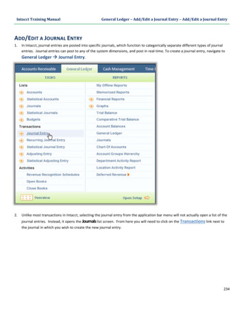
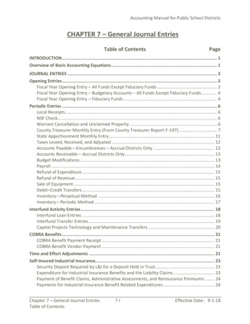
(2021) 28:79Pao and Tsai Journal of Biomedical pen AccessREVIEWThree decades of Cdk5Ping‑Chieh Pao1,2 and Li‑Huei Tsai1,2*AbstractCdk5 is a proline-directed serine/threonine protein kinase that governs a variety of cellular processes in neurons, thedysregulation of which compromises normal brain function. The mechanisms underlying the modulation of Cdk5, itsmodes of action, and its effects on the nervous system have been a great focus in the field for nearly three decades.In this review, we provide an overview of the discovery and regulation of Cdk5, highlighting recent findings revealingits role in neuronal/synaptic functions, circadian clocks, DNA damage, cell cycle reentry, mitochondrial dysfunction, aswell as its non-neuronal functions under physiological and pathological conditions. Moreover, we discuss evidenceunderscoring aberrant Cdk5 activity as a common theme observed in many neurodegenerative diseases.Keywords: Cdk5, p35, p25, Neurodegeneration, Alzheimer’s diseaseBackgroundDiscovery of Cdk5Cyclin-dependent kinase 5 (Cdk5) is a proline-directedserine/threonine protein kinase. Thirty years ago, Cdk5was first discovered on the basis of its close sequencehomology to the human cell division cycle protein 2(Cdc2, also known as Cdk1), a regulator of cell cycle progression [1–3]. The kinase activity of Cdc2 is detected inproliferating cells and activates prior to or during S-phase[4]. Cdk5 was originally inferred to function in cell cycleregulation based on its 60% sequence identity and similar substrate specificities to Cdc2 [1–3]. However, threedecades of research have demonstrated that Cdk5 playsmany key roles in post-mitotic neurons.Unlike other Cdks, Cdk5 activity is most apparent inadult mouse brain [5], where most of the cells are terminally differentiated. During brain development, theexpression and kinase activity of Cdk5 gradually increasefrom embryonic day 11 (E11) and peak at E17, which correlates with the main phase of neuronal differentiationin the developing mouse neocortex [5]. In contrast, theexpression pattern and kinase activity of Cdc2 decline*Correspondence: lhtsai@mit.edu1Picower Institute for Learning and Memory, Massachusetts Instituteof Technology, Cambridge, MA 02139, USAFull list of author information is available at the end of the articleas development proceeds [5]. These findings suggestfunctions for Cdk5 in brain development and neuronaldifferentiation.Regulation of Cdk5: regulatory subunitThe Cdk family comprises 21 members, most of whichdepend on the association with specific cyclin partnersto become constitutively active protein kinases [6]. Surprisingly, the subunit required for Cdk5 activity is a noncyclin protein named p35. p35 was identified as the Cdk5regulatory subunit and lacked the conserved amino acidsequences typically found in cyclins [7–9]. p35 physicallyassociates with Cdk5 in brain lysates and is capable ofactivating Cdk5 upon direct binding [7]. The expressionof Cdk5 and p35 transcripts overlaps spatially and temporally in the developing mouse neocortex, and p35 isprimarily expressed in the post-mitotic neurons [7]. Single-cell transcriptomic analysis of the adult mouse brainindicates that the expression of Cdk5 is not restrictedto neurons [10] (Fig. 1). Nonetheless, Cdk5r1, the geneencoding p35, shows marked expression predominatelyin the neuronal linage [10] (Fig. 1).Another Cdk5 regulatory subunit, p39, was later discovered in part by its high degree of sequence identity top35 [11]. While p39 shares many characteristics with p35including its high expression in the brain, direct bindingto Cdk5, and the ability to activate Cdk5 [11–13], there The Author(s) 2021. Open Access This article is licensed under a Creative Commons Attribution 4.0 International License, whichpermits use, sharing, adaptation, distribution and reproduction in any medium or format, as long as you give appropriate credit to theoriginal author(s) and the source, provide a link to the Creative Commons licence, and indicate if changes were made. The images orother third party material in this article are included in the article’s Creative Commons licence, unless indicated otherwise in a credit lineto the material. If material is not included in the article’s Creative Commons licence and your intended use is not permitted by statutoryregulation or exceeds the permitted use, you will need to obtain permission directly from the copyright holder. To view a copy of thislicence, visit http:// creat iveco mmons. org/ licen ses/ by/4. 0/. The Creative Commons Public Domain Dedication waiver (http:// creat iveco mmons. org/ publi cdoma in/ zero/1. 0/) applies to the data made available in this article, unless otherwise stated in a credit line to the data.
Pao and Tsai Journal of Biomedical Science(2021) 28:79Page 2 of 17Fig. 1 Single-cell transcriptomic analysis of the expression of Cdk5, Cdk5r1 (the gene encoding p35), and Cdk5r2 (the gene encoding p39) in youngand aged mouse brain. The data are based on Ximerakis et al., 2019, Nat. Neurosci., and the graphs were generated by Single Cell Portal website(https:// singl ecell. broad insti tute. org/ single cell/ study/ SCP263/ aging- mouse- brain). Astrocyte lineage: neural stem cells, astrocyte-restrictedprecursors and astrocytes. Ependymal cells: ependymocytes, hypendymal cells, tancytes, and choroid plexus epithelial cells. Immune cells:microglia, monocytes, macrophages, dendritic cells, and neutrophils. Neuronal lineage: neuronal-restricted precursors, immature neurons, matureneurons, and neuroendocrine cells. Vasculature cells: endothelial cells, pericytes, hemoglobin-expressing vascular cells, vascular smooth musclecells, vascular and leptomeningeal cells, and arachnoid barrier cellsare distinct properties between p35 and p39. Duringbrain development, the expression of p35 is high fromembryonic stage to postnatal stage, whereas that of p39starts increasing postnatally [14]. p35 and p39 exhibit different regional distribution patterns with p35 expressedmost prominently in the cerebral cortex and cerebellum while p39 highly expressed in the cerebellum, brainstem, and spinal cord [14]. Moreover, p39 protein displays a higher protein stability and lower binding affinityfor Cdk5, relative to p35 [15, 16]. Studies have identifiedfunctions that are preferentially regulated by Cdk5/p39but not Cdk5/p35, regardless of their redundant rolesin the nervous system. Deletion of p39 or Cdk5 in cultured neurons causes defective dendritic morphogenesis
Pao and Tsai Journal of Biomedical Science(2021) 28:79whereas no abnormality was observed in cultured neurons lacking p35 expression [17]. Cdk5/p39 also plays adominant role in Rac1-induced lamellipodia formation[18].Regulation of Cdk5: membrane targetingSubcellular fractionation assays reveal the enrichmentof p35 and p39 proteins in membrane-bound fractionsof cultured neurons [12, 19]. The membrane-associatedlocalization of p35 and p39 is promoted by the myristoylation signal on a conserved glycine located at position two of these two proteins [20]. Myristoylated p35and p39 recruits Cdk5 to cell membrane [20, 21], whereasharboring mutated myristoylation signal fails to retaintheir membrane-targeting distribution [20, 21]. Thus,under normal circumstances, active Cdk5 complexes primarily reside on the cell membrane.Page 3 of 17Neuronal migrationGene-targeting studies demonstrate a role for Cdk5 inbrain development, particularly in neuronal migration[38–40]. Cdk5 modulates neuronal migration throughmultiple pathways. Cdk5/p35 interacts with N-cadherinadhesion complex. Pharmacological inhibition of Cdk5enhances N-cadherin-mediated cell adhesion, whichhinders neuronal migration [41]. A recent study showsthat Cdk5 phosphorylates RapGEF2 at S1124, and subsequently activates Rap1, a key factor modulating neuronalmigration [42]. Moreover, semaphorin-3A (Sema3A)elicited neuronal migration requires Cdk5 phosphorylating Synapsin III at Ser 404 [43]. Additional substrates ofCdk5 functioning in neuronal migration include NUDEL,FAK, disrupted-in-Schizophrenia-1 (Disc1), doublecortin(Dcx), and Dix-domain containing 1 (Dixdc1), and p27[19, 44–49].Neurite outgrowthRegulation of Cdk5: post‑translational modificationsWhile binding to a regulatory subunit is obligatory forCdk5 activation, its activity can be further modulated bya variety of post-translational modifications. The phosphorylation of Cdk5 at two different residues within theATP-binding sites leads to opposing effects. Phosphorylation at Tyr 15 is stimulatory but inhibitory at Thr 14 [22,23]. Moreover, phosphorylation of Ser 159 in the T-loopof Cdk5 is critical for p35 binding [24], and acetylation atLys 33 impairs Cdk5 activity due to the loss of ATP binding [25]. Recent evidence indicates a role for S-nitrosylation in Cdk5 regulation. S-nitrosylation of Cdk5 at Cys83, a residue locating in the ATP-binding pocket, suppresses Cdk5 activity [26]. S-nitrosylation of p35 at Cys92 induces its degradation via the proteasome, which inturn reduces Cdk5 activity [27].Roles of Cdk5 in the nervous systemThere has been rapid progress in our understanding ofCdk5 function in the nervous system.Prior review articles on Cdk5 have provided a comprehensive coverage of its roles in neuronal migration, neurite outgrowth, axonal guidance, and synaptic plasticity. Inthis review, we focus on the recent findings of Cdk5, particularly its roles in neuronal/synaptic functions and circadian clock regulation under physiological conditions,as well as its pathological links to DNA damage, cell cyclereentry, mitochondrial dysfunction, and oxidative stress.The Cdk5 substrates discussed in this review and theirfunctional categories are listed in Table 1, and the readeris referred to several excellent reviews with a more comprehensive list of known Cdk5 substrates [28–37].On the basis of its unique expression and activity patternin the developing mouse brain, Cdk5 is believed to haveroles in neurogenesis. Indeed, Cdk5 is essential for neurite growth and axonal formation. Cdk5 and p35 show colocalization with actin filaments in axonal growth cones,and inactivation of Cdk5 in cultured neurons inhibitsneurite outgrowth [50]. Consistently, disrupting Cdk5function causes axon patterning defects in the Drosophilamodel [51], and p35-null mice show altered axonal anddendritic trajectories [52]. In contrast, co-expression ofCdk5 and p35 increases neurite length in cultured neurons [50]. Cdk5 can promote neurite outgrowth throughphosphorylating Pak1, a kinase that regulates the dynamics of actin and microtubule fibers [53], whereas neuritegrowth elicited by the serotonin 6 receptor (5-HT6R)requires receptor phosphorylation at Ser 350 by Cdk5[54]. Cdk5 also modulates axonal outgrowth by phosphorylating GRAB, a guanine nucleotide exchange factorfor Rab8 [55]. Recent findings suggest that Cdk5 regulates dendritic morphogenesis by an adaptor protein WDrepeat and FYVE domain-containing 1 (WDFY1) [17].Cdk5 can also enhance neurite outgrowth through theassociation with Cables and c-Abl complex [22], as wellas the phosphorylation of Map1b [56], a microtubulebinding protein, and Axin [57], a scaffold protein of theWnt pathway.Synaptic plasticityImpaired long-term depression and depotentiation oflong-term potentiation have been shown in p35-deficient mice [58], implying a role for Cdk5/p35 complex insynaptic plasticity. Synaptic plasticity reflects modification of the efficacy or strength of synaptic transmission
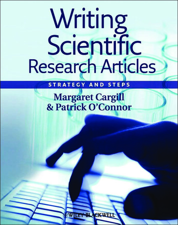
Pao and Tsai Journal of Biomedical Science(2021) 28:79Page 4 of 17Table 1 List of Cdk5 substrates discussed in this review and their functional categoriesSubstrates of Cdk5CategoryProtein/sitesFunctional outcomeReferencesMicrotubule andcytoskeleton-relatedfunctionAxin/T485Promoting axon formation[57]Synaptic functionCircadian clockNeurodegenerationDcx/S710Regulation of neuronal migration[49]Dixdc1/S250Increasing the association between DISC1 and NUDEL[48]FAK/S732Regulation of microtubule organization and neuronal migration[45]GRAB/S169, S180Regulation of neurite outgrowth[55]Map1bRegulation of neurite outgrowth[56]NUDEL/S198, T219, S231Regulation of neurite morphology and neurite outgrowth[19]p27/S10, S297Regulation of nuclear elongation in migrating neurons[49]PAK1/T212Regulation of neuronal migration and neurite outgrowth[53]RapGEF2/S1124Regulation of neuronal migration[42]Synapsin III/S404Regulation of neuronal migration[43]5-HT6R/S350Regulation of neurite outgrowth[54]Amphiphysin 1/S272, S276, S285Increasing clathrin-mediated endocytosis[61, 62]CaMKv/T345Reducing spine density[71]Dynamin 1/S774, S778Increasing clathrin-mediated endocytosis[62]Liprinα1/T701Enhancing excitatory synaptic[69]Munc18/S158Increasing vesicle release[60]NR2A/S1232Regulation of synaptic plasticity[65, 66]PSD-95/T19, S25, S35Regulation of synaptic plasticity[67, 69]SPAR/S1328Regulation of synaptic homeostasis[75][70]TrkB/S478Regulation of activity-dependent structural plasticityL-VDCC/S783Regulation of calcium influx[64]CaV2.2/S2013Regulation of calcium influx[63]CLOCK/T451, T461Promoting CLOCK nuclear translocalization and enhancing itsactivity[80]PER2/S394Stabilizing PER2 and promoting its nuclear translocalization[79]APP/T668Regulating of APP localization[103]ATM/S794Regulation of DNA damage response[115]Drp1/S616Promoting mitochondrial fission[124]Htt/S434Reducing Htt cleavage and aggregation[146]Parkin/S131Reducing Parkin ubiquitin ligase activity[137]Prx1/T90, Prx2/T89Reducing peroxidase activity[120, 135]STAT3/S727Increasing BACE1 gene expression via activating STAT3[101, 102]Tau/T181, S202, T205, T217, S235, S396, S404 Reducing microtubule bindingNon-neuronal function Coronin 1a/T418Facilitating actin polarization and migration of lymphocytes tochemokine signals[107, 108][181]DCL1/S120, S205, S422, S509Activating tumor suppressor DCL1[187]TRIM59/S308Promoting the degradation of tumor suppressive histone variantmacroH2A1[186]Paxillin/S244Promoting OPC maturation[177, 178]PPARγ/S273Inducing insulin resistance[161]Vimentin/S56Promotes melanoma cell extravasation[184]in response to neuronal activity and is integral tomemory formation. Regulation of synaptic plasticityoccurs through multiple mechanisms at both pre- andpost-synaptic levels [59]. Presynaptically, Cdk5 controlsexocytosis, endocytosis, and Ca2 influx. Phosphorylation of Munc-18 by Cdk5 results in the dissociation of
Pao and Tsai Journal of Biomedical Science(2021) 28:79Munc-18 from Syntaxin 1A, facilitating synaptic fusionand release [60]. Cdk5 also regulates endocytosis throughDynamin I and Amphiphysin I [61, 62], two componentsof Clathrin-mediated endocytosis. Cdk5 also induces Ca2 influx into the pre-synaptic cytoplasm via voltagedependent Ca2 channels (VDCCs), which increases theprobability of channel opening and facilitates the releaseof neurotransmitters [63, 64].Change in the numbers or properties of post-synapticreceptors is one of the mechanisms for modulating synaptic plasticity at the post-synapses. Cdk5 phosphorylatesthe NMDA receptor subunit NR2A at Ser1232, therebyenhancing NMDA receptor function [65, 66]. PSD-95 is apostsynaptic scaffold protein that tethers NMDA receptors to the cytoskeleton. Cdk5 catalyzes PSD-95 at threeresidues near the N-terminal domain, which causes areduction in co-clustering of PSD-95 and neuronal ionchannels [67]. Conversely, inhibition of Cdk5 functionenhances PSD-95 clustering [67]. Cdk5-mediated phosphorylation of PSD-95 increases the degradation of PSD95 by the ubiquitin-proteosome pathway [68]. Recentwork reveals that Cdk5 regulates activity-dependentdendritic spine remodeling through multiple mechanisms. Cdk5-mediated phosphorylation of scaffold protein Liprinα1 at Thr 701 declines in response to neuronalactivity, which is linked to enhanced excitatory synapticfunction by promoting the binding of Liprinα1to PSD-95and PSD-95 synaptic localization [69]. Moreover, Cdk5is crucial for BDNF-TrkB signaling. Phosphorylation ofTrkB on Ser 478 by Cdk5 increases activity-dependentstructural plasticity and spatial memory [70]. Conversely,Cdk5 impairs activity-dependent dendric spine maintenance through a pseudokinase CaMKv, as phosphorylation of CaMKv by Cdk5 at Thr345 is associated withreduced spine density [71].Synaptic homeostasisSynaptic homeostasis is a compensatory process thatallows neurons to adapt to altered levels of network activity. Neurons potentiate synaptic efficacy when inputsare dampened and downmodulate their firings wheninputs are heightened [72]. Thus, homeostatic mechanisms ensure that neurons maintain their firings withinan optimal range and protect network stability despiterecurrent alterations in input activity. Cdk5 is importantfor synaptic scaling, a principal mechanism underlyinghomeostatic plasticity. At presynaptic terminals, synapticvesicles recycle into recycling or resting pools. Recyclingpools are available for release upon neuronal activation,whereas resting pools remain silent [73]. Long-term suppression of neuronal activity reduces presynaptic Cdk5levels, and inhibition of Cdk5 is associated with unlockedPage 5 of 17resting vesicles and an increased pool of recycling vesicles [74], indicative of synaptic strengthening.At the postsynaptic levels, Cdk5 has been implicatedin depressing synaptic strength following heightenedneuronal activity [75]. Upon increased network activity,Cdk5 phosphorylates spine-associated Rap guanosinetriphosphatase-activating protein (SPAR), a postsynapticscaffold protein regulating actin dynamics and promotingthe growth of dendritic spines [76]. Priming phosphorylation of SPAR at S1328 by Cdk5 induces Plk2-mediatedphosphorylation of SPAR, leading to ubiquitin-dependent degradation of SPAR and synaptic weakening [75].Collectively, these observations underscore the role ofCdk5 in modifying synaptic scaling by regulating the partition of presynaptic vesicles and the degradation of postsynaptic SPAR scaffold protein.Circadian clocksCircadian clocks are oscillators that synchronize dailycycles of behavior and physiology. The suprachiasmaticnucleus (SCN) of the hypothalamus is the master circadian pacemaker in mammals and entrains the peripheralclocks across the body [77]. Circadian clocks are generated in a transcriptional autoregulatory feedback loopby the circadian machinery. The core circadian machinery consists of the transcriptional activators CLOCKand BMAL1 and the repressors PER1/2 and CRY1/2.CLOCK/BMAL1 heterodimer activates the transcription of a set of circadian clock genes, including Per1/2and Cry1/2. Newly synthesized PER and CRY proteinsheterodimerize, translocate into the nucleus, and inhibitCLOCK/BMAL1 activity through direct binding, resulting in the subsequent repression of downstream targetgenes [77]. Thus, circadian clock genes display an oscillatory expression pattern. Perturbation of circadian clockscompromises brain function, and circadian dysfunctionis a common symptom of various neurodegenerative diseases including Alzheimer’s disease (AD) [78].Cdk5 has been implicated in the regulation of circadian clocks [79, 80]. The running wheel test is a methodto record circadian rhythm, whereby wild-type micestart wheel running precisely at the beginning of thedark phase. Mice injected with adeno-associated virusexpressing shRNA against Cdk5 in the SCN show earlier onset of wheel running activity, which phenocopiesmice harboring Per2 silencing [79]. Moreover, rhythmicexpression of CLOCK target genes, including Per1 andPer2, is disturbed in p35 heterozygous knockout mice[80]. Cdk5 interacts with and phosphorylates CLOCKat residues T451 and T461. Cdk5-mediated phosphorylation causes nuclear translocation and transcriptionalactivation of CLOCK [80]. In addition, Cdk5 phosphorylates PER2 at S394 residue, which stabilizes PER2 protein
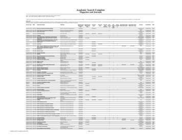
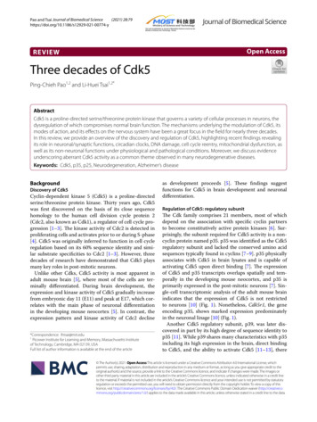
Pao and Tsai Journal of Biomedical Science(2021) 28:79and promotes its nuclear translocation [79]. Collectively,Cdk5 regulates circadian clocks by its phosphorylationon several components of the core clock machinery.Dysregulation of Cdk5: Calpain‑dependentproteolytic cleavage of p35 to p25Aberrant Cdk5 activity caused by p25 accumulation contributes to the pathogenesis of various neurodegenerative diseases [81]. p25 is a 208-residue carboxy-terminalfragment of p35. The mechanism underlying the cleavage of p35 to p25 has been well-characterized. Neurotoxic insults such as ischemia, the addition of hydrogenperoxide, glutamate or ionomycin, cause calcium influxand trigger the activation of a cysteine protease namedcalpain [82]. Calpain cleaves p35 at P he98/Ala99 sequenceand generates p25 in a calcium-dependent manner.Accordingly, increasing intracellular calcium level stimulates p25 generation, whereas removing calcium preventsp25 accumulation [82].p25 causes constitutive activation and mislocalizationof Cdk5. p25 activates Cdk5 through direct binding, andPage 6 of 17p25 has an approximately 5- to 10-fold longer proteinhalf-life compared to p35 [20], thereby prolonging Cdk5activation. Moreover, p25 lacks the myristoylation signalthat normally tethers Cdk5 to the membrane. Immunohistochemical and cell fractionation analysis demonstrate that p25 is enriched in the nuclear and perinuclearregions of the cell [20]. These findings suggest that p25promotes Cdk5 hyperactivation and redirects Cdk5 toa wider array of substrates under pathological contexts(Fig. 2).p25‑mediated neurotoxicity and neurodegenerationDysregulation of Cdk5 activity leads to neurotoxicity andneurodegeneration [83]. Cultured neurons overexpressing p25 exhibit morphological deterioration and apoptotic cell death, characterized by degenerated neuritesand fragmented nuclei [20]. To understand the consequence of Cdk5 hyperactivation in vivo, several groupshave generated mouse models of p25 overexpression [84,85]. Different genetic approaches converge on similarpathological phenotypes.Fig. 2 Activation of Cdk5 and its functions under physiological or pathological conditions
Pao and Tsai Journal of Biomedical Science(2021) 28:79CK-p25 mice are one such p25 overexpression model.CK-p25 mice overexpress an inducible human p25 underthe control of a forebrain-specific CamKII promoter,which is expressed predominantly in excitatory pyramidal neurons. The expression of p25 is induced upon theremoval of the tetracycline derivative doxycycline fromthe animal diet [85]. Following p25 induction in excitatory neurons, CK-p25 mice exhibit progressive neurodegenerative phenotypes. Immunohistochemical analysisin CK-p25 mice following acute p25 induction (2-week)reveals substantial DNA damage of double-strand breaks(DSBs) in p25-expressing neurons [86]. Neurons containing DSBs also express ectopic cell cycle markers [86],suggesting Cdk5 hyperactivation may lead to cell cyclereentry, which has been reported in post-mortem ADpatient brain samples [87]. Neuronal DNA damage isassociated with a dramatic morphological and transcriptional response in microglia, including structural remodeling of processes/cell bodies as well as upregulatedexpression of genes regulating cell division [85, 88].Aberrant Cdk5 activity is also found to trigger neuroinflammation. Neurons overexpressing p25 markedlyproduce and secrete a soluble lipid known as lysophosphatidylcholine, which activates glia and induces theexpression of cytokines and chemokines [89]. Prolongedp25 induction is accompanied by increased tau phosphorylation and elevated levels of Aβ peptide from thecleavage of APP [85]. CK-p25 mice also exhibit severeneuronal loss and reduced hippocampal LTP induction,together with behavioral alterations including locomotor hyperactivity, increased anxiety, and memory impairment [85, 90]. In Drosophila, dysregulated Cdk5 disruptsautophagy and augments the expression of anti-microbialpeptides. Anti-microbial peptides cause the hyperactivation of innate immune response and are linked to dopaminergic neuronal death in Drosophila [91]. In summary,overexpression of p25 in excitatory neurons triggers several AD-like pathological hallmarks.Cdk5 dysfunction in Alzheimer’s diseaseAD is the leading cause of senile dementia, which is pathologically characterized by the accumulation of amyloidplaques and neurofibrillary tangles (NFTs) [92]. Severallines of evidence support a strong association betweenCdk5 dysregulation and AD pathogenesis [93]. p25 accumulates in NFT-bearing neurons and in brain lysatesfrom AD patients [20, 94]. Relative to age-matched nonAD individuals, Cdk5 immunoprecipitated from ADbrain tissue displays a greater activity when histone H1was used as a substrate [20]. Similar observations weremade in cellular and mouse models of AD. IntroducingAβ42 peptide to cultured neurons leads to p25 generationand neuronal death [82], and accordingly, the Cdk5 smallPage 7 of 17molecule inhibitor butyrolactone or the calpain inhibitorcalpeptin alleviates Aβ-induced neuronal death [82, 95].A plethora of AD mouse models also reveal dysregulationof Cdk5 via p25 accumulation [96, 97]. Here, we discusspertinent aspects of how aberrant Cdk5 activity may contribute to AD progression.APP processing and Aβ productionDysregulation of Cdk5 activity promotes amyloid plaquedeposition by promoting amyloidogenic APP processing. The amyloidogenic APP pathway includes sequential cleavage by β- and γ-secretases, which ultimatelyproduce a secreted form of APP (sAPPβ), C-terminalfragments (CTF99 and CTF89) and Aβ peptides [98].Aβ peptides form fibrillar aggregates that form amyloid plaques [99]. The first observation linking aberrantCdk5 activity to Aβ production was made in the CK-p25mouse model [100]. ELISA analysis demonstrates anincrease in endogenous mouse Aβ levels after p25 induction. Immunolabeling of two antibodies recognizing Aβpeptide, 4G8 and 6E10, reveals intracellular accumulation of Aβ in neurons expressing p25 [100]. Studies indicate that Cdk5/p25 enhances Aβ production throughSTAT3-mediated transcriptional regulation of BACE1,a gene encoding β-secretase. Cdk5/p25 phosphorylatesSTAT3 at S727 residue and in turn activates BACE1 transcription [100–102]. Significant upregulation of BACE1immunoreactivity and increased β-secretase processingof APP are found in CK-p25 mouse brain [100]. In addition, Cdk5 phosphorylates APP at T668, which facilitatesthe BACE1 cleavage of APP to increase Aβ generation[103]. As discussed above, Aβ peptide is able to inducep25 accumulation and Cdk5 overactivation which canresult in a feed-forward reaction, whereby aberrant Cdk5activity in turn amplifies Aβ-associated pathologies. Inthe context of familial AD, it is likely that elevated Aβ levels might be the causal factor initiating this cascade. Onthe other hand, Cdk5/p25 is linked to Aβ-induced synaptic depression. Cdk5/p25 leads to DARPP-32 inhibitionand PP1/Calcineurin activation, which promotes AMPAreceptor subunit GluA1 (Ser 845) dephosphorylation andimpacts negatively on AMPAR endocytosis [104].A previous report established a knock-in mouse modeldeficient in p25 generation named Δp35KI mice [104]. InΔp35KI mice, a mutant p35 resistant to calpain cleavagewas designed by substituting an alaine [99] residue at thecleavage site with leucine and removing six amino acidresidues (A93NLSTF98) adjacent to the calpain-cleavagesite [104]. 5XFAD mice are an AD mouse model thatexhibit increased p25 levels, and the blockade of p25 generation by crossing with Δp35KI mice rescues AD pathology in 5XFAD, Δp35KI mice. Compared to 5XFAD mice,the 5XFAD, Δp35KI mice display a reduction in soluble
Pao and Tsai Journal of Biomedical Science(2021) 28:79Aβ peptide levels, plaque deposition, and inflammatorycytokine expression. Furthermore, the 5XFAD, Δp35KImice show an improvement in synaptic plasticity andmemory function relative to the 5XFAD mice [104]. Collectively, these findings highlight therapeutic potential oftargeting Cdk5 hyperactivation in AD.TauopathyTau is a microtubule-associated protein mainly distributed in axons. Tau stabilizes neuronal microtubules andregulates microtubule dynamics involved in axonal outgrowth and transport [105]. Tauopathy is a pathologicalfeature characterized by the deposition of abnormal tauaggregation in the brain, and is present in a wide varietyof neurodegenerative diseases such as AD and frontotemporal dementia (FTD). Hyperphosphorylation of tauis well known to enhance tau aggregation and form NFTs[105].Cdk5 is a tau protein kinase: Cdk5 co-purifies withtau and phosphorylates tau in vitro [106]. Overexpression of Cdk5/p25 increases tau phosphorylation in cultured neurons compared to overexpression of the Cdk5/p35 complex [20]. Emerging evidence suggests that Cdk5phosphorylates tau on many sites including T181, S202,T205, T212, T217, S235, S396, and S404. Notably, manyof the Cdk5 target sites are hyperphosphorylated in postmortem AD brain samples [107, 108].Mutations in the MAPT gene (the gene encoding tau)are associated with FTD and Parkinsonism linked tochromosome 17 (FTDP-17) [107]. Importantly, mutanttau protein shows higher propensity for phosphorylation and aggregation than the wild-type tau protein [105].Increased calpain activity, p25 protein accumulation, andCdk5 hyperactivity are observed in P301L and P301Stauopathy mouse models, which harbor FTD-associatedtau mutations [109, 110]. Crossing tau P301S mice withΔp35KI mice reduces tau phosphorylation at residuesT181 and S202 [110]. Compared to the tau P301S mice,brain extracts from the tau P301S, Δp35KI compoundmice display a reduction in tau seeding activity [110],which is believed to be critical for tau aggregation andpropagation observed in AD brains. Importantly, neuronal loss and synaptic dysfunction are also amelioratedrelative to the tau P301S mice [110]. Similarly, pharmacological inhibition of calpain by calpastatin reduces tauphosphorylation/aggregation and delays disease progressi
Regulation of Cdk5: post‑translational modications While binding to a regulatory subunit is obligatory for Cdk5 activation, its activity can be further modulated by a variety of post-translational modications. e phos-phorylation of Cdk5 at two dierent residues within the ATP-binding sites leads to opposing eects. Phosphoryl-