Transcription
ARTICLEReceived 14 Jun 2015 Accepted 24 Jun 2016 Published 5 Aug 2016DOI: 10.1038/ncomms12355OPENPTEN regulates EG5 to control spindle architectureand chromosome congression during mitosisJinxue He1,*, Zhong Zhang1,*, Meng Ouyang1, Fan Yang1,w, Hongbo Hao1, Kristy L. Lamb1, Jingyi Yang1, Yuxin Yin1,w& Wen H. Shen1Architectural integrity of the mitotic spindle is required for efficient chromosome congressionand accurate chromosome segregation to ensure mitotic fidelity. Tumour suppressor PTENhas multiple functions in maintaining genome stability. Here we report an essential role ofPTEN in mitosis through regulation of the mitotic kinesin motor EG5 for proper spindlearchitecture and chromosome congression. PTEN depletion results in chromosomemisalignment in metaphase, often leading to catastrophic mitotic failure. In addition,metaphase cells lacking PTEN exhibit defects of spindle geometry, manifested prominentlyby shorter spindles. PTEN is associated and co-localized with EG5 during mitosis. PTENdeficiency induces aberrant EG5 phosphorylation and abrogates EG5 recruitment to themitotic spindle apparatus, leading to spindle disorganization. These data demonstratethe functional interplay between PTEN and EG5 in controlling mitotic spindle structure andchromosome behaviour during mitosis. We propose that PTEN functions to equilibratemitotic phosphorylation for proper spindle formation and faithful genomic transmission.1 Department of Radiation Oncology, Weill Medical College of Cornell University,New York, New York 10065, USA. * These authors contributed equally to thiswork. w Present addresses: Tianjin Key Laboratory of Lung Cancer Metastasis and Tumor Microenvironment, Tianjin Lung Cancer Institute, Tianjin MedicalUniversity General Hospital, Tianjin 300052, China (F.Y.); Department of Pathology, Institute of Systems Biomedicine, School of Basic MedicalSciences, Peking University Health Science Center, Beijing 100191, China (Y.Y.). Correspondence and requests for materials should be addressed toY.Y. (email: yinyuxin@bjmu.edu.cn) or to W.H.S. (email: wes2007@med.cornell.edu).NATURE COMMUNICATIONS 7:12355 DOI: 10.1038/ncomms12355 www.nature.com/naturecommunications1
ARTICLECNATURE COMMUNICATIONS DOI: 10.1038/ncomms12355hromosome instability is a hallmark of cancer and oftenstems from loss of tumour suppressor genes. PTEN isone such gene that is frequently mutated or inactivatedin human cancers1,2. Loss of PTEN leads to structural andnumerical chromosome aberrations2–4, suggesting that PTENplays an important role in guarding the process of genetictransmission. We have previously shown that PTEN associateswith centromeres to protect chromosomal integrity4. Ourrecent work pointed to the interplay of PTEN with histones inchromatin remodelling5,6 and revealed PTEN function inDNA replication7 and decatenation8. Mitosis is the centralmachinery for genetic transmission, during which chromosomecongression advances segregation of each replicated pair ofsister chromatids. Although PTEN has been implicated in mitoticregulation4,9, direct involvement of PTEN in the mitoticmachinery remains largely unexplored.PTEN was originally identified as a lipid phosphatase thatantagonizes the PI3-kinase/Akt pathway10. Increasing evidencehas revealed a number of protein targets of PTEN phosphatasesuch as focal adhesion kinase FAK11, transcriptional factorsCREB1 (ref. 12) and IRF3 (ref. 13), and even PTEN itself14.These findings indicate the versatility of PTEN phosphatase inregulation of a variety of cellular processes.Fidelity of chromosome segregation relies on the action of themitotic spindle that uses dynamic microtubules plus multiplekinesin and dynein motors to generate forces required for mitoticchromosome movement15,16. EG5 is a member of the kinesin-5family of plus-end-directed microtubule-based motor proteins17.Kinesin-5 motors display a conserved, bipolar homotetramericorganization consisting of two motor dimers lying at oppositeends of a central rod, allowing them to crosslink microtubuleswithin the mitotic spindle and to coordinate their relative slidingduring spindle assembly, maintenance and elongation18–21. EG5is vertebrate kinesin-5 homologous to BimC in Aspergillusnidulans22,23, KLP61F in Drosophila24, Cut7 in fission yeast25,as well as Cin8 and Kip1 in budding yeast26. Phosphorylation ofEG5 on an evolutional conserved site (Thr926 of human EG5,Thr925 of mouse EG5, Thr937 of Xenopus EG5, Thr1006 ofBimC, Thr1011 of Cut7 and so on) in its C-terminal bimC boxregulates its association with kinetochore microtubules aswell as its function in spindle bipolarity and dynamics24,27–30.Current knowledge regarding functional relevance of EG5phosphorylation status is mainly from studies in lowerinvertebrate species such as Drosophila and Saccharomycescerevisiae, and reports on vertebrate species, particularlymammalian systems, are relatively limited. It is well known thatEG5 is phosphorylated on Thr926 in the conserved bimC box byp34cdc2 kinase27. However, very little is known with regards tohow phosphorylated EG5 can be dephosphorylated to achieve itsfunctional balance.In this report, we provide evidence that depletion of PTENimpairs chromosome congression leading to markedlyincreased incidences of unaligned chromosomes and resultantmitotic catastrophe, suggesting that PTEN plays an essential rolein mitotic chromosome stability. In addition, PTEN is found toco-localize and interact with the critical mitotic motor EG5during mitosis. We demonstrate that EG5 is a mitotic target ofPTEN phosphatase and PTEN deficiency induces EG5 phosphorylation. Elevated phosphorylation levels of EG5 impair itsassociation with microtubules and centrosomes, leading tospindle shortening and pole fragmentation. Phospho-dead EG5can rescue these spindle defects in Pten-deficient cells. These dataestablish a functional link between PTEN phosphatase and EG5motor protein in coordinating chromosome behaviour that isrequired for successful completion of mitosis and faithfulchromosome transmission.2ResultsDepletion of PTEN causes chromosome misalignment. PTENdeficiency can disrupt chromosome integrity and result inaneuploidy3,4, which may attribute to erroneous chromosometransmission during mitosis31. To understand the importanceof PTEN in chromosome inheritance during mitosis, weknocked down PTEN with two distinct shRNAs in HeLa cells(Fig. 1a; Supplementary Fig. 1a) and observed prominentmitotic arrest (Fig. 1b), which indicates an impeded processof chromosome transmission. To further document how PTENregulates chromosome behaviour during cell division, we usedtime-lapse microscopy to monitor mitotic progression in cellsexpressing fluorescent histone H2B (H2B-GFP). An increasednumber of PTEN-depleted cells fail to survive the 80-h phasesuccession analysis, dying during or after a lengthy mitosis(Fig. 1c; Supplementary Fig. 1b). Examination of mitotictiming reveals that PTEN knockdown cells require a markedlylonger time to complete chromosome congression, reflected by aprolonged metaphase (Fig. 1d). The majority of control cellscongress their chromosomes on the metaphase plate within50 min, and in contrast, over 20% of PTEN knockdown cellstake longer than 200 min to achieve full alignment. Moreover,persistent congression failure often leads to mitotic catastrophe(Fig. 1e). As summarized in Fig. 1f and Supplementary Fig. 1c,d,PTEN knockdown results in a marked increase in thefrequency of chromosome misalignment or catastrophic mitoticfailure. These data suggest that PTEN is necessary for properchromosome congression.Loss of PTEN impairs spindle architecture and pole integrity.Chromosome congression and subsequent segregation aremediated by the mitotic spindle, which is comprised of dynamicmicrotubules nucleated primarily at two centrosomes withintrinsic polarity15,32. It is thus plausible that disruption ofchromosome alignment in PTEN-depleted cells may be due to theimpairment of mitotic spindle organization. Indeed, multipolarand distorted spindles occur at a higher frequency and mitoticspindle poles in PTEN-depleted cells feature irregular shapes anddisplay pole fragmentation, even when spindle bipolarity isproperly established (Fig. 2a,b). These observations indicate thatmitotic spindle pole integrity is compromised due to PTEN loss.Spindle pole defects often cause kinetochore misattachment andkinetochore fibre (k-fibre) instability as geometrically defectivespindles reduce the force generated on kinetochore microtubulesand allow each kinetochore to interact with microtubules frommore than one spindle pole33,34. To determine whetherdepletion of PTEN may impair spindle fibre stability, weinvestigated the morphology of spindle microtubules and theirsensitivity to depolymerization at low temperatures. Much smallermetaphase spindles were observed in cells with shPTEN andfurther examination attributes the smaller-sized spindles observedin PTEN-depleted cells to a significantly reduced pole-to-poledistance (Fig. 2d; Supplementary Fig. 1e). However, the squatnessof metaphase spindles in PTEN-depleted cells is unlikely due tok-fibre instability, as cold treatment reduces spindle length in cellsboth with and without shPTEN to a similar extent (Fig. 2d). Todetermine whether the reduction of spindle size in PTEN-depletedcells results in an overall lower quantity of tubulin componentin metaphase spindles, we measured microtubule intensities.Surprisingly, PTEN knockdown cells display a significantlyincreased mean intensity of a-tubulin (Fig. 2e), indicative ofmicrotubule condensation in a smaller spindle area.To consolidate the geometric defects of the metaphasespindle observed in PTEN knockdown cells, we compared spindlemorphology in Pten þ / þ and Pten / MEFs and foundNATURE COMMUNICATIONS 7:12355 DOI: 10.1038/ncomms12355 www.nature.com/naturecommunications
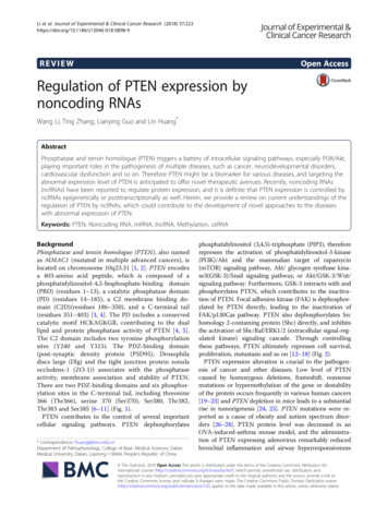
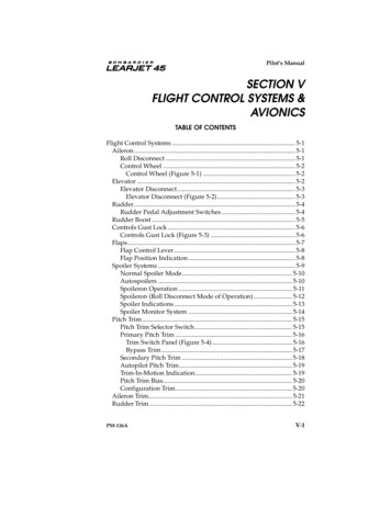
ARTICLENATURE COMMUNICATIONS DOI: 10.1038/ncomms12355cPTEN- 37GAPDH0*20406080shPTENMitotic index (%)15105300Pro*200AnaNS1000020406080Time shcsh coo200:00:00M. Catastrophe(%)eshPTENMet***shsh cooP 2sh TENcsh ooPT 2sh ENcsh ooPT 2ENb400Time in mitosis (min)kDa- 55dshcoo2shcoosh 2PTENaFigure 1 Depletion of PTEN leads to prolonged mitosis and defective chromosome congression. (a) Knockdown of PTEN. HeLa cells were transducedwith a lentiviral shPTEN plasmid and PTEN expression was evaluated by western blotting and compared with cells containing the shcoo2 control vector.(b) Mitotic arrest by PTEN depletion. HeLa cells with and without PTEN knockdown were immunofluorescent stained with phospho-histone H3 (Ser10).Mitotic index was determined by flow cytometry as percentage of phospho-histone H3-positive cells. Data from three independent experiments wereanalysed by two-tailed t-test and presented as means s.e.m. *Po0.05. (c) Prolonged mitosis in cells lacking PTEN. Time-lapse fluorescent microscopywas performed for 80 h for phase succession analysis in H2B-GFP cells with or without shPTEN. Interphase (blue) and mitosis (red) time lengths werescored in cells with and without shPTEN, showing 20 representative cells in each group. (d) Prominent metaphase delay upon PTEN knockdown. Mitotictiming was measured for different phases in cells of each group that completed mitosis during our analysis (n ¼ 100). Data are presented as means s.e.m.and processed by one-way analysis of variance (ANOVA) and Newman–Keuls multiple comparison test. *Po0.05; ***Po0.001; NS not significant(P40.05). (e) Comparison of mitotic progression in H2B-GFP cells with or without shPTEN shown by representative still frames of live cell microscopy.PTEN depletion results in frequent chromosome misalignment (middle panel) and subsequent mitotic catastrophe (bottom panel). Arrowheads point tochromosomes that fail to congress at the metaphase plate. (f) Summary of the frequency of chromosome misalignment (upper) and mitotic catastrophe(lower) in PTEN-depleted cells as compared to control cells (n490). Data were analysed by two-tailed t-test. **Po0.01; ***Po0.001.consistent spindle architectural abnormalities in Pten null cells.Cells lacking Pten display extra pericentric foci and fragmentedspindle poles (Fig. 2f,g), and exhibit a significant reduction ofspindle length (Fig. 2f,h) in metaphase as compared with wild-typecells. Pten deletion also results in condensed microtubules, likelydue to smaller mitotic spindles (Fig. 2i). Altogether, these resultsillustrate that PTEN maintains normal spindle architecture toensure faithful chromosome transmission during mitosis. As thecontrol of spindle length represents a mitotic force-generatingmechanism35, shorter spindle lengths in PTEN knockdown cellsreflect a reduction of forces generated on chromosomes. Spindlepole fragmentation may also weaken centrosome-directedmicrotubule forces, leading to spindle shortening. Therefore,PTEN may sustain force generation through multiplemechanisms to promote proper spindle–chromosome interactionand efficient chromosome congression.Previous studies showed that nuclear PTEN interacts withCENP-C to maintain centromere integrity, and PTEN also regulatesthe mitotic ubiquitin ligase complex APC/C in a phosphataseindependent manner4,9. We therefore tested the possible role ofCENP-C and APC3 (an APC/C component) in mediating themitotic function of PTEN. As shown in Supplementary Fig. 2a,metaphase spindle length remains unaffected by knockdown ofeither CENP-C or APC3, suggesting that PTEN maintains spindlesize in a manner independent of its interaction with CENP-C orAPC3. We next examined spindle pole integrity and found no effectof CENP-C depletion. In contrast, APC3 knockdown causes mitoticarrest and spindle pole fragmentation, whereas ectopic expressionof APC3 can rescue spindle pole instability in Pten-deficient cells(Supplementary Fig. 2b,e). However, despite the significantalterations of spindle pole integrity caused by different APC3status, the overall spindle morphology remains normal and stableNATURE COMMUNICATIONS 7:12355 DOI: 10.1038/ncomms12355 www.nature.com/naturecommunications3
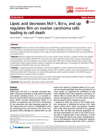
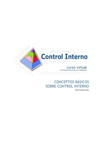
ARTICLENATURE COMMUNICATIONS DOI: N65CRESTColdshcoo211α-tubulinControl12shsh cooPT 2ENshPTENSpindle pole fragmentation (%)shcoo2bPericentrin / α-tubulin / DNAade***shcoo2***012345Mean intensity of α-tubulin25***201023Pt1enPt / en–/–0Spindle length (μm)321Loss of spindle poleintegrity –***2015105Mean intensity of α-tubulingPten / 5*43210enPt / en–/–f14Pt81012Spindle length (μm)en6Pt4***shPTENPt / en–/–shcoo2shPTENshcoo2ColdshPTENControlFigure 2 PTEN depletion impairs mitotic spindle geometry. (a) Spindle pole fragmentation in PTEN knockdown cells. PTEN knockdown and control cellswere immunofluorescent stained for mitotic spindle (a-tubulin, green) and spindle poles (pericentrin, red). Arrows point to fragmented spindle poles.Scale bar, 5 mm. (b) Summary of the frequency of mitotic cells exhibiting spindle pole fragmentation in cells with and without shPTEN. (c) Shortening ofmitotic spindles by PTEN knockdown. HeLa cells with and without shPTEN were left at 37 C or placed at 4 C for 10 min before immunofluorescence ofmicrotubules (a-tubulin, green) and kinetochores (CREST, red) with DAPI counterstaining of chromosomes. Scale bar, 5 mm. (d) Box-and-Whisker plotshowing the distribution of spindle lengths in PTEN knockdown cells and control cells with and without cold treatment. Data (n450 in each condition)were analysed by one-way analysis of variance (ANOVA) followed by Bonferroni’s multiple comparisons. ***Po0.001. (e) Summary of a-tubulin meanintensity within the spindle area in cells with and without PTEN knockdown. (f) Immunofluorescence of mitotic spindles (a-tubulin, green) and spindlepoles (pericentrin, red) in Pten þ / þ and Pten / MEFs. Scale bar, 5 mm. (g–i) The frequency of spindle pole fragmentation (g), spindle pole distance(h) and a-tubulin mean intensity (i) is compared in Pten þ / þ and Pten / cells. Data are presented as means s.e.m. and two-tailed t-test was used fordata analysis. ***Po0.001.following depletion of endogenous APC3 or ectopic APC3expression (Supplementary Fig. 2a,f). These data suggest thatspindle pole fragmentation in PTEN-deficient cells may notcontribute to spindle shortening. Therefore, PTEN controls twoindependent mitotic functions, namely metaphase spindle lengthand spindle pole integrity, likely through distinct mechanisms. Thisnotion was further confirmed by decoupling of the two phenotypesin a rescue experiment using a nuclear-excluded PTEN mutant(K13E;K289E)36. While PTENK13E;K289E retains the PTEN functionin preventing spindle pole fragmentation, this mutant fails torestore spindle length in Pten null cells. More interestingly, it evenexacerbates spindle shortening (Supplementary Fig. 2g,h). Thesedata highlight the importance of PTEN nuclear localization to its4mitotic function, which is predominantly reflected in themaintenance of mitotic spindle length.EG5 is a PTEN-associated mitotic protein. To gain furtherinsight into the PTEN mitotic function, we sought to identifypotential cellular factors in the PTEN pathway that controlmitotic spindle architecture. Using an affinity purificationapproach, we identified EG5 as a potential PTEN-interactingprotein (Fig. 3a,b; Supplementary Fig. 3a). EG5 plays a criticalrole in the dynamic assembly and function of the mitotic spindleby cross-linking and sliding adjacent microtubules20,21. Inaddition, EG5 function also contributes to spindle poleNATURE COMMUNICATIONS 7:12355 DOI: 10.1038/ncomms12355 www.nature.com/naturecommunications
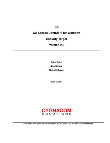
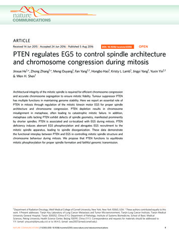
ARTICLENATURE COMMUNICATIONS DOI: 10.1038/ncomms12355cb250130-EG59575dePrometMetAna 1Ana 2PTEN ��551aa552–750aa751–1,05755-PTEN55-IgGNC2- 1–403N:aa1–185C:aa186–403C2:aa186–351FL N C C2N. EN. EN. EN.EPT PT PT PT- FLAG-EG5- GST-PTEN.FLGST-PTEN.CGST-PTEN.NGST-PTEN.C2hHead Neck Tail1 C2gkDa130InputMappingEGEG5.FLEG5.heEG5.n adeEG5.ta ck5. il1EG tail2EG5.FL5EG .heEG5.n adeEG5.ta ck5. il1tail2InterInpMascotResidues PeptidePeptide 1032 801.434429 R.GINTLER.S733–739 895.410935 R.FCALEEK.C298–305 899.544035 R.VITALVER.T319–327 957.524340 R.ILQDSLGGR.T112–119 969.422615 K.TFTMEGER.S182–189 1036.464811 R.LQMFDDPR.N377–384 1067.477128 K.EYTEEIER.L499–508 1146.613250 K.LLNTVEETTK.D703–712 1176.544565 K.QCGNLTEDLK.T147–157 1209.58773K.LTDNGTEFSVK.V909–918 1278.627845 K.LNCFLEQDLK.L222–234 1385.660964 R.TTAATLMNAYSSR.S478–491 1627.746441 K.EEYITSALESTEEK.L330–355 2829.553613 R.TSIIATISPASLNLEETLSTLEYAHR.A(kDa)utIP:IgIP G:PTENCLaddeon rtPT rolco ENmplexesa- EG5- Head- Tail2- GST-PTENFigure 3 PTEN colocalizes and interacts with EG5 during mitosis. (a) Identification of EG5 as a component of PTEN-associated protein complexes bypull-down assay. HeLa cells containing FLAG-HA-tagged PTEN or a control vector were synchronized by releasing from double thymidine block for 8 hbefore tandem affinity purification and separation on SDS-PAGE gel. A 125-kD band was analysed by mass spectrometry (MS) revealing EG5 as a potentialPTEN-associated protein. (b) A list of matched peptides from MS analysis. (c) Validation of the interaction between PTEN and EG5 in vivo. HeLa cell lysateswere immunoprecipitated with an anti-PTEN monoclonal antibody followed by detection of EG5 by Western blotting. The same blot was probed with apolyclonal PTEN antibody. (d) Co-localization between PTEN and EG5 during mitosis. HeLa cells were fixed for immunofluorescent staining of PTEN(green in overlay images) and EG5 (red) before confocal microscopic analysis of spatial relationship between PTEN and EG5 during the cell cycle. DNA wascounterstained with DAPI. Scale bar, 10 mm. (e) GST-tagged PTEN constructs for mapping the EG5-binding domains. (f) In vitro binding assay withFLAG-tagged full-length EG5 and GST-tagged different domains of PTEN as indicated. (g) FLAG-tagged full-length EG5 and different fragments wereconstructed for mapping the PTEN-binding region. (h) EG5 and its fragments with a FLAG tag as indicated in g were expressed in HEK293T cells (leftpanel) for in vitro interaction with Sf9-expressed His-PTEN. Ni-NTA purified protein complexes were subjected to FLAG and PTEN immunoblotting (rightpanel).organization18. As a plus-end-directed microtubule motor,kinesin 5 mediates spindle–kinetochore interaction to controlchromosome behaviour during congression and segregation37–39.To validate the pull-down results, we examined the interaction ofendogenous PTEN with EG5 and detected PTEN-bound EG5 inboth HeLa cells and Pten þ / þ MEFs, but not in Pten null cells(Fig. 3c; Supplementary Fig. 3b). Further analysis usingsynchronized samples defines a mitosis-dependent interactionof PTEN with EG5 (detectable only during a period of 6–10 hafter release from double thymidine block, SupplementaryFig. 3c). Moreover, an in vitro binding assay using purifiedrecombinant proteins reveals a direct interaction between PTENand EG5 (Supplementary Fig. 3d).We next examined PTEN localization during mitosis to obtainan overall image of the spatial and temporal relationshipbetween PTEN and EG5 (Fig. 3d). Interphase PTEN exhibits adiffuse staining in the cytoplasm but shows speckle-like foci inthe nucleus. These nuclear foci denote kinetochores onchromosomes, as reported previously4. In contrast, EG5 isprimarily localized in the cytoplasm during interphase. WhenNATURE COMMUNICATIONS 7:12355 DOI: 10.1038/ncomms12355 www.nature.com/naturecommunications5
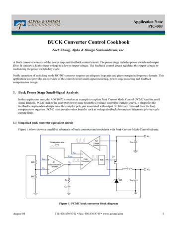
ARTICLENATURE COMMUNICATIONS DOI: 10.1038/ncomms12355cells enter prophase, the majority of PTEN is found on condensedchromosomes while some PTEN signals begin to appear aroundthe centrosome region. Simultaneously, EG5 starts to accumulateon the centrosome and exhibit an overlap with PTEN. Startingfrom prometaphase, a very similar pattern of localizationwas observed for both PTEN and EG5. These molecules areenriched in the separating centrosomes at prometaphase, coverthe whole spindle region during metaphase, accumulate at thespindle crest in anaphase 1, distribute to both the poles and themidbody in anaphase 2, and remain in the cleavage furrow bytelophase (Fig. 3d). The only slight difference is that EG5 stainingoutlines the spindle microtubules, showing more detailedmicrotubule fibres, whereas PTEN only highlights the overallshape of the spindle, likely constituting the spindle matrix.These results clearly illustrate that PTEN is co-localized with EG5during mitosis and further support their physical interaction,likely in a dynamic and mitosis-dependent manner. Further,domain mapping analysis showed that EG5 interacts with boththe N-terminal phosphatase domain and the C2 domainof PTEN (Fig. 3e,f). Interestingly, we did not detect theinteraction between EG5 and the tail-containing region ofPTEN C terminus, which implies a possible interference by thePTEN C-terminal tail. Mapping of EG5 binding domains withPTEN revealed that PTEN interacts with either the N-terminalhead domain or the C-terminal tail of EG5 (Fig. 3g,h).PTEN deficiency leads to increased phosphorylation of EG5.EG5 function in binding mitotic spindles and mediatingmicrotubule-kinetochore attachment is regulated by mitoticspecific phosphorylation27,28,38. To determine whether themitotic-specific association of PTEN with EG5 may represent afunctional regulatory relationship, we employed a massspectrometric phosphoproteome approach to identify potentialphosphorylation sites of EG5 that are regulated by PTEN(Fig. 4a). Incubation of EG5 with PTEN results indownregulation of multiple phosphorylation sites of EG5,among which Thr926 exhibits the highest basal level ofphosphorylation indicative of physiological importance(Supplementary Table 1). Thr926 is an evolutionary conservedresidue within the C-terminal tail domain that can bephosphorylated by Cdc2 (ref. 27). Mutations of this site impairnormal EG5 localization and cause various spindle defects leadingto erroneous chromosome segregation27,29,30,38. These studiesdemonstrate the importance of EG5 phosphorylation for itsmitotic functions, although they have been carried out mostly inyeast or Xenopus. Evidence from mammalian systems is thereforenecessary for understanding how EG5 phosphorylation isregulated and how EG5 deregulation affects spindle andchromosome behaviour in mitosis.PTEN significantly reduces EG5 phosphorylation at Thr926(Fig. 4b–d), suggesting PTEN may target this EG5 site fordephosphorylation. We therefore focused on EG5Thr926 forfurther investigation. We first examined Pten knockout MEFsand observed a higher level of EG5 phosphorylation but steadylevels of total EG5 in Pten null cells as compared with wild-typecells (Fig. 4e), suggesting that PTEN targets EG5 to preferentiallyregulate its phosphorylation. We next analysed thymidinesynchronized HeLa cells with or without PTEN shRNA. Similarkinetics of mitotic-specific EG5 phosphorylation were observedwith or without PTEN knockdown. When comparing eachspecific time point, we found a consistent elevation of EG5phosphorylation in PTEN-depleted cells as compared withcontrol cells (Fig. 4f). The expression levels of EG5 exhibit aslight increase during 6–8 h of DTB release independent of PTENstatus. These data suggest that PTEN regulates EG5 activity bycounterbalancing its phosphorylation.6PTEN dephosphorylates EG5. To characterize EG5 as a mitotictarget of PTEN phosphatase, we employed an in vitro phosphatase assay to determine whether PTEN could directly dephosphorylate EG5. Gradually increased amounts of PTEN result in aprogressive reduction of EG5 phosphorylation at Thr926 withoutaffecting the total levels of EG5 (Fig. 4g). To evaluate theimportance of PTEN phosphatase activity in regulation of EG5phosphorylation, we also examined the ability of two phosphatase-deficient PTEN mutants to dephosphorylate EG5. Incubationwith PTENG129E, which lacks lipid phosphatase activity, results ina significant reduction of EG5 phosphorylation, whereas thedouble-phosphatase-deficient His-PTENC124S mutant fails to doso (Fig. 4h). These results are consistent with our hypothesis thatEG5 is a mitotic target of PTEN phosphatase and highlight theimportance of the protein phosphatase activity of PTEN fordephosphorylation of EG5.To further assess the role of PTEN phosphatase activity insuppression of EG5 phosphorylation in vivo, we examined thelevel of EG5 phosphorylation in Pten null cells with and withoutectopic expression of wild-type PTEN or phosphatase-deficientPTEN mutants PTENC124S and PTENG129E. EG5 phosphorylation is increased in Pten null cells and the introduction of wildtype PTEN significantly reduces EG5 phosphorylation (Fig. 4i).However, phosphorylation of EG5 remains at a high level incells transfected with PTENC124S, suggesting the requirement ofPTEN protein phosphatase activity for dephosphorylating EG5.In contrast, the lipid phosphatase-deficient mutant PTENG129Eretains the ability to suppress EG5 phosphorylation. Nevertheless,we noticed a remaining amount of EG5 phosphorylation inPTENG129E-expressing cells, implying a possible involvement ofthe PI3-kinase/Akt pathway in regulating EG5 phosphorylation.To test this possibility, we treated both Pten þ / þ and Pten / cells with LY294002, a PI3-kinase inhibitor, before evaluation ofEG5 phosphorylation. While LY294002 blocks Akt phosphorylation in the presence and absence of Pten, this inhibitor reducesphospho-EG5 only in Pten / cells. In the presence of PTEN, apredominant EG5 regulator during mitosis, alterations ofPI3-kinase activity no longer affect EG5 phosphorylation(Supplementary Fig. 4). Therefore, the suppression of EG5phosphorylation by PTEN is unlikely dependent on its activityin antagonizing PI3-kinase.PTEN maintains EG5 interaction with the mitotic spindle. Thespatial and temporal dynamics of EG5 ensure its motor functionin driving spindle formation and chromosome segregation. AsPTEN regulates EG5 phosphorylation, we hypothesized thatPTEN loss may affect the spatial relationship between EG5 andthe mitotic spindle. Indeed, we observed aberrant EG5distribution in PTEN knockdown cells. In prophase cells lackingPTEN, EG5 often fails to enrich at the centrosome region and themean intensity of EG5 on centrosomes is significantly reduced(Fig. 5a,b). In the absence of PTEN, EG5 loses the spindleoutlining property and the staining gradient from the spindle poleto the metaphase plate. With an overall reduction of the signalintensity on the metaphase spindle, EG5 often forms clusters nearthe spindle pole region (Fig. 5a,c). These observations suggest thatPTEN is necessary for proper interaction between EG5 and themitotic spindle apparatus.Loss of PTEN results in elevation of the EG5 phosphorylationlevel, shown in the biochemistry assays (Fig. 4). To monitor andcompare the localization changes of the Thr926 phosphorylatedspecies of EG5 during mitosis, we performed phospho-EG5immunofluorescence in wild type and PTEN knockdown cells. Asa functionally active form of EG5 on the mitotic spindle,phospho-EG5 exhibits a similar overall distribution pattern inNATURE COMMUNICATIONS 7:12355 DOI: 10.1038/ncomms12355 www.nature.com/naturecommunications
ARTICLEThr926efkDa130- EG5130gUntreated1510PTENtreated026.0 26.5 27.0 27.5 28.0 28.5Time (min)His-PTEN130- pEG5- EG5shPTEN0 6 8 10 12 14 16 0 6 8 10 12 14 16 DTB release (h)- pEG5- EG5- β-actin37hkDakDa1305shcoo2- pEG5- GapdhT926 (phospho)20kDai ––– –– – – –– EG5PTENC124SG129E55- PTEN130- pEG5130- pEG5130- EG5
These data demonstrate the functional interplay between PTEN and EG5 in controlling mitotic spindle structure and . Peking University Health Science Center, Beijing 100191, China (Y.Y.). Correspondence and requests for materials should be addressed to . the mitotic ubiquitin ligase complex APC/C in a phosphatase-independent manner4,9. We .