Transcription
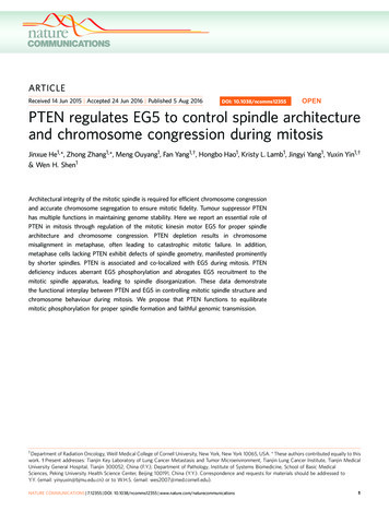
Li et al. Journal of Experimental & Clinical Cancer Research (2018) IEWOpen AccessRegulation of PTEN expression bynoncoding RNAsWang Li, Ting Zhang, Lianying Guo and Lin Huang*AbstractPhosphatase and tensin homologue (PTEN) triggers a battery of intracellular signaling pathways, especially PI3K/Akt,playing important roles in the pathogenesis of multiple diseases, such as cancer, neurodevelopmental disorders,cardiovascular dysfunction and so on. Therefore PTEN might be a biomarker for various diseases, and targeting theabnormal expression level of PTEN is anticipated to offer novel therapeutic avenues. Recently, noncoding RNAs(ncRNAs) have been reported to regulate protein expression, and it is definite that PTEN expression is controlled byncRNAs epigenetically or posttranscriptionally as well. Herein, we provide a review on current understandings of theregulation of PTEN by ncRNAs, which could contribute to the development of novel approaches to the diseaseswith abnormal expression of PTEN.Keywords: PTEN, Noncoding RNA, miRNA, lncRNA, Methylation, ceRNABackgroundPhosphatase and tensin homologue (PTEN), also namedas MMAC1 (mutated in multiple advanced cancers), islocated on chromosome 10q23.31 [1, 2]. PTEN encodesa 403-amino acid peptide, which is composed of aphosphatidylinositol-4,5-bisphosphate-binding domain(PBD) (residues 1–13), a catalytic phosphatase domain(PD) (residues 14–185), a C2 membrane binding domain (C2D)(residues 186–350), and a C-terminal tail(residues 351–403) [3, 4]. The PD includes a conservedcatalytic motif HCKAGKGR, contributing to the duallipid and protein phosphatase activity of PTEN [4, 5].The C2 domain includes two tyrosine phosphorylationsites (Y240 and Y315). The PDZ-binding domain(post-synaptic density protein (PSD95), Drosophiladiscs large (Dlg) and the tight junction protein zonulaoccludens-1 (ZO-1)) associates with the phosphataseactivity, membrane association and stability of PTEN.There are two PDZ-binding domains and six phosphorylation sites in the C-terminal tail, including threonine366 (Thr366), serine 370 (Ser370), Ser380, Thr382,Thr383 and Ser385 [6–11] (Fig. 1).PTEN contributes to the control of several importantcellular signaling pathways. PTEN dephosphorylates* Correspondence: lhuang@dmu.edu.cnDepartment of Pathophysiology, College of Basic Medical Sciences, DalianMedical University, Dalian, Liaoning 116044, People’s Republic of Chinaphosphatidylinositol (3,4,5)-triphosphate (PIP3), thereforerepresses the activation of phosphatidylinositol-3-kinase(PI3K)/Akt and the mammalian target of rapamycin(mTOR) signaling pathway, Akt/ glycogen synthase kinase3(GSK-3)/Snail signaling pathway, or Akt/GSK-3/Wnt/signaling pathway. Furthermore, GSK-3 interacts with andphosphorylates PTEN, which contributes to the inactivation of PTEN. Focal adhesion kinase (FAK) is dephosphorylated by PTEN directly, leading to the inactivation ofFAK/p130Cas pathway. PTEN also dephosphorylates Srchomology 2-containing protein (Shc) directly, and inhibitsthe activation of Shc/Raf/ERK1/2 (extracellular signal-regulated kinase) signaling cascade. Through controllingthese pathways, PTEN ultimately represses cell survival,proliferation, metastasis and so on [12–18] (Fig. 2).PTEN expression alteration is crucial to the pathogenesis of cancer and other diseases. Low level of PTENcaused by homozygous deletions, frameshift, nonsensemutations or hypermethylation of the gene or destabilityof the protein occurs frequently in various human cancers[19–23] and PTEN depletion in mice leads to a substantialrise in tumorigenesis [24, 25]. PTEN mutations were reported as a cause of obesity and autism spectrum disorders [26–28]. PTEN protein level was decreased in anOVA-induced-asthma mouse model, and the administration of PTEN expressing adenovirus remarkably reducedbronchial inflammation and airway hyperresponsiveness The Author(s). 2018 Open Access This article is distributed under the terms of the Creative Commons Attribution 4.0International License (http://creativecommons.org/licenses/by/4.0/), which permits unrestricted use, distribution, andreproduction in any medium, provided you give appropriate credit to the original author(s) and the source, provide a link tothe Creative Commons license, and indicate if changes were made. The Creative Commons Public Domain Dedication o/1.0/) applies to the data made available in this article, unless otherwise stated.
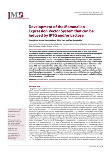
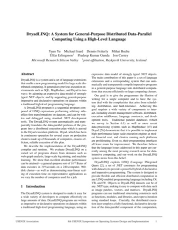
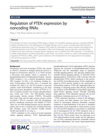
Li et al. Journal of Experimental & Clinical Cancer Research (2018) 37:223Page 2 of 12Fig. 1 The Structure of PTEN. PTEN encodes a 403-amino acid peptide, which is composed of a phosphatidylinositol-4, 5-bisphosphate-bindingdomain (PBD) (residues 1–13), a catalytic phosphatase domain (PD) (residues 14–185), a C2 membrane binding domain (C2D)(residues 186–350),and a C-terminal tail (residues 351–403). The PD includes a conserved catalytic motif HCKAGKGR. The C2 domain includes two tyrosine phosphorylationsites (Y240 and Y315). There are two PDZ-binding domains (PDZ-BD) and six phosphorylation sites in the C-terminal tail. PDZ, post-synaptic density protein(PSD95), Drosophila discs large (Dlg) and the tight junction protein zonula occludens-1 (ZO-1)[29]. However, high level of PTEN either contributes topathological processes. Elevated PTEN expression was observed in endothelium of atherosclerotic brachial arteriesfrom hemodialysis patients. PTEN overexpression stimulated the thrombosis formation of arteriovenous graft byinducing endothelial dysfunction [30]. PTEN negativelyregulates neuron survival, and PTEN downregulationshowed neuroprotective effects in mouse models ofneuron death and Parkinson’s disease [31, 32]. Inhibitionof PTEN rescued synaptic function and cognition in cellular and animal models of Alzheimer’s disease, whereasPTEN transgenic mice displayed synaptic depression [33].In brief, abnormal PTEN expression level is associated tomultiple diseases. Understanding the regulation mechanisms of PTEN expression and maintaining the homeostasis of PTEN should be beneficial.The expression and activity of PTEN is modulated byseveral upstream molecules. P53 binds PTEN promoterand induces its transcription [34]. PDZK1 (PDZ-containing 1) induces PTEN dephosphorylation through bindingthe PDZ-binding domain in the PTEN C-terminal domain, which promotes the anti-oncogenic function ofFig. 2 The schematic representation of the major signaling pathways in which PTEN is involved. CK2, casein kinase II; PDZK1, PDZ-containing 1;GSK3, Glycogen synthase kinase3; FAK, Focal adhesion kinase; Rac, Ras-related C3 botulinum toxin substrate; SHC, Src homology 2-containingprotein; MEK, MAPKK(mitogen-activated protein kinase kinase); ERK1/2, Extracellular signal-related kinase 1/2; PIP3, Phosphatidylinositol (3,4,5)trisphosphate (PtdIns(3,4,5)P3); Akt, Protein kinase B (PKB); MDM2, Mouse double minute 2 homolog; TSC2, Tuberous Sclerosis Complex 2; mTORC,Mammalian target of rapamycin complex; CSCs, Cancer stem cells
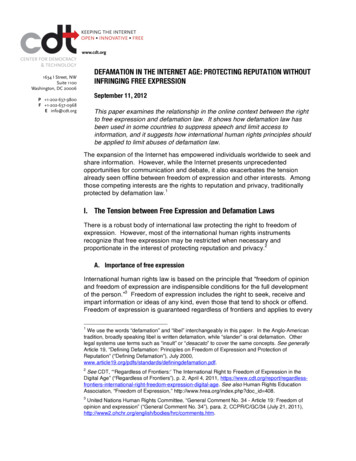
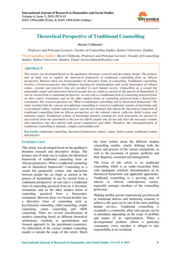
Li et al. Journal of Experimental & Clinical Cancer Research (2018) 37:223PTEN. Protein kinase CK2 (formerly casein kinase II) interacts with and phosphorylates PTEN C-terminal tail,which contributes to maintain PTEN stability [35]. Src inhibits PTEN activity to promote the post-ischaemic contractile recovery in apelin-induced cardioprotection [36].Recently, with the development of the study on noncodingRNAs (ncRNAs), the control of PTEN expression byncRNAs attracted more attention. Herein, we focus on theregulation of PTEN expression by ncRNAs, which is supposed to provide a reference for the coming laboratoryand clinical studies on PTEN regulation (Fig. 3).More than 98% DNAs that do not encode proteins arecalled ncRNAs [37, 38]. In general, ncRNAs are classifiedinto two groups as small ncRNAs ( 200 nt) and longncRNAs (lncRNAs) ( 200 nt). MicroRNAs (miRNAs) ( 18 to 24 nt) are an important group of small ncRNAs,which epigenetically or posttranscriptionally control theexpression of the target mRNAs by pairing to them, leading to the alteration of transcription, mRNA stability ortranslation [39–42]. LncRNAs take up a great proportionin the “transcriptome”, which play vital gene regulatoryroles in the chromatin modification, transcriptional regulation, posttranscriptional regulation and so on [43, 44].Emerging evidence indicates that PTEN functions in adosage-dependent manner during tumor development[24, 45]. NcRNAs are key regulators of PTEN dosage, including miRNAs and lncRNAs, which delicately modulatethe PTEN expression (Tables 1 and 2).MiRNAs modulate PTEN expressionAltering PTEN expression by directly targeting PTEN mRNAMiR-21 is one of the first identified mammalian microRNAs. The human miR-21 gene is located at chromosomePage 3 of 1217q23.2 within a coding gene TMEM49 (also called vacuole membrane protein), which is highly conserved [46].Early lineage tracing studies demonstrated that miR-21was upregulated in various diseases, including acute pancreatitis [47], Myelodysplastic syndromes [48], severesteroid-insensitive allergic airway disease [49], liver cancer[50] and lung cancer [51].PTEN is one of the important targets negatively regulated by miR-21. The 3′UTR of human PTEN contains aputative region that is able to pair to the seed sequence ofmiR-21 (Fig. 4). The exosomal miR-21 transferred frommacrophages downregulated the PTEN level in gastriccancer cells, which resulted in the suppression of cellapoptosis and activation of PI3K/AKT signaling pathway[52]. Inhibition of miR-21 reversed EMT by increasingPTEN protein level in head and neck squamous cell carcinoma (HNSCC), resulting in the suppression of cell proliferation and motility [53]. MiR-21 was able to directlytarget the 3′UTR of PTEN, increasing clear-cell renal cellcarcinoma (CCRCC) cell migration, invasion both in vitroand in vivo [54].Expression of miR-130 family members has been recently reported to be correlated inversely to PTEN expression in bladder cancer, breast invasive carcinoma,lung adenocarcinoma and colon adenocarcinoma [55,56]. Overexpression of miR-130a increased cell proliferation and motility via repression of PTEN expression, associated with the upregulation of FAK and Aktphosphorylation [55–57]. MiR-130a decreases the PTENlevel to activate PI3K/Akt/eNOS (endothelial nitricoxide synthase) signaling pathway, promoting humancoronary artery endothelial cells (HCAECs) injury andinflammatory responses [57]. Exogenous expression ofFig. 3 The regulation of PTEN expression. PTEN expression is dynamically regulated by various events, including genomic mutation or deletion,transcriptional, epigenetic, posttranscriptional, and posttranslational modulation. Noncoding RNAs epigenetically or posttranscriptionally regulatePTEN expression
Li et al. Journal of Experimental & Clinical Cancer Research (2018) 37:223Page 4 of 12Table 1 Regulation of PTEN expression by miR-21Downdirectly targeting PTEN mRNAGastric cancer, HNSCC, CCRCC[52–54]miR-130Downdirectly targeting PTEN mRNABladder cancer, Breast invasive carcinoma,HCAECs injury, Inflammatory responses, PD,Lung adenocarcinoma, Colon adenocarcinoma[55–58]miR-130Updirectly targeting PTEN mRNANSCLC[59]miR-451Updirectly targeting PTEN mRNALung cancer, Ovarian cancer[60, 61]miR-221 /222Downdirectly targeting PTEN mRNANSCLC,HCC, TRAIL-induced cell death[62]miR-301aDowndirectly targeting PTEN mRNABreast cancer, Neuronal death, Ewing’s carcoma,Melanoma, Insulin resistance[63–67]miR-214Downdirectly targeting PTEN mRNATumorigenesis, Immunology, Cardiac injury[68–71]miR-494Downdirectly targeting PTEN mRNAIschemia/Reperfusion -induced myocardial injury[72, 73]miR-155-5p/130b/616/19/92a/10a/106a/429/26a /486-5pDowndirectly targeting PTEN mRNAHCC, NSCLC, Breast cancer, Lung cancer,Colorectal Cancer, Chronic myeloid leukemia,Intestinal cancer, Acute T-cell lymphoblastic leukemia[74–84]miR-29Upinducing the hypomethylation of PTENpromoter by inhibiting DNMT1, DNMT3band SET1A expressionLiver fibrosis[87, 88]miR-101Upinducing the hypomethylation of PTENpromoter by inhibiting DNMT3A expressionLung cancer[89, 90]miR-185Upinducing the hypomethylation of PTENpromoter by inhibiting DNMT1 expressionHCC[91]miR-130a prevented midbrain dopaminergic (mDA)neuron degeneration in Parkinson’s disease (PD) by suppressing the synthesis of PTEN [58].Controversially, miR-130 was also found to be downregulated and positively correlated to PTEN levels innon-small cell lung cancer (NSCLC) tissue samples. Theupregulation of miR-130 significantly increased PTENexpression, inhibited NSCLC cell growth and enhancedcell apoptosis both in vitro and in vivo [59]. Even thesame pairing sequence of miR-130 and PTEN 3’UTRwere used, opposite results were obtained in dual luciferase reporter assays from two reports. The relative activity of the luciferase harboring PTEN 3’UTR waspromoted in A549 cells but repressed in 293 T cells bymiR-130 [56, 59]. Although the mechanisms remain obscure, a tissue-specific pattern is possible for the regulation of PTEN by miR-130. MiR-130 might regulatePTEN expression through different ways according tothe cellular context. PTEN protein was found to beslightly increased after the pre-miR-451-transfection inlung cancer cells [60]. Both miR-451 and PTEN expression level was reported to be significantly reduced inovarian cancer [61].Over the past decade, mountains of results show thatthe interaction of PTEN with miRNAs related to differentdiseases. MiR-221 and miR-222 were reported to be upregulated in aggressive NSCLC and hepatocarcinoma(HCC) cells, and conferred resistance to TNF-relatedapoptosis-inducing ligand (TRAIL)-induced cell death bytargeting PTEN [62]. MiR-301a mediates the tumorigenesis of breast cancer, Ewing’s carcoma and melanoma, prevents neuronal death, and contributes to insulin resistancevia decreasing PTEN protein level [63–67]. MiR-214 induces tumorigenesis, stimulates immunology, and protectscardiac injury through inhibiting PTEN expression [68–71]. MiR-494 targets PTEN and activates Akt pathway,leading to protect against ischemia/reperfusion -inducedmyocardial injury [72, 73]. There are also many othermiRNAs directly targeting PTEN, such as miR-155-5p[74], miR-130b [75], miR-616 [76], miR-19 [77], miR-92a[78], miR-10a [79], miR-106a [80], miR-429 [81], miR-26a[82, 83] and miR-486-5p [84]. Consistent with miR-21,these miRNAs directly bind to the 3′UTR of humanPTEN, and inhibit PTEN expression.Upregulating PTEN expression by targeting DNAmethyltransferases (DNMTs)DNA methyltransferases (DNMTs) are the enzymes forDNA methylation, transferring a methyl group to thecytosine residues of DNA. DNA methylation of a genepromoter typically represses the gene transcription. Thepromoter region of the PTEN gene consists of threemethylation sites. Overexpression of DNMT1 led toPTEN downregulation due to the CpG island methylation in promoter, which promoted tumorigenesis ofbreast cancer, ovarian cancer and acute myeloidleukemia (AML) [85, 86]. MiRNAs targeting DNMTs increase the PTEN expression. MiR-29a was found to
Li et al. Journal of Experimental & Clinical Cancer Research (2018) 37:223Page 5 of 12Table 2 Regulation of PTEN expression by lncRNAsLncRNAPTEN expression MechanismMiRNADiseaseReferencePTENP1Upacting as ceRNAsmiR-21, miR-17, miR-214,miR-19, miR-20, miR-93,miR-106b, miR-26CCRCC, OSCC, HCC,Gastric cancer[54, 96–101]HOTAIRUpacting as ceRNAsmiR-19Cardiac hypertrophy[105]Linc-USP16Upacting as ceRNAsmiR-21,miR-590-5pHCC[106]LncRNA-BGL3Upacting as ceRNAsmiR-17, miR-20a, miR-20b,miR-93, miR-106aChronic myeloid leukemia[80]CASC2Upacting as both ceRNAsmiR-21, miR-181aand downregulators of miRNAsOsteosarcoma, Glioma,Cervical cancer[107–109]MEG3Upacting as both ceRNAsmiR-1297, miR-19a, miR-21and downregulators of miRNAsBreast cancer, Glioma, CAD[111–113]lncRNA GAS5Upacting as both ceRNAsmiR-21, miR-103, miR-196a, HER2-positive breast cancer,[114–120]and downregulators of miRNAs miR-205, miR-32-5pHCC, NSCLC, Cardiac fibrosis,Endometrial cancer, Cervical cancer,Pancreatic cancerXISTUpacting as both ceRNAsmiR-181a, MiR-494and downregulators of miRNAsHCC, Spinal Cord Injury[121, 122]NBAT1Upacting as both ceRNAsmiR-21and downregulators of miRNAsOsteosarcoma[123]lnc-2 /lnc-6Upacting as both ceRNAsmiR-26aand downregulators of miRNAsProstate cancer, Glioma[126, 127]FER1L4Upacting as both ceRNAsmiR-106a-5pand downregulators of miRNAsColon cancer, Gastric cancer[130, 131]lincRNA-p21Upacting as both ceRNAsmiR-181band downregulators of miRNAsLiver fibrosis[132]PTENP1 asRNA β Upincreasing the stabilityand miRNA sponge activityof PTENP1––[133]HOTAIRenhancing PTEN methylationvia miRNA spongingmiR-29bLiver Fibrosis, LSCC[134, 135]enhancing PTEN methylationvia the recruitment of DNMT3aand EZH2––[133]DownPTENP1 asRNA α Downinhibit DNMT1, DNMT3b and SET domain containing1A (SET1A) expression, resulting in elevated PTEN expression and decreased offibrogenic activities in hepaticstellate cells (HSCs) [87]. Curcumin treatment suppressed liver fibrosis by inducing miR-29b expression inHSCs, which led to the low expression of DNMT3b andPTEN hypomethylation [88] (Fig. 5). Bioinformatics anddual luciferase reporter assays demonstrated thatDNMT3A is a target of miR-101 [89]. Introduction ofmiR-101 inhibitor increased the protein level ofDNMT3A instead of the mRNA expression. Overexpression of miR-101 or silencing of DNMT3A induced thehypomethylation of PTEN promoter which was verifiedby a methylation specific PCR assay [90]. The expressionof miR-185 was inhibited in cultured human HCC cells[91]. Introduction of miR-185 mimics significantly reduced DNMT1 expression, decreased PTEN promotermethylation and increased the protein level of PTEN.MiR-185 overexpression decreased the reporter activityof the luciferase with DNMT1 3’UTR, and forced expression of DNMT1 reversed the loss of PTEN promotermethylation mediated by miR-185.LncRNAs modulate PTEN expression indirectlyLncRNAs have multiple important functions in cellularand developmental processes. LncRNAs may carry outFig. 4 A Predicted miR-21 binding site within the 3’UTR of PTEN mRNA. By Target Scan Human Release 7.0 (http://www.targetscan.org)
Li et al. Journal of Experimental & Clinical Cancer Research (2018) 37:223Page 6 of 12Fig. 5 MiR-29a upregulates PTEN expression by targeting DNMTs. MiR-29a could repress DNMTs at posttranscriptional level, resulting in adecrease of CpG island methylation of the PTEN promoter. DNMTs, DNA methyltransferasesboth gene inhibition and activation through diversemechanisms [43, 44]. The studies on the lncRNAs associated with PTEN suggest that lncRNAs modulate PTENexpression by altering either the related miRNAs or promoter methylation.Acting as competing endogenous RNAs (ceRNAs)LncRNAs can act as competing endogenous RNAs (ceRNAs) to indirectly regulate mRNAs through the sharedmiRNAs. LncRNAs compete the seed sites of miRNAswith their target mRNAs, leading to block the effects ofmiRNAs on the mRNA targets [92–95].PTENP1, located on chromosome 9p21, is a highly conserved pseudogene of PTEN. Gan Yu et al. reported thelow expression of PTENP1 due to methylation in CCRCCtissues and cell lines. Both PTEN and PTENP1 expressionis inversely correlated with miR-21 expression. In miR-21overexpressing cells, PTENP1 introduction suppressedcell proliferation and metastasis, and increased the cellsensitivity to cisplatin and gemcitabine, restoring thephenotypes induced by PTEN in vitro and in vivo [54].Activation of PTENP1 partially inhibited the suppressionof PTEN by miR-21 in oral squamous cell carcinoma(OSCC) tumor xenografts [96]. Evidences have revealedthat PTENP1 expression level is positively related toPTEN transcript, and PTENP1 protects PTEN mRNAthrough serving as a decoy for miRNAs, such as miR-21,miR-17, miR-214, miR-19, miR-20, miR-93, miR-106b andmiR-26 families [5, 54, 97–101] (Fig. 6).Homeobox (HOX) transcript antisense RNA (HOTAIR)is encoded within the HoxC gene cluster on chromosome12, which silences the expression of HoxD genes and numerous tumor and metastasis suppressors [102, 103] byinteracting with chromatin-remodeling enzymes [104]. Onthe contrary, HOTAIR regulates PTEN expression as aceRNA. HOTAIR expression decreased notably in sustained cardiac hypertrophy mouse models, in whichmiR-19 expression was increased and inversely correlatedwith HOTAIR expression. HOTAIR has a binding site formiR-19 seed sequence, and HOTAIR overexpressionFig. 6 PTENP1 works as a ceRNA to promote PTEN expression. PTENP1 recruits miRNAs such as miR-181a and miR-21, therefore impairs themiRNAs binding PTEN
Li et al. Journal of Experimental & Clinical Cancer Research (2018) 37:223restored the inhibition of luciferase activity with PTEN3’UTR mediated by miR-19 [105].Linc-USP16 acted as a ceRNA for miR-21 andmiR-590-5p, promoting PTEN expression to repress thegrowth and stimulate apoptosis in HCC in vivo and invitro [106]. LncRNA-BGL3 worked as a ceRNA formiR-17, miR-93, miR-20a, miR-20b, miR-106a andmiR-106b, rescuing the repression of PTEN expressionto inhibit Bcr-Abl-induced cellular transformation [80].Acting as both ceRNAs and downregulators of miRNAsLncRNAs can also decrease the expression level ofmiRNA as well as being sponges, leading to suppress theeffects of miRNAs on their mRNA targets.Cancer susceptibility candidate 2 (CASC2), mapped tochromosome 10q26, encodes a lncRNA which acts as aceRNA of miR-21 or miR-181a and exerts biological effects by increasing the expression of PTEN [107, 108].The expression of CASC2 is significantly downregulatedin glioma, osteosarcoma or cervical cancer tissues and celllines, and CASC2 expression level is negatively correlatedto miR-181a level in glioma tissues. CASC2 overexpression significantly repressed cell proliferation, and amplified temozolomide- or cisplatin-induced repression of cellproliferation in vitro, which was associated with the downregulation of miR-181a and miR-21. CASC2 overexpression upregulated PTEN level, which was partially restoredby miR-181a and miR-21 mimics. In addition, CASC2 wasfound to interact directly with miR-181a and miR-21 indual-luciferase reporter assays [108, 109].Maternally expressed gene 3 (MEG3), encoding alncRNA, is located at the chromosome 14q32. In testiculargerm cell tumor (TGCT) tissues, lncRNA MEG3 level issignificantly decreased, while PTEN protein but notmRNA levels are notably downregulated [110]. Bioinformatics analyses showed that miR-1297 bound not only3’UTR of PTEN mRNA but also MEG3 [111]. MEG3overexpression disturbed the binding of miR-1297 to3’UTR of PTEN mRNA and remitted the reduction ofPTEN induced by miR-1297. MEG3 downregulation andmiR-19a upregulation were reported in malignant gliomatissues and cell lines, and luciferase results verified thecomplementary binding between miR-19a and MEG3.MiR-19a overexpression repressed the expression ofPTEN and promoted glioma cell proliferation, migration,and invasion [112]. Moreover, in the coronary artery disease (CAD) tissues, MEG3 level declines, and miR-21 expression has negative correlation with MEG3 expression.Overexpression of MEG3 suppressed miR-21 expression,promoted the expression of PTEN, and suppressed theproliferation of endothelial cells [113].LncRNA growth arrest specific transcript 5 (lncRNAGAS5) is downregulated in NSCLC, breast cancer andHCC tissues, and lncRNA GAS5 knockdown repressedPage 7 of 12cell viability. lncRNA GAS5 competes with PTEN tobind miR-21, and depletion or overexpression of lncRNAGAS5 could increase or decrease miR-21 expression,resulting in the downregulation or upregulation ofPTEN level in these tumor cells [114–116]. A low expression of lncRNA GAS5 and an upregulation ofmiR-21 are reported in cardiac fibrosis. The downregulation of PTEN expression mediated by miR-21 mimicswas reversed by overexpressing lncRNA GAS5 in cardiacfibroblast cells [117]. LncRNA GAS5 could also inducePTEN expression by inhibiting miR-103 [118], miR-196aand miR-205 [119], and miR-32-5p [120].The lncRNA X inactivate-specific transcript (XIST) directly interacts with miR-181a, and they repress the expression of each other. XIST overexpression restored thePTEN downregulation induced by miR-181a mimics, andtransfection with XIST siRNA significantly enhanced theproliferation and invasion of liver cancer cells togetherwith a decreased PTEN level [121]. Neuronal apoptosisand lncRNA XIST expression level were found to be promoted in an spinal cord injury model. XIST acts as a sinkfor miR-494, leading to derepression of PTEN. MiR-494expression was upregulated with XIST knockdown,whereas was downregulated with XIST overexpression.AntagomiR-494 treatment reversed the protective effectsof XIST depletion on spinal cord injury through blockingthe PTEN/PI3K/AKT signaling pathway [122].The low expression of LncRNA neuroblastoma associated transcript 1 (NBAT1) in osteosarcoma tissues andcells was closely correlated to clinical stages, lymph nodemetastasis and poor prognosis [123]. NBAT1 bindsmiR-21, and suppresses miR-21 expression. NBAT1 overexpression downregulated osteosarcoma growth and metastasis via acting as a ceRNA against miR-21, which wasassociated with PTEN upregulation in vitro and in vivo.The expression of lnc-2 and lnc-6 showed positive correlation with PTEN in prostate cancer cohorts [124,125]. Knockdown of lnc-2 or lnc-6 led to a significantdecrease in PTEN expression at both protein and mRNAlevels and a significant increase in cell proliferation. Onthe contrary, depletion of PTEN reduced the expressionof both lnc-2 and lnc-6,and the reduction of PTEN expression by overexpressing known PTEN-regulatingmiRNAs could be rescued by overexpressing lnc-2sub-sequences [126]. PTEN and lnc-6 are downregulatedwhile miR-26a is upregulated in human glioma. Lnc-6introduction into glioma cells resulted in a decrease ofmiR-26a expression [127].Microarray and real time PCR results showed thatlncRNA fer-1-like family member 4 (FER1L4) was downregulated in gastric cancer, endometrial carcinoma andcolon cancer tissues or cell lines [128]. Enforced expression of FER1L4 increased PTEN expression at bothmRNA and protein levels, which might contribute to cell
Li et al. Journal of Experimental & Clinical Cancer Research (2018) 37:223cycle arrest and apoptosis [129]. In colon cancer celllines, FER1L4 expression is inversely correlated withmiR-106a-5p expression [130]. Luciferase assay resultssuggested direct interactions between miR-106a-5p andFER1L4 or PTEN. Knockdown of FER1L4 increasedmiR-106a-5p expression level and decreased the levels ofPTEN mRNA and protein [130, 131].Fujun Yu et al. reported a novel lincRNA-p21-miR181b-PTEN signaling cascade in liver fibrosis [132].LincRNA-p21 overexpression significantly suppressedthe isolated rat HSC activation and the expression ofextracellular matrix (ECM) proteins, which was reversedby depletion of PTEN. MiR-181b binds lincRNA-p21,and miR-181b level was reduced by exogenouslincRNA-p21, while the effects of lincRNA-p21 onPTEN expression and HSC activation were inhibited bymiR-181b mimics.Increasing the stability of lncRNAsPTENP1, also encodes antisense RNAs (asRNAs), whichhas two isoforms, α and β. PTENP1 asRNA β interactswith PTENP1 through an RNA:RNA pairing interaction,and the stability of PTENP1 was decreased when theinteraction was interfering using U6 encoded ssRNAs orPTENP1 asRNA β was knocked down. Thus PTENP1asRNA β upregualtes PTEN level via increasing the stability and miRNA sponge activity of PTENP1 [133].Prompting the methylation of PTEN promoterHOTAIR expression is upregulated in HSCs during liver fibrosis. HOTAIR knockdown suppressed HSC proliferationand activation in vitro and in vivo, increasing PTEN level,with the loss of DNA methylation mediated by miR-29b[134]. HOTAIR levels were significantly higher in humanlaryngeal squamous cell cancer (LSCC), and bisulfite sequencing of the PTEN promoter addressed that PTENCpG islands were unmethylated in HOTAIR siRNA-transduced cells and PTEN methylation was significantly reduced [135]. Collectively, HOTAIR might contribute toPTEN promoter methylation via sponging miR-29b.The expression of PTEN and PTENP1 asRNA α isnegatively correlated in cell lines, and the α depletion resulted in the increase of PTEN transcript. PTENP1asRNA α binds the PTEN promoter, and epigeneticallydownregulates PTEN transcription by the recruitment ofDNMT3a and Enhancer of zeste homolog 2 (EZH2) toenhance the methylation of PTEN promoter. PTENP1asRNA α knockdown induces cell cycle arrest and sensitizes cells to doxorubicin, suggesting the biological function for the PTENP1 asRNAs [133, 136].Conclusions and future directionsDue to the essential physiological function of PTEN, thencRNAs controlling PTEN expression play crucial roles inPage 8 of 12various biological activation, such as autophagy and cellstemness. PTEN induces autophagy through repressingPI3K/Akt pathway, while miR-21 elevation was found inhuman degenerative nucleus pulposus tissues, which inhibits autophagy and induces ECM degradation viarepressing PTEN expression [137]; Human aortic smoothmuscle cell-derived exosomal miR-221/222 suppressedthe autophagy in human umbilical vein endothelial cellsby regulating PTEN/Akt signaling pathway in a co-culturesystem [138]; MiR-21-5p significantly increases cell stemness of keloid keratinocytes, mediated by PTEN repressionand AKT activation, which may account for the invasionand recurrence of keloids [139]. MiR-10b promotes cellular self-renewal and expression of stemness markers inbreast cancer stem cells through negative regulation ofPTEN and sustained activation of AKT [140].Actually, therapeutic strategies for multiple diseasesfocus on PI3K/Akt pathway inhibitors. However, thetherapeutic benefit is modest due to the network complexities [141,
17q23.2 within a coding gene TMEM49 (also called vacu-ole membrane protein), which is highly conserved [46]. Early lineage tracing studies demonstrated that miR-21 was upregulated in various diseases, including acute pan-creatitis [47], Myelodysplastic syndromes [48], severe steroid-insensitive allergic airway disease [49], liver cancer