Transcription
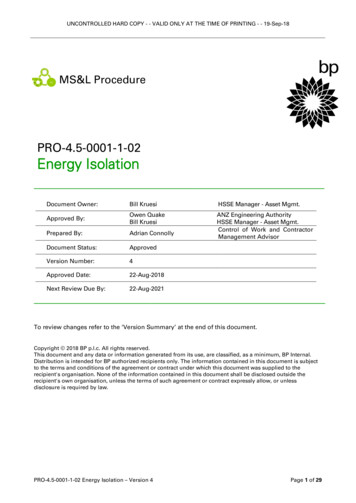
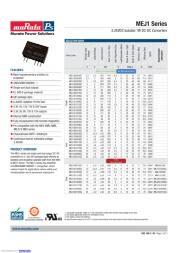
Carnino et al. Respiratory Research(2019) IEWOpen AccessIsolation and characterization ofextracellular vesicles from Broncho-alveolarlavage fluid: a review and comparison ofdifferent methodsJonathan M. Carnino†, Heedoo Lee† and Yang Jin*AbstractExtracellular vesicles (EVs) are cell-derived membranous vesicles secreted by cells into the extracellular space, whichplay a role in cell to cell communication. EVs are categorized into 3 groups depending on their size, surface marker,and method of release from the host cell. Recently, EVs have become of interest in the study of multiple diseaseetiologies and are believed to be potential biomarkers for many diseases. Multiple different methods have beendeveloped to isolate EVs from different samples such as cell culture medium, serum, blood, and urine. Onceisolated, EVs can be characterized by technology such as nanotracking analysis, dynamic light scattering, andnanoscale flow cytometry. In this review, we summarize the current methods of EV isolation, provide details intothe three methods of EV characterization, and provide insight into which isolation approaches are most suitable forEV isolation from bronchoalveolar lavage fluid (BALF).Keywords: Extracellular vesicle (EV), Bronchoalveolar lavage fluid (BALF), Exosome, Microvesicle, Apoptotic bodyIntroductionExtracellular vesiclesExtracellular vesicles (EVs) are membrane bound vesicles which play a role in cell to cell communication. EVsare released from host cells into extracellular space andhave been found in many bodily fluids: urine, sputum,blood, saliva, breast milk, BALF, and more [1]. EVs contain and carry diverse materials such as lipids, proteins,RNA, glycolipids, and metabolites which originate fromthe host cells they are generated from [2, 3]. All categories of EVs have a lipid bilayer which encases the innermaterials; this creates a stable internal environment andprotects EVs from degradation by enzymes [4]. WhenEVs were first discovered, EVs were simply thought tobe involved in the cellular excretion of byproducts, andwere not given attention or studied very extensively [5].Due to the similar characteristics of the major groups of* Correspondence: yjin1@bu.edu†Jonathan M. Carnino and Heedoo Lee contributed equally to this work.Division of Pulmonary and Critical Care Medicine, Department of Medicine,Boston University Medical Campus, 72 E Concord St. R304, Boston, MA02118, USAEVs, the process of isolating and characterizing each typeis difficult to do effectively [6]. Recently, it has become apparent that EV secretion, as well as EV-mediated pathways, are important in both normal biological processesand in several diseases processes [7]. Despite the increasedinterest and research into EV regulatory roles in diseasepathology, the inconsistency in methodology for the collection, isolation, and analysis of EVs has posed a majorbarrier in further development of the field [8]. To combatthis, the International Society for Extracellular Vesiclesrecently published a position statement offering guidelinesto researchers in order to prevent variations across thestudies of EVs [9].EV categoriesBased on their mechanism of development, EVs are classified into three major groups: microvesicles, exosomes,or apoptotic bodies [10]. Figure 1. Microvesicles rangein size from 100 to 1000 nm, and are formed from theoutward budding of the plasma membrane of the hostcell [11]. The membrane of microvesicles are known tocontain larger amounts of cholesterol, diacylglycerol, and The Author(s). 2019 Open Access This article is distributed under the terms of the Creative Commons Attribution 4.0International License (http://creativecommons.org/licenses/by/4.0/), which permits unrestricted use, distribution, andreproduction in any medium, provided you give appropriate credit to the original author(s) and the source, provide a link tothe Creative Commons license, and indicate if changes were made. The Creative Commons Public Domain Dedication o/1.0/) applies to the data made available in this article, unless otherwise stated.
Carnino et al. Respiratory Research(2019) 20:240Page 2 of 11Fig. 1 Schema of Each Major Category of EV. Schema highlighting the key difference in size and method of production between the threecategories of EVs: Microvesicles, Exosomes, and Apoptotic Bodies. MBV: membrane-bound nanovesiclesphosphatidylserine; and the main protein markers forthis category of EVs are integrins, selectins, and CD40 [12].Exosomes range in size from 30 to 150 nm, and are formedwithin the cell as multivesicular bodies, then eventually released into extracellular space after fusion with the cellmembrane [11]. Exosome membranes are known to contain cholesterol, sphingomyelin, phosphatidylinositol, ceramide, and lipid rafts; and contain protein markersincluding CD63, CD9, CD81, and CD82, flotillin, TSG101,Alix, HSP60, HSP70, HSPA5, CCT2, and HSP90 [12].Dying cells produce apoptotic bodies, which range from 50to 5000 nm in size [13]. Apoptotic bodies contain exposedphosphatidylserine on their membranes, and their majorprotein markers include histones, TSP, and C3b [14]. Anotable distinction between apoptotic bodies and the othertwo major EV groups is that apoptotic bodies also containfragmented DNA and cell organelles from their hostcell [15, 16].EVs as a potential biomarkerImmune cells, along with many other cell types, use EVsas a mode of cell to cell communication by transferringprotein and genetic material, which exerts a regulatoryrole in the physiology and pathology of the cells in whichthey target [17]. This ability of EVs to transfer regulatory“messages” to other cells make them worthy of study aspotential biomarkers [6]. MicroRNAs (miRNAs) havebeen extensively studied as they are known to playregulatory roles and serve as biomarkers in many diseases;therefore, the study of EV-containing miRNAs is understandably of specific interest [18, 19]. Development ofbodily fluid-extracted biomarkers would be extremelybeneficial as it would limit the need for collection of tissuesamples and other invasive procedures [4]. Although, onedisadvantage and barrier for now is that bodily fluids contain large amounts of soluble proteins and aggregateswhich pose contamination issues during EV isolationmethods [7]. The isolation of highly pure EVs is essentialto ensure the analysis of the results are not misleadingdue to contamination by viruses, lipoproteins, proteins, orother aggregates [18]. BALF, serum, and pleural fluid areall potentially good specimens which EVs can be isolatedfrom to detect disease biomarkers in the future.Emerging evidence displays that BALF EVs play anessential role in the pathogenesis of various lung diseases[20–35]. For example, BALF EVs have been reported tofunction as carriers of signaling mediator WNT5A, contributing to the pathogenesis of idiopathic pulmonaryfibrosis [22]. Furthermore, BALF EVs generated by sarcoidosis patients have been reported to display proinflammatory effects [32]. Additional studies uncoveringpotential roles of EVs in many different disease processes can be found in Table 1.In this review, we will cover a variety of EV isolationmethods, and discuss the pros and cons of each methodfor isolating EVs from BALF and serum.
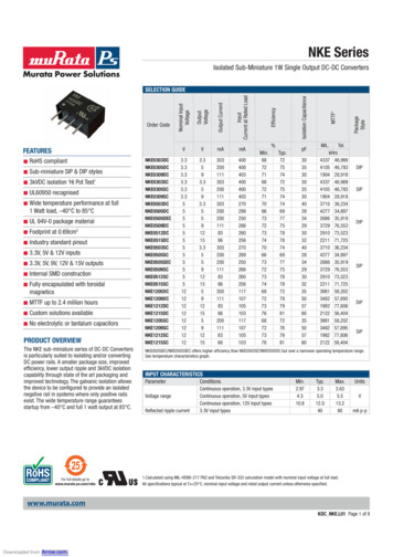
Carnino et al. Respiratory Research(2019) 20:240Page 3 of 11Table 1 Partial Current Literature on BALF-EVs in Lung DiseasesDiseases/processesMain conclusionAuthor/JournalIdiopathic Pulmonary Fibrosis (IPF)Increased BALF-EVs function as carriers for WNT5A,and contribute to the pathogenesis of IPFMartin-Medina et al.;AJRCCM 2018, Jul 25.Asthma/COPDBronchoconstrictionMediate leukotriene conversion LTC4-LTD4Lukic et al.;J Lipid Res 2016;57:1659–69Allergic AsthmaLeukotriene/cytokine productionTorregrosa Paredeset al.; Allergy. 2012Jul;67(7):911–9Allergy and vaccinationEVs can potentially induce tolerancePrado et al.;J. Immunology2008. 181AsthmaEV-lipid profile as a biomarkerHough et al.;Sci Report, 201810,340COPDEVs from PMN regulate the pathogenesisGenschmer et al.Cell. 2019 Jan 10SarcoidosisBALF EVs from sarcoidosis patients carrypro-inflammatory effects.Qazi et al.;Thorax, 2010; 65Lung transplantAcute rejectionThe BALF EV profiles are altered in patientswith acute rejectionGregson et al.;AJRCCM 2015, Dec.15Lung CancerBALF EVs contribute to lung cancer growthYang et al.: Frontier inOncology 2019; April 12Early stage Lung CaBALF EVs as a diagnostic markerKim et al.: Chest2016 Oct Vol 150–4Lung CancerBiomarker of cancer growthYang et alARDSBALF-EV-miRNAs mediate inflammation and ALISheller et al.:J Infectious Dis.2019. Jan. 19thLung InjuryBALF-EV-miRNAs mediate sterile stimuli-associated ALI.Lee et al.:J Immunology 2018Pneumonia/sepsisMacrophage-derived EVs regulate inflammation.Soni et al.Thorax.2016 June 10PulmonaryHypertensionExosomal 15-LO2 mediates hypoxia-mediated HTNZhang et al.Cell Death Dis.2018 Oct 3;9(10):1022Current methods to isolate EVsDifferential centrifugationDifferential centrifugation is a conventional method whichuses centrifugal force to separate contaminants from samples containing EVs. This separation technique involvesseparating and removing components other than EVsfrom a solution in a stepwise manner [36]. First, cell culture media or body fluids should be centrifuged at 300 gfor 10 min at 4 degree Celsius to pellet dead cells and debris [5, 37]. The remaining supernatant is then centrifugedat 2000 g for 10 min at 4 degree Celsius to pellet ABs, andnext, the remaining supernatant can be centrifuged at 10,000 g for 30 min at 4 degree Celsius to pellet MVs [37].Lastly, the remaining supernatant is centrifuged oncemore at 100,000 g for 70 min at 4 degree Celsius to pelletExos; the remaining pellet of ABs/MVs/Exos can be resuspended in PBS [5, 37]. The major advantages to thismethod are the low processing cost, the ability to workwith large quantities of solution and isolate a large quantity of EVs at once, and the absence of additional chemicals needed for the technique [10, 38]. The need forultracentrifugation equipment, the complexity of the stepwise technique, and that fact that efficiency of the technique is dependent on the type of rotor used are alldisadvantages to differential centrifugation [10, 14]. Differential centrifugation can take between 140 and 600 min tocomplete [5, 38]. The sample volume parameters aredependent on the centrifuge rotary tubes used. Samplesizes can range from 1.5 mL to 25 mL depending onthe availability of centrifuge and rotary tubes. Due tothe ability to process large sample sizes at once, ultracentrifugation is likely a useful method for isolationof EVs from human samples. Additionally, for the isolation of EVs from BALF, ultracentrifugation has beenproven to be a consistent method to isolate EVs frommouse BALF [39, 40].
Carnino et al. Respiratory Research(2019) 20:240Density gradient centrifugationDensity gradient centrifugation isolates EVs into specificlayers based on their buoyant density in solutions of either sucrose, iohexol, or iodixanol [41]. It is known thatthis method can successfully separate subcellular components such as peroxisomes, mitochondria, and endosomes into distinct layers within the density gradientsolution [14]. Most density gradient protocols serve tofurther isolate EVs which have previously been partiallyisolated by centrifugation methods. One established protocol for density gradient centrifugation starts with loading 4mL of Tris/sucrose/D2O solution to the bottom of a SW28 tube, then carefully adding 25 mL of PBS containingpartially isolated EVs to the top of this sucrose cushion,and subsequently centrifuging for 75 min at 100,000 g at4 C [42]. Next, 3.5 mL of the Tris/sucrose/ D2O cushioncan be removed from the centrifuged tube and transferredto a new centrifuge tube [42]. This mixture can then be diluted with 60 mL of PBS, and centrifuged for 70 min at100,000 g at 4 C [42]. The resulting pellet contains the isolated EVs and should be resuspended in 50–100 μL of PBS[42]. The advantages to this method include: pure preparation, no contamination with viral particles, and absence ofadditional chemicals for the technique [14]. The disadvantages include: complexity, the need for ultracentrifugationequipment, and loss of sample during isolation [10, 14].Density gradient centrifugation can be a time-consumingprocedure, taking between 250 min to 2 days to complete[14, 42]. Similar to ultracentrifugation, sample size fordensity gradient centrifugation is mostly dependent on sizeof the centrifuge and rotary tubes available. This meanssample volume parameters can potentially be within 1.5mL and 25 mL, however, layering of gradients for thismethod may be difficult at low volumes and therefore alarger volume may be preferred. This is a suitable methodfor EV isolation from mouse BALF, however, due to thesample size may result in a lengthy processing time.Size-exclusion chromatographySize exclusion chromatography makes use of porousbeads to separate biomolecules based on their hydrodynamic radius [43]. This involves the filtration of a solution through a column of porous beads with radiismaller than the EV of interest [44]. During this process,fractions of solution will be eluted in order of decreasingsize, and the fraction containing biomolecules with thesize of the EVs of interest can be selectively isolated[14]. In one protocol, first 12 mL of Sepharose CL-2B isstacked into a 20 mL column, then rinsed and equilibrated with PBS [45]. Once the column is set up, 2 mLof cell culture media can be loaded into the column, andusing PBS as an elution buffer, twenty 0.5 mL fractionsshould be collected from the column [45]. One clearissue with this technique is that there will likely bePage 4 of 11contamination of the sample by other molecules of similar size which elute at the same rate. The purity of preparation, preservation of vesicle integrity, and preventionof EV aggregation are notable advantages for using sizeexclusion chromatography [45]. Also, due to the sizeoverlap between categories, it is difficult to entirely isolate samples of EVs by their category. Additionally, thismethod allows for EVs to be isolated by their 3 respective categories based on their size differences. The disadvantages include: limitations on sample volume, theneed for specialized equipment and a column, and complexity of the technique [45]. The processing time forsize-exclusion chromatography is relatively much fasterthan most methods of EV isolation, taking 1 min per mLof solution [45]. It is recommended to use a sample volume of around 2–5% of the column volume, so samplevolume is limited by the size of the column used for thisprotocol. This method is suitable for the rapid isolation ofEVs from mouse BALF, however, if isolation of each category is desired, an alternative method should be utilizedbecause of size crossover between ABs, MVs, and Exos.Commercial kits for polymer precipitationCommon commercial kits for EV isolation by Polyethyleneglycol (PEG) precipitation are: ExoQuick (System Biosciences), Total Exosome Isolation Reagent (Invitrogen), ExoPrep (HansaBioMed), Exosome Purification Kit (NorgenBiotek), exoEasy (Qiagen), and miRCURY Exosome Isolation Kit (Exiqon) [14]. These kits all use solutions of superhydrophilic polymers, or PEGs, in order to decrease thesolubility of EVs, forming a pellet precipitate. A pellet isformed by mixing the sample with a solution of PEGs, thencentrifuging at low speed (about 1500 g) [44]. The pellet,consisting of EVs and some proteins contaminants, canthen be resuspended in PBS and further analyzed. Commercial kits are relatively fast and have easy to follow protocols. Each kit is slightly different, however, most contain aPEG-based solution and utilize centrifugation as well. Theadvantages to this method are that it is a simple procedureand there is no need for additional equipment [18]. However, there are disadvantages as well, in that the kits areusually costly, may not be good for large samples of EVs,and there is a high concentration of impurities from isolation with these kits [44]. Another problem is that these kitscannot differentiate the three types of EVs, and therefore,during analysis we cannot identify which category of EVcontained any packaged miRNA or protein cargo. Consequently, this method has a significant limitation if used todevelop potential biomarkers, such as markers related toEV-cargo miRNAs. The run time for these commercial kitscan be between 30 and 60 min or sometimes overnightdepending on the kit used [14, 46]. Sample volume forthese kits can range from 63 μL to 10 mL depending on thekit used and the type of sample processed. These kits are
Carnino et al. Respiratory Research(2019) 20:240most commonly used for isolation of EVs from cell culturemedia, serum, or urine. These may be suitable for isolationfrom BALF as well, depending on the kit used and samplevolume required. However, if isolation of EVs by categoryis required then alternative methods should be used.Precipitation with chemicalsPrecipitation of EVs can be done with organic solvents,PEGs, sodium acetate, or protamine [47]. If using organicsolvents such as acetone, chloroform, trichlo- roacetic acid,the ion-pairing effect can provide high efficiency whenusing these solutions to precipitate out EVs [48]. Precipitation by solutions of PEGs, as mentioned earlier, allows EVsand proteins to precipitate out of sample solution into apellet, which can be further analyzed separately. Thismethod tends to have many protein contaminants due tosimilar solubility. Using sodium acetate as a precipitationsolution takes advantage of EVs negatively charged phosphatidylserine [49]. This method disrupts the hydration ofEVs, leading to aggregation by the hydrophobic effect andforming a precipitate pellet [14]. A solution of protamine, apositively charged molecule, can be used to interact withand aggregate EVs because all EVs are known to be negatively charged [50]. After centrifugation, the mixture is gelfiltered in order to remove the protamine and other impurities [14]. A common protocol for isolation of EVs by PEGprecipitation is to combine cell culture media with PEG solution to create an 8% solution, followed by an ultracentrifugation wash at 100,000 g [51]. The resulting washed EVpellet can then be resuspended in sterile PBS [51]. Lowcost, the simplicity of the procedure, and the ability toprocess samples of large volumes are all advantages tomethods which use chemicals to precipitate out EVs [49,51]. The overall disadvantages to these methods are thecontamination issues with non-EV proteins, retention ofchemicals or polymers, and the long processing time forsome of these techniques [14]. Isolation with chemicalscan be relatively quick depending on which solution is used(60–120 min), or overnight incubation may be necessary[18]. This method of EV isolation by precipitation withchemicals is able to be used on a wide range on samplevolumes. For example, in EV isolation with PEG, it is onlyrequired that the final volume is 5–8% PEG. After incubation the mixture should then be centrifuged, thereforesample volume will be dependent on both size of sampleand rotary tubes available, usually between 1.5 mL and 25mL. For studies which don’t require isolation of EVs bycategory, this method is suitable for isolation from mouseBALF. However, if it is required to isolate each category ofEV separately, alternative methods should be used.ImmunoprecipitationImmunoprecipitation can be used to take advantage ofEV surface protein markers such as CD63, CD9, CD8 [6,Page 5 of 1152]. In this method, sample solution is run through magnetic beads, which are coated with antibodies for common EV surface proteins [6]. This method allows forhigh selectivity, however, some types of EVs may elutewith the solution and not be isolated if they do not contain the surface protein markers selected for. One protocol for EV isolation by immunoprecipitation involvesrunning a resuspended EV pellet through a column containing beads coated in antibodies for CD63, CD9, andCD8 [52]. After this affinity-based isolation, the antibodybeads are then washed to elute the EVs isolated [52].This method is most commonly used for further isolation of EVs after a centrifugation method has been utilized. The overall advantages for this method seem to bethe purity of isolated EVs and the high selectivity [53].This method also allows for separation of different EVsbased on their respective protein markers. The disadvantages are that selectivity may be too high, high cost,some difficulties with detachment of antibodies fromEVs, and analysis of intact vesicles [6]. This methodtakes about 240 min to isolate EVs from a sample solution [6, 18]. Sample volume for EV isolation by immunoprecipitation is dependent on the amount of antibodycoated beads used. For large sample volumes, highamounts of beads will be required, and vice versa forsmall sample sizes. Based on volume parameters, thismethod is also suitable for the isolation of EVs frommouse BALF. However, do to extremely high selectivityand possible issues in purity, this method may not bepreferred. Additionally, it may require a large amount ofantibody beads to process samples from mouse BALF.UltrafiltrationUltrafiltration uses porous membranes to trap moleculesor particles of a specific size, allowing smaller moleculesand particles to flow through the membranous filter[54]. This method is usually done in successive steps toisolate EVs of precisely the desired size [38]. Ultrafiltration is based on the particles size and mass, whichmeans it is likely for proteins and other unwanted contaminants to be filtered with the desired EVs. One established protocol begins by concentrating 150 mL of cellculture media to 500 μL with a Centricon Plus-70 Centrifugal Filter (Ultracel-PL Membrane, 100 kDa) deviceusing centrifugation at 3500 g at 4 C [55]. Followingthis, the concentrate can be recovered with a reversespin at 1000 g for 2 min [55]. The Centricon filtershould then be washed with 30 mL of 70% ethanol bycentrifugation at 3500 g, and then rinsed with 30 mL ofPBS by centrifugation at 3500 g [55]. The simplicity ofthe procedure, the ability for concurrent processing ofmany samples, and the lack of limitations on sample volume are all notable advantages to this method [14, 54].The disadvantages include: filter plugging which results
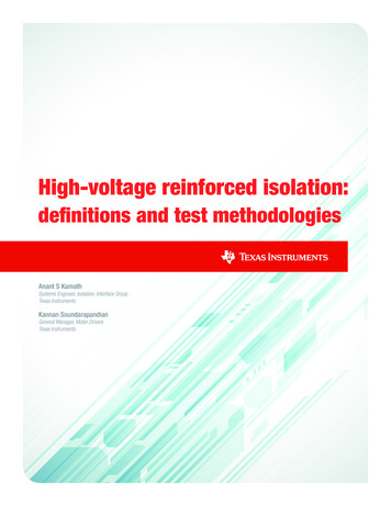
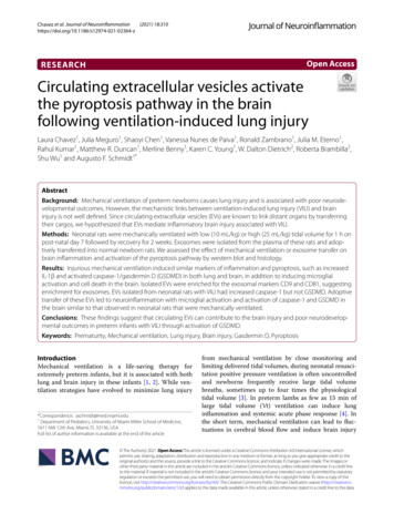
Carnino et al. Respiratory Research(2019) 20:240in loss of sample, and sample contamination by proteins[38]. The technique of ultrafiltration for isolation of EVsusually takes about 130 min [38, 55]. This method doesnot have established limitations on sample volume, however, large sample sizes may lead to long processingtimes. Additionally, larger sample size will increase thelikelihood of filter plugging, which will result in lowyield. This method is suitable for the isolation of EVsfrom BALF, however, due to risks of low yield withhigher sample volumes, this method may lead to difficulties in EV yield.Microfluidic technologiesBeing relatively new technology, microfluidic devices directthe flow of liquids within small, micro-sized channels,which is able to separate and purify samples much moreefficiently than any other sample separation method [42,56]. These devices specifically capture and separate EVs byeither immunoaffinity methods or by the entrapmentwithin porous structures [57]. One microfluidic isolationprotocol which can process samples up to 400 μL utilizes amicrofluidic device with a straight flow channel of 19 mmin width, 20 μm in depth and 4.5 cm long with herringbonegroves on its ceiling that are 50 μm wide and 10 μm deep[42]. For this protocol, cell culture media should beinjected into the device at 16 μL/min for 25 min, thenrinsed with PBS at 30 μL/min for 6 min [55]. EVs shouldadhere to the inner surface of the microfluidic device during initial injection, and be washed out by the followingPBS injection. The resulting solution consists of the isolated EVs. Immunoaffinity methods involves the binding ofparticles by using antibodies which bind to surface proteins. The speed of processing with microfluidic technologyis nearly instant. The advantages to this method include:rapidness of processing, sample purity, and processing efficiency [14, 18]. The high complexity of necessary devices,need for additional equipment, and high cost are all disadvantages to using microfluidic technologies [14]. The sample size required for this method is dependent on thelength of the flow channel. Additionally, the rate of injection of sample size is low, and therefore for large samplesizes there will be a lengthy processing time. This methodmay be suitable for EV isolation from mouse BALF, however depending on the sample size, processing times maybe lengthy. Due to the relatively large sample amount frommouse BALF, this method may end up becoming timeconsuming and complex. Figure 2 and Table 2.EV isolation from BALFThis review focuses specifically on the isolation of EVsfrom bronchoalveolar lavage fluid (BALF). The mainissue for BALF is the limited amount of specimen.Therefore, some techniques which require large volumecannot be used to isolate EVs from BAL.Page 6 of 11Isolation and identification of BALF-derived EVs isstill at the very initial, or “concept” stage, and protocolsare not yet well established. According to the International Society of Extracellular Vesicles (ISEV), andmany other published papers, three main subgroups ofEVs (ABs, MVs, and Exos) can be enriched by 2000–3000 g (AB), 10,000–16,000 g (MVs), and 100,000–120,000 g (Exos) force of sequential centrifugation. We haveshown that the MV population is the main type of BALFEV and falls into the size range of 100–400 nm using sequential centrifugation [39, 40]. On the other hand, Exosare 50–150 nm sized BALF EVs [39, 40]. Notably, in ourprevious study [58], we found that the EV proteinmarkers were differentially expressed among the ABs,MVs, and Exos. Especially TSG101, which is a criticalprotein for generating MVBs, was highly expressed inthe Exosome population. On the other hand, caveolin-1,which is a central component in lipid-raft microdomains,was predominantly expressed in MV population, suggesting that the BALF EV isolation using sequential centrifugation technique is a reliable and convincing method.During the processes of BALF EV isolation using UCand PEG precipitation, there are several basic advanceswhich should be reported. To begin with, it would beideal if the EV purification is performed immediatelyafter the BALFs are obtained. We monitored the criticalEV aggregation and size modification when the EVs werepurified from frozen BALF samples, and it is very hard torecuperate their unique original character. Secondly, wesuggest delicate sonication of the purified EVs utilizing awater-bath sonicator before EV analyses are conducted. Itsignificantly helps to disperse the EV aggregates, whichare possibly generated during the sequential centrifugationor EV freezing/thawing step, and get accurate and consistent results. Finally, long-term storage of the isolated EVsis not recommended. We found that remarkable destruction or loss of EV components, including proteins andRNAs, occurs during the long-term storage of the EVs.Characterization of EVsIsolated samples of EVs also often contain a mixture ofcontaminants consisting of small organelles, lipids, cholesterol, and other undesired microparticles [58]. It is essential to verify the purity of isolated EV samples inorder to validate the accuracy of the experimental resultsderived from processing of the samples. It is possiblethat contamination of isolated EVs may lead to abnormal or misleading data, therefore, checking the samplepurity is a crucial step in properly analyzing EVs.Additionally, characterizing the category of EV (Exo,MV, or AB) may be important for the analysis and interpretation of results from EVs. A reason for this is because some compositions (RNA or protein) may existmore in certain categories of EVs than others. For
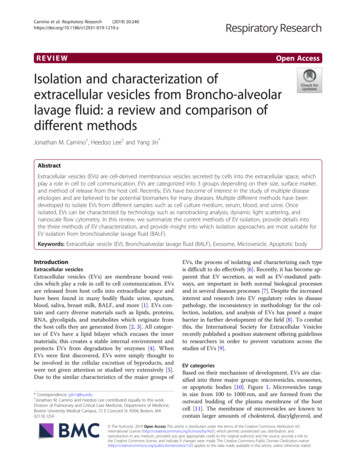
Carnino et al. Respiratory Research(2019) 20:240Page 7 of 11Fig. 2 Flowchart of EV isolation methods. Summary of multiple different protocols for the isolation of EVs. a: Differential centrifugation, b: Densitygradient centrifugation, c: Size-exclusion chromatography, d: Commercial kits for polymer precipitation, e: Precipitation with chemicals, f:Immunoprecipitation, g: Ultrafiltration, and h: Microfluidic technologiesexample, it has been previously reported that serum exosomes contain a very small amount of miRNAs per Exo,and therefore are unlikely to possess a biological purpose[59]. Exosomes are much smaller (30–150 nm) andformed by endosomal origin, whereas MVs are muchlarger (100–1000 nm) and formed by the outward budding of the lipid membrane [11, 60]. Due to this distinction in quantity of contents, MVs may play a greaterrole in communicating cell injury and could be a morevaluable prospect for future studies.Another important characteristic of EVs which shouldbe analyzed is the integrity of the isolated microparticle.In order for EVs to have a future potential use particularly in the development of drug delivery, it is criticalthat EVs maintain their integrity and efficacy after multiple cycles of being frozen and thawed in order to havethe ability to be developed into a pharmaceutical product for the future [61]. Isolated EVs from different cells,which preserve both their integrity and effectivenessafter many freeze-thaw cycles, are good candidates to beused for drug delivery in the future.Additionally, EVs can be characterized to determinethe cell type from which the EV originated from basedon detection of EV surface antigens that are identical tothe surface antigens found on its cell of origin [58]. Thisinformation is useful for study as we ca
they target [17]. This ability of EVs to transfer regulatory "messages" to other cells make them worthy of study as potential biomarkers [6]. MicroRNAs (miRNAs) have been extensively studied as they are known to play regulatory roles and serve as biomarkers in many diseases; therefore, the study of EV-containing miRNAs is under-