Transcription
Fluids, Electrolytesand Acid-Base BalanceTodd A. Nickloes, DO, FACOSAssistant Professor of SurgeryDepartment of SurgeryDivision of Trauma/Critical CareUniversity of Tennessee Medical Center - Knoxville
Objectives Define normal ranges of electrolytesCompare/contrast intracellular, extracellular,and intravascular volumesOutline methods of determining fluid andacid/base balanceDescribe the clinical manifestations of variouselectrolyte imbalances.
Normal Plasma Ranges ofElectrolytes Cations SodiumPotassiumCalciumMagnesium Concentration Anions ChlorideBicarbonatePhosphateSulfateOrganic Acids (Lactate)Total Protein 135-145 mEq/L3.5-5.0 mEq/L8.0-10.5 mEq/L1.5-2.5 LmEq/LmEq/LmEq/L
Normal Ranges of Electrolytes Sodium (Na )Range 135 - 145 mEq/L in serum Total body volume estimated at 40 mEq/kg 1/3 fixed to bone, 2/3 extracellular and available fortrans-membrane exchange Normal daily requirement 1-2 mEq/kg/day Chief extracellular cation
Normal Ranges of Electrolytes Potassium (K )Range 3.5 - 5.0 mEq/L in serum Total body volume estimated at 50 mEq/kg 98% intracellular concentration of 150 mEq/L extracellular concentration of 70 mEq/LNormal daily requirement 0.5 – 0.8 mEq/kg/day Chief intracellular cation
Normal Ranges of Electrolytes Intracellular v Extracellular Electrolyte composition is different -K , Mg , PO4-, SO4-, and proteinsNa , Ca , Mg , Cl-, HCO3- and lactateCompositions of ions are maintained IntracellularExtracellularselective permeability of cell membranesactive ion pumpsMovement of water is passive colloid osmotic gradients intravascular v interstitial spaces(extracellularly)osmolar gradients intracellularly v extracellularly
Fluid Balance Calculation of OsmolarityOsm 2 x [Nas] [glu / 18] [BUN / 2.8] Normal osmolarity is 280-300 mOsm/L Na resorption and excretion are the drivingforces for osmolarity Calculating TBW deficit TBWD males [(140 – SNa ) x 0.6 x IBW (kg)]/ 140 TBWD females [(140 – SNa ) x 0.5 x IBW (kg)]/ 140
Fluid Balance Here’s a trick For every 3.5 mEq the Na is over 140, there is anestimated free water deficit of 1 L.
Normal Physiology Total body water 60% IBW of males50-55% IBW of females directly related to musclemass (70% water)inversely related to fatcontent (10% water)This is why witches floatCompartments IntracellularExtracellularInterstitial
10
Body Fluid Compartments 2/3(65%) of TBW is intracellular (ICF) 1/3 extracellular water 25% interstitial fluid (ISF) 5- 8 % in plasma (IVF intravascular fluid) 1- 2 % in transcellular fluids – CSF, intraocularfluids, serous membranes, and in GI, respiratoryand urinary tracts(third space)11
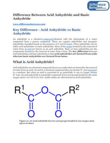
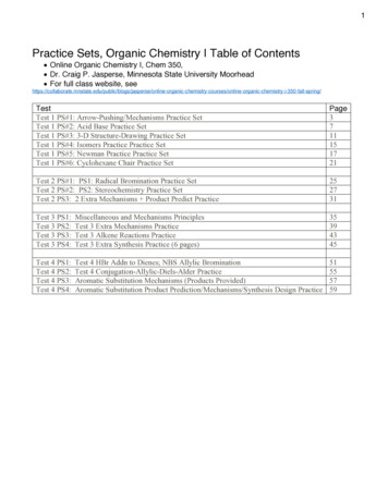
Normal Physiology8%25%67%IntravascularwaterInterstitial waterIntracellularwater
13
Normal Physiology Two main compartments Intracellular 2/3 of TBW40% body weight8%Extracellular Intravascular andInterstitial compartments1/3 of TBW20% body weight25%67%IntravascularwaterInterstitial waterIntracellularwater
15
Fluid compartments are separated bymembranes that are freely permeable to water.Movement of fluids due to: hydrostatic pressure osmotic pressureCapillary filtration (hydrostatic) pressureCapillary colloid osmotic pressureInterstitial hydrostatic pressureTissue colloid osmotic pressure16
17
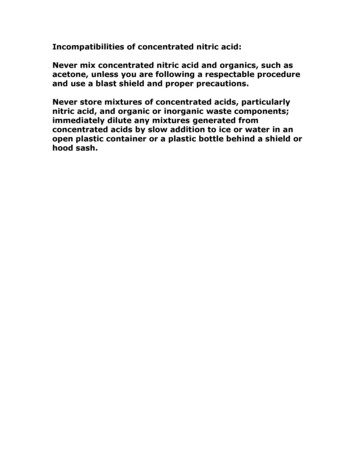
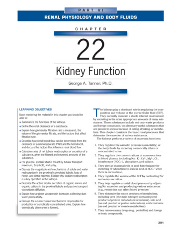
Cell in ahypertonicsolution18
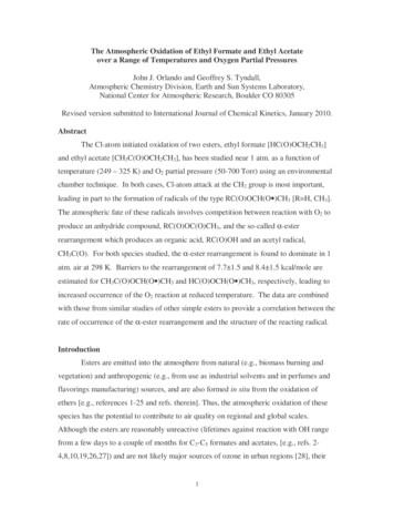
Cell in ahypotonicsolution19
20
Balance Fluid and electrolyte homeostasis is maintainedin the bodyNeutral balance: input output Positive balance: input output Negative balance: input output 21
Solutes – dissolved particles Electrolytes – charged particles Cations– positively charged ions Na , K , Ca , H Anions – negatively charged ions Cl-, HCO3- , PO43 Non-electrolytes - Uncharged Proteins,urea, glucose, O2, CO222
Body fluids are: Electrically neutral Osmotically maintained Specific number of particles per volume offluid23
Homeostasis maintained by: Ion transportWater movementKidney function24
Basic DefinitionsMW (Molecular Weight) sum of the weights ofatoms in a moleculemEq (milliequivalents) MW (in mg)/ valencemOsm (milliosmoles) number of particles in asolution25
Movement of body fluids“ Where sodium goes, water follows.”Diffusion – movement of particles down aconcentration gradient.Osmosis – diffusion of water across aselectively permeable membraneActive transport – movement of particles upa concentration gradient ; requires energy26
27
Normal Physiology Na resorption secondary to aldosteroneOccurs in distal convoluted tubules Active exchange for K and H ions Maintains extracellular volume and osmolarity Water resorption secondary to antidiuretichormoneOccurs in collecting ducts Modulated by intracranial osmoreceptors and atrialstretch receptors
Normal PhysiologyNa modulationRenal perfusion decreasesRenin secretion ceasesJG apparatus secretes reninRenin cleaves angiotensinogento angiotensin IAngiotensin I converted toII by ACERenal perfusion increasesExtracellular volumeexpansion as waterfollows Na Angiotensin II stimulates aldosteronesecretion from adrenal cortexAldosterone increases Na resorption in exchange for K inDCT
30
Normal PhysiologyNa modulationResult:increased water consumptionincreased water conservationIncreased water in bodyincreased volume anddecreased Na concentration31
Dysfunction and/or TraumaLeads to:Decreased amount of water in bodyIncreased amount of Na in the bodyIncreased blood osmolalityDecreased circulating blood volume32
Regulation of body water ADH – antidiuretic hormone thirstDecreased amount of water in body Increased amount of Na in the body Increased blood osmolality Decreased circulating blood volume Stimulate osmoreceptors in hypothalamusADH released from posterior pituitaryIncreased thirst33
Normal PhysiologyFree H2O modulationIntracranial osmoreceptors detectincreased plasma osmolarityAdenohypophysis (posteriorpituitary) secretes ADHPlasma osmolarity increasesRenal collecting ducts becomemore permeable to waterRenal collecting ducts becomeless permeable to waterAdenohypophysis ceases ADHsecretionPlasma osmolarity decreasesIntracranial osmoreceptorsdetect decreased osmolarity
Acid/Base Balance The management of Hydrogen ionsmeasured as pH maintained at 7.4 /- 0.05 Three mechanisms (differing effective intervals)buffering systems in plasma ventilatory changes for CO2 excretion Renal tubular excretion of Hydrogen ions
Acid/Base Balance Henderson-HasselbalchRemember H2O CO2 H2CO3 HCO3- H pH 6.1 log[HCO3-]/0.03 x PaCO2or[H ] 24 x PaCO2/ [HCO3-]
Acid/Base BalanceExtracellular RegulationPulmonary regulation of PaCO2 and renal tubularregulation of HCO3- are important determinants ofextracellular pH.Basically, the pH is determined by the ratio of[HCO3-/H2CO3]Normally 20:1 (7.40) As one increases, the other increases to re-establishthe 20:1 ratio
Acid/Base BalanceIntracellular Regulation Intracellular buffering Excessive CO2 retention or excretion 50% of fixed acid loads (lactate)95% of hydrogen ion changesReciprocal K ion exchange Alkalosis Acidosis H moves extracellularlyK moves intracellularlyH moves intracellularlyK moves extracellularlyThis can have significant clinical effect, especially regardingmyocardial function
Fluids and Electolytes in thePostoperative Period MaintenanceResuscitationReplacement of Losses
Fluids and Electolytes in thePostoperative Period Maintenance Normal daily outputs Urine 12-15 cc/kg Stool 3 cc/kg Sweat 1.5 cc/kg Respiratory and Skin insensible losses 10 cc/kg Increased by 8%/degree F for feverNormal daily endogenous input Oxidation of carbohydrates and fat 3 cc/kg Standard nomograms estimate daily requirements
41
42
Fluids and Electolytes in thePostoperative Period Third Space Fluid Losses Fluids sequestered into extracellular and intersitial spaces PeritonitisIntestinal obstructionSoft tissue inflammation/edemaTraumatic lossesEvidence of diminished volume Hemodynamic changes TachycardiaNarrowed pulse pressuresHypotension
EdemaThe accumulation of fluid within the interstitialspaces.Leads to:increased hydrostatic pressurelowered plasma osmotic pressureincreased capillary membrane permeabilitylymphatic channel obstruction44
Hydrostatic pressure increasesVenous obstruction:thrombophlebitis (inflammation of veins)hepatic obstructiontight clothing on extremitiesprolonged standingSalt or water retentioncongestive heart failurerenal failure45
Decreased plasma osmoticpressure plasma albumin (liver disease orprotein malnutrition)plasma proteins lost in :glomerular diseases of kidneyhemorrhage, burns, open woundsand cirrhosis of liver46
Increased capillary permeability:Inflammationimmune responsesLymphatic channels blocked:surgical removalinfection involving lymphaticslymphedema47
Electrolyte imbalances: Sodium Hypernatremia(high levels of sodium) PlasmaNa 145 mEq / L Due to Na or water Water moves from ICF ECF Cells dehydrate48
49
Hypernatremia Causes HypertonicIV soln. Oversecretion of aldosterone Loss of pure water Long term sweating with chronic fever Respiratory infection water vapor loss Diabetes – polyuria Insufficient intake of water (hypodipsia)50
Clinical manifestationsof Hypernatremia ThirstLethargyNeurological dysfunction due to dehydration ofbrain cellsDecreased vascular volume51
Treatment of Hypernatremia Lower serum Na Isotonic salt-free IV fluid Oral solutions preferable52
Hyponatremia Overall decrease in Na in ECFTwo types: depletional and dilutionalDepletional HyponatremiaNa loss: diuretics, chronic vomiting Chronic diarrhea Decreased aldosterone Decreased Na intake53
Dilutional Hyponatremia:Renal dysfunction with intake of hypotonic fluids Excessive sweating increased thirst intake ofexcessive amounts of pure water Syndrome of Inappropriate ADH (SIADH) oroliguric renal failure, severe congestive heart failure,cirrhosis all lead to: Impaired renal excretion of water Hyperglycemia– attracts water54
Clinical manifestations ofHyponatremia Neurological symptoms Lethargy, headache, confusion, apprehension, depressedreflexes, seizures and coma Muscle symptoms Gastrointestinal symptoms Cramps, weakness, fatigueNausea, vomiting, abdominal cramps, and diarrheaTx – limit water intake or discontinue meds55
Hypokalemia Serum K 3.5 mEq /LBeware if diabetic Insulin gets K into cell Ketoacidosis – H replaces K , which is lostin urine β– adrenergic drugs or epinephrine56
Causes of Hypokalemia Decreased intake of K Increased K loss Chronic diuretics Acid/base imbalance Trauma and stress Increased aldosterone Redistribution between ICF and ECF57
Clinical manifestations ofHypokalemia Neuromuscular disorders Weakness, flaccid paralysis, respiratory arrest,constipationDysrhythmias, appearance of U wavePostural hypotensionCardiac arrestOthers – table 6-5Treatment Increase K intake, but slowly, preferably by foods58
Hyperkalemia Serum K 5.5 mEq / LCheck for renal diseaseMassive cellular traumaInsulin deficiencyAddison’s diseasePotassium sparing diureticsDecreased blood pHExercise causes K to move out of cells59
Clinical manifestations ofHyperkalemia Early – hyperactive muscles , paresthesiaLate - Muscle weakness, flaccid paralysisChange in ECG patternDysrhythmiasBradycardia , heart block, cardiac arrest60
Treatment of Hyperkalemia If time, decrease intake and increase renalexcretionInsulin glucoseBicarbonateCa counters effect on heart61
Calcium Imbalances Most in ECFRegulated by: Parathyroid hormone Blood Ca by stimulating osteoclasts GI absorption and renal retention Calcitonin from the thyroid gland Promotes bone formation renal excretion62
Hypercalcemia Results from:Hyperparathyroidism Hypothyroid states Renal disease Excessive intake of vitamin D Milk-alkali syndrome Certain drugs Malignant tumors – hypercalcemia of malignancy Tumor products promote bone breakdown Tumor growth in bone causing Ca release 63
Hypercalcemia Usually also see hypophosphatemiaEffects:Many nonspecific – fatigue, weakness, lethargy Increases formation of kidney stones and pancreaticstones Muscle cramps Bradycardia, cardiac arrest Pain GI activity also common Nausea, abdominal cramps Diarrhea / constipation Metastatic calcification64
Hypocalcemia Hyperactive neuromuscular reflexes and tetanydifferentiate it from hypercalcemiaConvulsions in severe casesCaused by:Renal failure Lack of vitamin D Suppression of parathyroid function Hypersecretion of calcitonin Malabsorption states Abnormal intestinal acidity and acid/ base bal. Widespread infection or peritoneal inflammation 65
Hypocalcemia Diagnosis:Chvostek’s sign Trousseau’s sign TreatmentIV calcium for acute Oral calcium and vitamin D for chronic 66
?
Normal Ranges of Electrolytes Intracellular v Extracellular Electrolyte composition is different Intracellular - K , Mg , PO 4-, SO4-, and proteins, and proteins Extracellular - Na , Ca , Mg , Cl-, HCO, HCO 3-and lactateand lactate Compositions of ions are maintained selective permeability of cel