Transcription
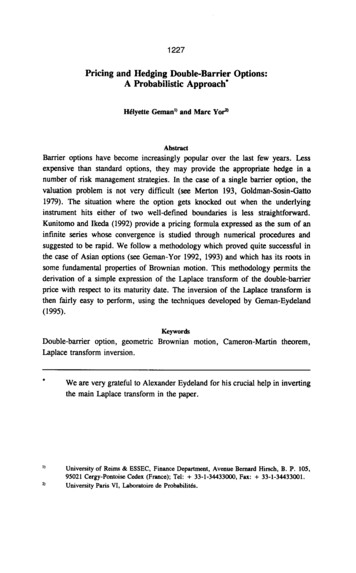
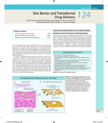
MEDICAL THERAPY SECTION 19Skin Barrier and TransdermalDrug Delivery124Mark R Prausnitz, Peter M Elias, Thomas J Franz, Matthias Schmuth, Jui-Chen Tsai,Gopinathan K Menon, Walter M Holleran and Kenneth R FeingoldSTRUCTURE AND ORIGIN OF THE SKIN BARRIERChapter ContentsStructure and origin of the skin barrier . . . . . . . . . . . . . . . . . . . . . . . . 2065Stratum Corneum Structure and OrganizationParameters affecting skin permeability . . . . . . . . . . . . . . . . . . . . . . . . 2068The stratum corneum is a composite material made of proteins andlipids structurally organized as “bricks and mortar ” (Fig. 124.1; Table124.1)2. Instead of being uniformly dispersed, the highly hydrophobiclipids in normal stratum corneum are sequestered within the extracellular spaces, where this lipid-enriched matrix is organized into lamellarmembranes that surround the corneocytes3. Hence, rather than stratumcorneum thickness, variations in number of lamellar membranes( lipid weight %), membrane structure, and/or lipid compositionprovide the structural and biochemical basis for site-related variationsStrategies to enhance transdermal drug delivery . . . . . . . . . . . . . . 2070The skin provides the largest interface between the human bodyand the external environment. Therefore, one of its most importantfunctions is to regulate what enters the body via the skin, as well aswhat exits. In general, the skin is designed to let very little enter, sinceother tissues, such as the permeable epithelia of the gastrointestinaltract and lung, provide the primary means of regulated entry into thebody. Likewise, the skin must prevent excessive loss of water and otherbodily constituents.The skin’s remarkable barrier properties are due in large part tothe stratum corneum, which represents the thin outer layer of theepidermis1. In contrast to other tissues in the body, the stratumcorneum consists of corneocytes (composed primarily of aggregatedkeratin filaments encased in a cornified envelope) that are surroundedby an extracellular milieu of lipids organized as multiple lamellar bilayers. These structured lipids prevent excessive loss of water from thebody and likewise block entry of most topically applied drugs, otherthan those that are lipid-soluble and of low molecular weight. Thisposes a significant challenge to administering medications via the skineither for local cutaneous effects or as systemic therapy following theirentry into superficial dermal capillaries.FEATURES OF THE STRATUM CORNEUM Primary barrier to drug absorption into skin Two-compartment organization: “bricks and mortar ” Microheterogeneity within extracellular spaces: “ There’s more to the mortarthan lipid” Persistent metabolic activity: dynamic changes in cytosol, cornified envelope,and interstices from inner to outer stratum corneum Homeostatic links to the nucleated cell layers: barrier function regulatesepidermal DNA and lipid synthesis Pathophysiologic links to deeper skin layers: barrier abrogation and/orepidermal injury initiates epidermal hyperplasia and inflammation Stratum corneum as a biosensor: changes in external humidity alone regulateproteolysis of filaggrin, epidermal DNA/lipid synthesis, and initiation ofinflammationTable 124.1 Features of the stratum corneum.TWO-COMPARTMENT "BRICKS AND MORTAR" SYSTEM AND "PORE" PATHWAYA"Bricks and mortar"BHydrophobic lipids in extracellularspace mortar"Pore" pathway within the stratum corneumDiscontinuous,non-permeable lacunar system:basal conditionsCorneocyte brickFig. 124.1 Two-compartment “bricks and mortar”system and “pore” pathway. A The stratum corneumis a unique two-compartment system, analogous to abrick wall. Whereas lipids are sequesteredextracellularly within the stratum corneum, thecorneocyte is lipid-depleted but protein-enriched.B The degradation of corneodesmosomes results indiscontinuous lacunar domains, which represent thelikely aqueous “pore” pathway. These lacunae canenlarge and extend, forming a continuous butcollapsible network under certain conditions, e.g.occlusion, prolonged hydration, ilicextracellular space0133-ch0124-9780723435716.indd 2065HydrophobicmembranesContinuous,permeable lacunar system20655/21/2012 11:01:21 AM
SECTIONMEDICAL THERAPY19Fig. 124.2 Lamellar body secretion delivers notonly lipid precursors, but also several hydrolyticenzymes to extracellular domains. These organellesalso release antimicrobial peptides, including humanβ-defensin 2 and LL-37 (the carboxy-terminalCohesionfragment of human cathelicidin). As a result, theHydrationantimicrobial barrier is intimately linked to thepermeability barrier. In atopic dermatitis, forBarrier functionexample, there is both an impaired permeabilityAntimicrobial defense barrier and reduced expression of antimicrobialpeptides, explaining in part the predisposition toChemical defensecolonization with Staphylococcus aureus.LAMELLAR BODY SECRETION DELIVERS LIPID PRECURSORS AND HYDROLYTIC ENZYMESTO EXTRACELLULAR DOMAINSLipid spholipids,sphingomyelinConversion into non-polar lipid products(lipases, glucosidases)GlucosylceramidesCeramides 1–9SphingomyelinCeramides c enzymesProteases, lipases,acid phosphatase,glycosidases1. Degradation of corneodesmosomes(proteases)2. Degradation of other non-lipidextracellular species(acid phosphatase, glycosidases,proteases)FFA Free fatty acidsLamellar bilayers within lamellar bodyLamellar bilayers within the extracellular spaceCornified envelopeKeratin filaments within corneocytesCornified envelopeFACTORS AFFECTING HOW STRATUM CORNEUM LIPIDS MEDIATEBARRIER FUNCTION Extracellular localization: only intercellular lipids play a roleAmount of lipid (lipid weight %)Elongated, tortuous pathway: increases diffusion lengthOrganization into lamellar membrane structuresHydrophobic composition: absence of polar lipids and presence of very-longchain saturated fatty acids Correct molar ratio: approximately 1 : 1 : 1 of three key lipids: ceramides,cholesterol and free fatty acids Unique molecular structures (e.g. acylceramides)Table 124.2 Factors affecting how stratum corneum lipids mediate barrierfunction.2066in permeability4. It follows, then, that the extracellular, lipid-enrichedmatrix of the stratum corneum comprises not only the structure thatlimits transdermal delivery of hydrophilic drugs, but also the so-calledstratum corneum “reservoir ”5, within which lipid-soluble drugs, suchas topical corticosteroids, can accumulate and be slowly released.Human stratum corneum is typically comprised of about 20 corneocyte cell layers, which differ in their thickness, packing of keratin filaments, filaggrin content, and number of corneodesmosomes, dependingon body site. Corneocytes are surrounded by a highly cross-linked,resilient sheath, the cornified envelope, while the cell interior is packedwith keratin filaments embedded in a matrix composed mainly of filaggrin and its breakdown products (the latter are also referred to as“natural moisturizing factors”). As noted above, individual corneocytes,in turn, are surrounded by a lipid-enriched extracellular matrix, organized largely into lamellar membranes, which derive from secretedlamellar body precursor lipids (Fig. 124.2). Following secretion, lamellarbody contents fuse end-to-end, forming progressively elongated membrane sheets3, a sequence requiring the action of a battery of lipolytic“processing” enzymes (see below). Although corneocytes play a roleboth as spacers and as a scaffold for the extracellular matrix, transdermal drug delivery strategies have focused primarily on manipulationsof the extracellular lipid milieu6,7. Lastly, the existence of aqueous poreswithin the extracellular matrix8 not only adds further complexity to theextracellular pathway (see Fig. 124.1), but also provides additionalopportunities for novel delivery strategies.The exceptionally low permeability of normal stratum corneum towater-soluble drugs is the consequence of several characteristics ofthe lipid-enriched, extracellular matrix (Table 124.2), including its0133-ch0124-9780723435716.indd 2066Aqueous "pore"formationDesquamationKeratin filamentsorganization into a highly convoluted and tortuous extracellularpathway imposed by geometrically arrayed corneocyte “spacers”9. Moreover, not only the paired-bilayer arrangement of extracellular lipids, butalso their extreme hydrophobicity and the composition and distributionof the three key species (ceramides, cholesterol and free fatty acids) ina critical (1 : 1 : 1) molar ratio are further characteristics that provide forbarrier function.Ceramides account for approximately 50% of the total stratumcorneum lipid mass10,11, and are crucial for the lamellar organizationof the stratum corneum barrier12. Of the nine ceramide classes, acylceramides or ceramides 1, 4 and 7 (which contain ω-hydroxy-linked,essential fatty acids in an ester linkage) are epidermis-unique compounds, known to be important for the barrier13. Cholesterol, thesecond most abundant lipid by weight in the stratum corneum, promotes the intermixing of different lipid species and regulates its “phase”behavior14. Free fatty acids, which account for 10–15% of stratumcorneum lipids, consist predominantly of very-long-chain, saturatedspecies with 18 carbon atoms10. A decrease in the concentrations ofany of these critical lipid species compromises barrier integrity, byaltering the molar ratio of the membranes that mediate normal barrierfunction.The “domain-mosaic model” advocates a meandering, polar (pore)pathway for water transport through lamellar boundaries within thelipid mosaic15, adding potential complexity to the already tortuous,extracellular pathway. An alternative model is based upon the presenceof lacunar domains embedded within the lipid bilayers8 (see Fig. 124.1).These lacunae correspond to sites of subjacent corneodesmosomedegradation (see Fig. 124.2), and presumably they contain the hydrophobic degradation products of corneodesmosomes16. Whereas theselacunae are scattered and discontinuous under basal conditions, following certain types of permeabilization (e.g. occlusion, prolonged hydration, sonophoresis, iontophoresis), they are thought to expand untilthey interconnect, forming a continuous “pore pathway ” (see Fig.124.1). The pore pathway can revert back to its original, discontinuousstate once the permeabilizing stimulus disappears.Epidermal Lipid Metabolism and the Skin BarrierBiosynthetic activitiesEpidermal differentiation is a vectorial process that is accompaniedby dramatic changes in lipid composition, including loss of phospholipids with the emergence of ceramides, cholesterol and free fatty acidsin the stratum corneum11,13 (see Fig. 124.2). Although epidermallipid synthesis is both highly active and largely autonomous from5/21/2012 11:01:21 AM
ine terolFASMalonylCoASphinganineSqualeneFatty acidsGlcCer'aseGCSCeramideacetyl CoA carboxylaseGCSGlcCer'aseceramide synthaseSPTfatty acid synthasefarnesol pyrophosphate -glucocerebrosidaseserine palmitoyl transferasepH REGULATES SEQUENTIAL ENZYMATIC STEPS THAT LEAD TO FORMATION OF MATURESTRATUM CORNEUM LAMELLAR MEMBRANESStratum granulosum/stratum corneumRelease of polar lipidcontents from lamellar body7.3Lower stratum corneumSecretory phospholipase A2Steroid sulfataseMid-stratum corneum124Fig. 124.4 pH regulates sequential enzymatic stepsthat lead to formation of mature stratum corneumlamellar membranes. The process begins at thestratum granulosum–stratum corneum interface. Ofnote, in atopic dermatitis, a higher pH is observed.β-glucocerebrosidaseAcidic sphingomyelinasepHsystemic influences, it can be regulated by external influences, i.e.changes in the status of the permeability barrier17. Acute perturbationsof the permeability barrier stimulate a characteristic recovery sequencethat leads to restoration of normal function over about 72 hours inyoung skin (the cutaneous stress test). This sequence includes anincrease in cholesterol, free fatty acid and ceramide synthesis that isrestricted to the underlying epidermis, and attributable to a priorincrease in mRNA and enzyme activity/mass for each of the keysynthetic enzymes (Fig. 124.3). Furthermore, synthesis of each of thethree key lipids is required for normal barrier homeostasis, i.e. topicallyapplied inhibitors of the key enzymes in each pathway produce abnormalities in permeability barrier homeostasis17.Lamellar body secretionThe unique two-compartment organization of the stratum corneumis attributable to the secretion of lamellar body-derived lipids andco-localized hydrolases at the stratum granulosum–stratum corneuminterface3. Under basal conditions, lamellar body secretion is slow,but sufficient to provide for barrier integrity. Following acute barrierdisruption, calcium is lost from the outer epidermis, and much of thepreformed pool of lamellar bodies in the outermost cells of the stratumgranulosum is quickly secreted18–20. Calcium is an important regulatorof lamellar body secretion, with the high levels of Ca2 in the stratumgranulosum restricting lamellar body secretion to low, maintenancelevels21.Extracellular processingExtrusion of the polar lipid contents of lamellar bodies at the stratumgranulosum–stratum corneum interface is followed by the processingof those lipids into more hydrophobic species that form mature, lamellar membranes8 (Fig. 124.4). The extracellular processing of glucosylceramides, phospholipids and cholesterol sulfate with accumulationof ceramides, free fatty acids and cholesterol in the stratum corneum0133-ch0124-9780723435716.indd 2067Fig. 124.3 The major synthetic pathways that leadto the generation of the three key barrier lipids ofthe stratum corneum. The rate-limiting enzymes ineach pathway are shown. Applications of specific,conduritol-type inhibitors of β-glucocerebrosidase tointact skin lead to a progressive abnormality inbarrier function. In both a transgenic murine modelof Gaucher disease (GD) (produced by targeteddisruption of the β-glucocerebrosidase gene) and inthe severe, type 2 neuronopathic form of GD, infantspresent with a barrier abnormality. This wasattributable to accumulation of glucosylceramides,depletion of ceramides, and persistence of immaturelamellar bodies within the interstices of the stratumcorneum.Skin Barrier and Transdermal Drug DeliveryTHE MAJOR SYNTHETIC PATHWAYS FOR THE THREE KEY BARRIER LIPIDSOF THE STRATUM CORNEUM 5.0is attributable to the co-secretion of a set of hydrolytic enzymes3 (seeFig 124.2).Extracellular processing of glucosylceramides plays a key role inbarrier homeostasis (see legend to Fig. 124.3). In addition, phospholipidhydrolysis, catalyzed by one or more secretory phospholipases (e.g.sPLA2), generates a family of non-essential free fatty acids, which arerequired for barrier homeostasis22–24. Since applications of eitherbromphenacylbromide or MJ33 (chemically unrelated sPLA2 inhibitors)modulate barrier function in intact skin, sPLA2 appears to play a criticalrole in barrier homeostasis22–24. Moreover, applications of either inhibitor to perturbed skin sites delay barrier recovery.Sphingomyelin hydrolysis by acidic sphingomyelinase generatestwo of the nine ceramides required for normal barrier homeostasis (seeFig 124.2). Moreover, patients with mutations in the gene encodingacidic sphingomyelinase (Niemann–Pick, Type A) that lead to lowenzyme activity display an ichthyosiform dermatosis, and transgenicmice with an absence of acidic sphingomyelinase also demonstrate abarrier abnormality. Finally, applications of nonspecific inhibitors ofacidic sphingomyelinase to perturbed skin sites lead to a delay inbarrier recovery25.Just as with glucosylceramides and sphingomyelin, cholesterol sulfatecontent increases during epidermal differentiation, and then decreasesprogressively as the latter is desulfated during passage from the innerto the outer stratum corneum26. Both cholesterol sulfate and its processing enzyme, steroid sulfatase, are concentrated in membrane domainsof the stratum corneum. Of note, the content of cholesterol sulfate inthese sites increases by approximately 10-fold26 in recessive X-linkedichthyosis (see Ch. 57). Not only is recessive X-linked ichthyosischaracterized by a barrier defect27, but also repeated applications ofcholesterol sulfate to intact skin produce a barrier abnormality28. Inboth cases, the barrier abnormality is attributable to cholesterol sulfateinduced phase separation in lamellar membrane domains27. But thebarrier defect may also be, in part, attributed to a reduction in20675/21/2012 11:01:21 AM
SECTION19MEDICAL THERAPYTHE IMPACT OF DECREASED FILAGGRIN PRODUCTION ON THE FUNCTIONS OF THE STRATUM CORNEUMPyrrolidonecarboxylic acidGlutamineHydration-NH2Permeability teaseLL-37 carboxyterminal fragment of human cathelicidinSP serine proteaseTrans-urocanic acid g. 124.5 The impact of decreased filaggrin production on the functions of the stratum corneum. UVB, ultraviolet B.cholesterol content, since cholesterol sulfate is a potent inhibitor ofHMG-CoA reductase (see Fig. 124.3).In addition to lipid precursors and hydrolytic enzymes (e.g. steroidsulfatase, acidic sphingomyelinase), lamellar bodies contain proteasesand antiproteases that orchestrate the orderly digestion of corneodesmosomes, allowing corneocyte shedding. These organelles also releaseantimicrobial peptides, including human β-defensin 2 and LL-37(the carboxy-terminal fragment of human cathelicidin). As a result, theantimicrobial barrier is intimately linked to the permeability barrier. Inatopic dermatitis, for example, there is both an impaired permeabilitybarrier and reduced expression of antimicrobial peptides, explainingin part the predisposition to colonization with Staphylococcus aureus(see Table. 12.3). Inherited abnormalities in serine protease/antiproteaseexpression and filaggrin production are also observed in patients withatopic dermatitis and the impact of reduced filaggrin production onvarious functions of the stratum corneum is depicted in Figure 124.5.AcidificationThe fact that the stratum corneum displays an acidic external pH (“acidmantle”) is well documented, but its origin is not fully understood.Extraepidermal mechanisms (including surface deposits of eccrine- andsebaceous gland-derived products as well as metabolites of microbialmetabolism), endogenous catabolic processes (e.g. phospholipid-to-freefatty acid hydrolysis, deamination of histidine to urocanic acid; see Fig.124.5), and local generation of protons within the lower stratumcorneum (by sodium-proton antiporters [NHE1] inserted into theplasma membrane29,30) could actively acidify the extracellular space.These mechanisms would explain not only the pH gradient across theinterstices of the stratum corneum (see Fig. 124.4), but also selectiveacidification of membrane microdomains within the lower stratumcorneum.The concept that acidification is required for permeability barrierhomeostasis is supported by the observation that barrier recoveryis delayed when acutely perturbed skin sites are immersed in neutralpH buffers31, or when either the sodium–proton exchanger/antiporteror sPLA2-mediated phospholipid catabolism to free fatty acids isblocked29. Acidification appears to impact barrier homeostasis throughregulation of enzymes involved in extracellular processing, such asβ-glucocerebrosidase and acidic sphingomyelinase, which exhibit acidicpH optima (see Fig. 124.4).PARAMETERS AFFECTING SKIN PERMEABILITY2068As discussed in greater detail in the following sections, the skin is anattractive site for drug delivery6,7. However, normal skin provides a0133-ch0124-9780723435716.indd 2068significant barrier to drug absorption. Understanding the parametersthat affect the permeability of this barrier is essential for achievingsuccessful drug therapy via the skin. While local cutaneous effects aregenerally achieved by dissolving or suspending the drug in a vehiclethat is applied topically as a semi-solid formulation (e.g. cream or ointment)32, administration of systemic therapy via the skin is typicallyaccomplished through the use of a patch. In either situation, drug issupplied at the surface of the skin for diffusion across the stratumcorneum, with the goal of reaching therapeutic targets within the skinand/or systemic uptake via superficial dermal capillaries.Parameters Controlling AbsorptionConventional transdermal drug delivery is a passive process governedby Fick’s law, that is, the rate of absorption or flux (J) of any substanceacross a barrier is proportional to its concentration difference acrossthat barrier33,34. For topically applied drugs, the concentration differencecan be simplified as the concentration of drug in the vehicle, Cv, andthe proportionality constant relating flux to concentration is the permeability coefficient, Kp (equation 1). Kp is composed of factors that relateto both drug and barrier, as well as their interaction. These factors are:Km, the partition coefficient; D, the diffusion coefficient; and L, thelength of the diffusion pathway (equation 2). Thus, four factors controlthe kinetics of percutaneous drug absorption (equation 2); however, itis of great practical importance that two of the four (Cv, Km) are highlydependent on one additional factor, the vehicle.J K pCvJ DK m CvL (1)(2)Role of the VehicleThe vehicle is an important link between drug potency and therapeuticeffectiveness, since extensive pharmaceutical research has shown thatthe composition of the vehicle can profoundly influence the rate andextent of absorption (bioavailability). As illustrated by the potencyranking scale for glucocorticoids35, the same drug appears in differentpotency classes when formulated in different vehicles (Table 124.3). Itwas once axiomatic that ointments were more potent than creams.Though true for the early glucocorticoid products, it is no longer generally applicable. Greater understanding of the science underlying topicalformulations has allowed creams, gels, solutions and foams to bespecifically formulated equipotent to ointments (see Table 124.3).In the rational design of dermatologic vehicles that maximizebioavailability, two factors are of critical importance: (1) solubilizingthe drug in the vehicle (Cv); and (2) maximizing movement (partitioning) of drug from vehicle to stratum corneum (Km). The partition5/21/2012 11:01:22 AM
CHAPTERPotency class250BETAMETHASONE DIPROPIONATE112235CLOBETASOL PROPIONATE Temovate ointment 0.05% Temovate cream 0.05% Temovate gel 0.05% Temovate E cream 0.05% Olux foam 0.05% Temovate Scalp Application 0.05%111112FLUOCINONIDE Lidex ointment 0.05% Lidex cream 0.05% Lidex gel 0.05% Lidex solution 0.05% Lidex E cream 0.05%22223TRIAMCINOLONE ACETONIDE Aristocort A ointment 0.1% Kenalog cream 0.1% Kenalog lotion 0.1% Aristocort cream 0.1%3456Table 124.3 Effect of vehicle on potency. *Generic name in header, followedby trade names.coefficient describes the ability of a drug to escape from the vehicle andmove into the outermost layer of the stratum corneum. It is defined asthe equilibrium solubility of drug in the stratum corneum (sc) relativeto its solubility in the vehicle (Km Csc/Cv).Drug ConcentrationThe driving force for percutaneous absorption is the concentrationof soluble drug in the vehicle. Many older topical drug products weremarketed with the expectation that higher concentrations were morepotent. Although true for some products, e.g. tretinoin gels and creams(0.01–0.1%) in which the drug is completely solubilized at all concentrations, for others it is not. Hydrocortisone 1% and 2.5% in a creamformulation have been shown to be of equal potency, as have triamcinolone acetonide 0.025%, 0.1% and 0.5% creams35. One of the majoradvances in formulating glucocorticoids, as first shown with fluocinonide, came when it was discovered that the addition of propyleneglycol to the vehicle could completely solubilize the drug. This led tocorticosteroid products with greater potency, as demonstrated in thevasoconstrictor assay. Newer products are now tested during the development process to ensure that increased drug concentration results inincreased bioavailability. However, excess non-dissolved drug can sometimes be advantageous, especially in transdermal patches worn forprolonged periods of time (e.g. up to a week). In this situation, asdissolved drug is absorbed into the body, non-dissolved drug canthen become dissolved in order to maintain an equilibrium, therebymaintaining a constant dissolved drug concentration over time andproviding a constant rate of delivery36.Partition CoefficientIn general, topically applied drugs are poorly absorbed because only asmall fraction partitions into the stratum corneum. Most of the drugremains on the skin surface, subject to loss from a multitude of factors(exfoliation, sweating, wash-off, rub-off, adsorption onto clothing, andchemical or photochemical degradation). Even 10–12 hours followingdosing, a drug that has not been lost by exfoliation or rub-off remains0133-ch0124-9780723435716.indd 2069200Flux (mcg/cm2/h) Diprolene ointment 0.05% Diprolene gel 0.05% Diprolene cream AF 0.05% Diprosone ointment 0.05% Diprosone cream 0.05% Diprosone lotion 0.05%10% Lidocaine in DMSO1% Lidocaine in DMSO2.5% Lidocaine in EMLA15010050Skin Barrier and Transdermal Drug DeliveryEFFECT OF VEHICLE ON POTENCYCorticosteroid*124LIDOCAINE ABSORPTION THROUGH HUMAN SKININ VITRO00246810Time (hours)12141618Fig. 124.6 Lidocaine absorption through human skin in vitro. Incorporationof DMSO as a co-solvent with ethanol results in both increased drug solubility(Cv) and partitioning (Km). At 10% drug concentration, the maximum flux is10-fold greater than that achieved in an emulsion formulation (eutectic mixtureof lidocaine 2.5% and prilocaine 2.5% [EMLA]). At 1% drug concentration inDMSO the maximum flux is twofold greater than 2.5% drug in EMLA. Reproducedfrom Mallory SB, et al. Topical lidocaine for anesthesia in patients undergoing pulsed dye laser treatmentfor vascular malformations. Pediatr Dermatol. 1993;10:370–5.largely on the skin surface, and it is easily removed by a simple soapand water wash37. In the case of patches worn for several days, as muchas half of the original amount of drug may still be present in the patchwhen it is removed, and this can pose a safety hazard upon disposal,especially with potentially dangerous drugs such as fentanyl38.A number of physical and chemical factors can improve partitioning.Hydration of the skin due to occlusion, either from a topical formulation or a patch, expands the reservoir volume available to drugs withinthe stratum corneum; this can increase absorption as much as fiveto tenfold39. Common excipients such as ethanol and propylene glycolcan also alter barrier structure so as to increase partitioning40. In addition, many excipients have good solvent properties and, as a result,positively affect Cv as well as Km. The use of high concentrationsof propylene glycol to maximize bioavailability has become pervasiveamong the super- and high-potency corticosteroids, but at a price.Adverse events such as burning and stinging are common when suchpreparations are applied to fissured or eroded skin, and contact dermatitis may occur.A number of other compounds have been identified as enhancers.Dimethylsulfoxide (DMSO), the archetypical enhancer, exemplifies theeffects that can be achieved (Fig. 124.6). As with ethanol and propyleneglycol, both Cv and Km are affected. Because DMSO is a superb solvent,higher drug concentrations can be achieved than with other solvents,but it also expands the stratum corneum barrier, permitting increaseddrug uptake and possibly an increased rate of diffusion (D) through thebarrier. However, the use of powerful enhancers such as DMSO isconstrained by excessive skin irritation or toxicity41.Regional VariationAll body sites are not equally permeable42. Variations in stratumcorneum thickness, the number of sebaceous glands, and hydrationstatus can all affect absorption. Current data and clinical experiencesuggest that one can crudely rank regional permeability as follows: nail palm/sole trunk/extremities face/scalp scrotum.20695/21/2012 11:01:22 AM
SECTIONMEDICAL THERAPY19STRATEGIES TO ENHANCE TRANSDERMALDRUG DELIVERYDespite the significant permeability barrier of the stratum corneum,drug delivery via the skin is a very attractive option and is widelyemployed for both local and systemic therapy (Table 124.4; Fig. 124.7)6,7.Topical treatment of cutaneous disorders obviously targets the site ofdisease, thereby minimizing adverse side effects elsewhere within thebody. Delivery of systemic therapies via the skin avoids degradation ofthe medication within the gastrointestinal tract and first-pass metabolism by the liver, both of which are associated with oral administrationof drugs, in addition to evading the pain and safety issues associatedwith injections. Transdermal delivery of drugs, especially via longacting patches, enables infrequent dosing and maintenance of steadystate drug levels.Many dermatologic medications can be applied topically to the skinbecause the required dosage is often exceedingly small and thereforethey can be effective even in the setting of highly inefficient absorption.In addition, a number of skin disorders are associated with compromised barrier function, which leads to enhanced drug uptake in sitesof involvement43.In contrast, systemic dr
mal drug delivery strategies have focused primarily on manipulations of the extracellular lipid milieu 6,7. Lastly, the existence of aqueous pores within the extracellular matrix 8 not only adds further complexity to the extracellular pathway (see Fig. 124.1 ), but also provides additional opportunities for novel delivery strategies.