Transcription
The Spine Journal 14 (2014) 2525–2545Review ArticleLumbar disc nomenclature: version 2.0Recommendations of the combined task forces of the North AmericanSpine Society, the American Society of Spine Radiologyand the American Society of NeuroradiologyDavid F. Fardon, MDa, Alan L. Williams, MDb, Edward J. Dohring, MDc,d,*,F. Reed Murtagh, MDe, Stephen L. Gabriel Rothman, MDf, Gordon K. Sze, MDgaDepartment of Orthopaedics, Midwest Orthopaedics at Rush, Rush University Medical Center, Third Floor, 1611 W. Harrison, Chicago, IL 60612, USAbMedical College of Wisconsin, 9200 West Wisconsin Ave., Milwaukee, WI 53226, USAcMidwestern University School of Medicine, 19389 N 59th Ave, Glendale, AZ 85308, USAdSpine Institute of Arizona, 9735 N. 90th Pl., Scottsdale, AZ 85258, USAeMoffitt Cancer Center and Research Institute, University of South Florida College of Medicine, 3301 USF Alumni Dr., Tampa, FL 33612, USAfKeck School of Medicine of the University of Southern California, 1975 Zonal Ave., Los Angeles, CA 90089, USAgDepartment of Radiology, Yale University School of Medicine, 20 York St., New Haven, CT 06510, USAReceived 23 July 2013; revised 17 March 2014; accepted 14 April 2014AbstractBACKGROUND CONTEXT: The paper ‘‘Nomenclature and classification of lumbar disc pathology, recommendations of the combined task forces of the North American Spine Society, the American Society of Spine Radiology and the American Society of Neuroradiology,’’ was published in 2001in Spine (Ó Lippincott, Williams & Wilkins). It was authored by David Fardon, MD, and Pierre Milette, MD, and formally endorsed by the American Society of Spine Radiology (ASSR), American Society of Neuroradiology (ASNR), and North American Spine Society (NASS). Its purpose was topromote greater clarity and consistency of usage of spinal terminology, and it has served this purposewell for over a decade. Since 2001, there has been sufficient evolution in our understanding of the lumbar disc to suggest the need for revision and updating of the original document. The revised documentis presented here, and it represents the consensus recommendations of contemporary combined taskforces of the ASSR, ASNR, and NASS. This article reflects changes consistent with current conceptsin radiologic and clinical care.PURPOSE: To provide a resource that promotes a clear understanding of lumbar disc terminologyamongst clinicians, radiologists, and researchers. All the concerned need standard terms for the normal and pathologic conditions of lumbar discs that can be used accurately and consistently and thusbest serve patients with disc disorders.STUDY DESIGN: This article comprises a review of the literature.METHODS: A PubMed search was performed for literature pertaining to the lumbar disc. Thetask force members individually and collectively reviewed the literature and revised the 2001 document. The revised document was then submitted for review to the governing boards of the ASSR,ASNR, and NASS. After further revision based on the feedback from the governing boards, the article was approved for publication by the governing boards of the three societies, as representativeof the consensus recommendations of the societies.FDA device/drug status: Not applicable.Author disclosures: DFF: Nothing to disclose. ALW: Consulting:Zyga Technology (B). EJD: Royalties: Stryker (D, Paid directly to institution). FRM: Nothing to disclose. SLGR: Nothing to disclose. GKS:Scientific Advisory Board/Other Office: Guerbet Pharmaceuticals (B); Research Support (Investigator Salary): Siemens (B), Paid directly to institution); Research Support (Staff Materials): Siemens (B), Paid directly toinstitution).The disclosure key can be found in the Table of Contents and at www.TheSpineJournalOnline.com.No funds were received in support of this work, and there are no ascertainable conflicts of interest or associated biases in the text of the consensus manuscript.The authors wish to thank Andrea Gasten, MSc, Ms. Katherine Huffmanand Donna Lahey, RNFA, for their expertise and contributions in preparationof the manuscript. The authors also extend gratitude to Chadi Tannoury,MD, for creating the original artwork for the figures in this publication.* Corresponding author. Spine Institute of Arizona, 9735 North 90thPlace, Scottsdale, AZ 85258, USA. Tel.: (602) 953-9500; fax: (602) 953-1782.E-mail address: EdDohring@gmail.com (E.J. .0221529-9430/Ó 2014 The North American Spine Society, The American Society of Spine Radiology and The American Society of Neuroradiology. Publishedby Elsevier Inc. This is an open access article under the CC BY-NC-ND license ).
2526D.F. Fardon et al. / The Spine Journal 14 (2014) 2525–2545RESULTS: The article provides a discussion of the recommended diagnostic categories pertainingto the lumbar disc: normal; congenital/developmental variation; degeneration; trauma; infection/inflammation; neoplasia; and/or morphologic variant of uncertain significance. The article provides aglossary of terms pertaining to the lumbar disc, a detailed discussion of these terms, and their recommended usage. Terms are described as preferred, nonpreferred, nonstandard, and colloquial. Updated illustrations pictorially portray certain key terms. Literature references that provided the basisfor the task force recommendations are included.CONCLUSIONS: We have revised and updated a document that, since 2001, has provided awidely acceptable nomenclature that helps maintain consistency and accuracy in the descriptionof the anatomic and physiologic properties of the normal and abnormal lumbar disc and that servesas a system for classification and reporting built upon that nomenclature. Ó 2014 The NorthAmerican Spine Society, The American Society of Spine Radiology and The American Societyof Neuroradiology. Published by Elsevier Inc. This is an open access article under the CC BYNC-ND license ).Keywords:Annular fissure; Annular tear; Disc bulge (bulging disc); Disc degeneration; Disc extrusion; Disc herniation;Disc nomenclature; Disc protrusion; High intensity zone; Lumbar intervertebral discPrefaceThe nomenclature and classification of lumbar disc pathology consensus, published in 2001, by the collaborative effortsof the North American Spine Society (NASS), the AmericanSociety of Spine Radiology (ASSR) and the American Societyof Neuroradiology (ASNR), has guided radiologists, clinicians, and interested public for over a decade [1]. This document has passed the test of time. Responding to an initiativefrom the ASSR, a task force of spine physicians from theASSR, ASNR, and NASS has reviewed and modified thedocument. This revised document preserves the format andmost of the language of the original, with changes consistentwith current concepts in radiologic and clinical care. The modifications deal primarily with the following: updating and expansion of Text, Glossary, and References to meetcontemporary needs; revision of Figures to provide greaterclarity; emphasis of the term ‘‘annular fissure’’ in place of ‘‘annular tear’’; refinement of the definitions of ‘‘acute’’ and‘‘chronic’’ disc herniations; revision of the distinction betweendisc herniation and asymmetrically bulging disc; eliminationof the Tables in favor of greater clarity from the revised Textand Figures; and deletion of the section of Reporting and Coding because of frequent changes in those practices, which arebest addressed by other publications. Several other minoramendments have been made. This revision will update aworkable standard nomenclature, accepted and used universally by imaging and clinical physicians.Introduction and historyPhysicians need standard terms for normal and pathologic conditions of lumbar discs [2–5]. Terms that can beinterpreted accurately, consistently, and with reasonableprecision are particularly important for communicating impressions gained from imaging for clinical diagnostic andtherapeutic decision-making. Although clear understandingof the disc terminology between radiologists and cliniciansis the focus of this work, such understanding can be critical,also to patients, families, employers, insurers, jurists, socialplanners, and researchers.In 1995, a multidisciplinary task force from the NASSaddressed the deficiencies in commonly used terms defining the conditions of the lumbar disc. It cited several documentations of the problem [6–11] and made detailedrecommendations for standardization. Its work was published in a copublication of the NASS and the AmericanAcademy of Orthopaedic Surgeons [9]. The work had notbeen otherwise endorsed by major organizations and hadnot been recognized as authoritative by radiology organizations. Many previous [3,7,9–19] and some subsequent [20–25] efforts addressed the issues, but were of more limitedscope and none had gained a widespread acceptance.Although the NASS 1995 effort was the most comprehensive at the time, it remained deficient in clarifying somecontroversial topics, lacking in its treatment of some issues,and did not provide recommendations for standardization ofclassification and reporting. To address the remaining needs,and in hopes of securing endorsement sufficient to result inuniversal standardizations, joint task forces (Co-Chairs David Fardon, MD, and Pierre Milette, MD) were formed bythe NASS, ASNR, and ASSR, resulting in the first versionof the document ‘‘Nomenclature and classification of lumbar disc pathology’’ [1]. Since then, time and experiencesuggested the need for revisions and updating of the originaldocument. The revised document is presented here.The general principles that guided the original documentremain unchanged in this revision. The definitions are basedon the anatomy and pathology, primarily as visualized onimaging studies. Recognizing that some criteria, under somecircumstances, may be unknowable to the observer, the definitions of the terms are not dependent on or imply the valueof specific tests. The definitions of diagnoses are not intended to imply external etiologic events such as trauma,they do not imply relationship to symptoms, and they donot define or imply the need for specific treatment.
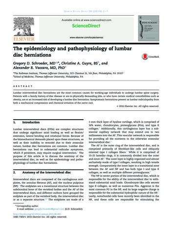
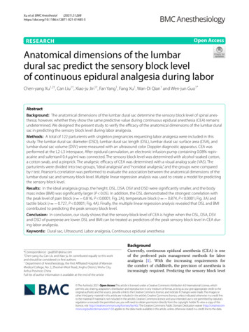
D.F. Fardon et al. / The Spine Journal 14 (2014) 2525–2545The task forces, both current and former, worked from amodel that could be expanded from a primary purpose ofproviding understanding of reports of imaging studies.The result provides a simple classification of diagnosticterms, which can be expanded, without contradiction, intomore precise subclassifications. When reporting pathology,degrees of uncertainty would be labeled as such rather thancompromising the definitions of the terms.All terms used in the classifications and subclassifications are defined and those definitions are adhered tothroughout the model. For a practical purpose, some existing English terms are given meanings different from thosefound in some contemporary dictionaries. The task forcesprovide a list and classification of the recommended terms,but, recognizing the nature of language practices, discussand include in the Glossary, commonly used and misusednonrecommended terms and nonstandard definitions.Although the principles and most of the definitions ofthis document can be easily extrapolated to the cervicaland dorsal spine, the focus is on the lumbar spine. Althoughclarification of terms related to posterior elements, dimensions of the spinal canal, and status of neural tissues isneeded, this work is limited to the discussion of the disc.While it is not always possible to discuss fully the definition of anatomical and pathologic terms without somereference to symptoms and etiology, the definitions themselves stand the test of independence from etiology, symptoms, or treatment. Because of the focus on anatomy andpathology, this work does not define certain clinical syndromes that may be related to lumbar disc pathology [26].Guided by those principles, we have revised and updateda document that, since 2001, has provided a widely acceptable nomenclature that is workable for all forms of observation, that addresses contour, content, integrity, organization,and spatial relationships of the lumbar disc; and that serves asystem of classification and reporting built upon thatnomenclature.Diagnostic category & subcategory recommendationsThese recommendations present diagnostic categoriesand subcategories intended for classification and reportingof imaging studies. The terminology used throughout theserecommended categories and subcategories remains consistent with detailed explanations given in the Discussion andwith the preferred definitions presented in the Glossary.The diagnostic categories are based on pathology. Eachlumbar disc can be classified in terms of one, and occasionallymore than one, of the following diagnostic categories: normal;congenital/developmental variation; degeneration; trauma; infection/inflammation; neoplasia; and/or morphologic variantof uncertain significance. Each diagnostic category can be subcategorized to various degrees of specificity according to theinformation available and purpose to be served. The data available for categorization may lead the reporter to characterizethe interpretation as ‘‘possible,’’ ‘‘probable,’’ or ‘‘definite.’’2527Note that some terms and definitions discussed beloware not recommended as preferred terminology, but areincluded to facilitate the interpretation of vernacular and,in some cases, improper use. Terms may be defined as preferred, nonpreferred, or nonstandard. Nonstandard terms byconsensus of the organizational task forces should not beused in the manner described.NormalNormal defines discs that are morphologically normal,without the consideration of the clinical context and not inclusive of degenerative, developmental, or adaptive changesthat could, in some contexts (eg, normal aging, scoliosis,spondylolisthesis), be considered clinically normal (Fig. 1).Congenital/developmental variationThe congenital/developmental variation category includes discs that are congenitally abnormal or that haveundergone changes in their morphology as an adaptationof abnormal growth of the spine, such as from scoliosisor spondylolisthesis.DegenerationDegenerative changes in the discs are included in abroad category that includes the subcategories annular fissure, degeneration, and herniation.Annular fissures are separations between the annular fibers or separations of annular fibers from their attachmentsto the vertebral bone. Fissures are sometimes classified bytheir orientation. A ‘‘concentric fissure’’ is a separation ordelamination of annular fibers parallel to the peripheral contour of the disc (Fig. 2). A ‘‘radial fissure’’ is a vertically,horizontally, or obliquely oriented separation of (or rent in)annular fibers that extends from the nucleus peripherally toor through the annulus. A ‘‘transverse fissure’’ is a horizontally oriented radial fissure, but the term is sometimes usedin a narrower sense to refer to a horizontally oriented fissurelimited to the peripheral annulus that may include separationof annular fibers from the apophyseal bone. Relatively wideannular fissures, with stretch of the residual annular margin,at times including avulsion of an annular fragment, havesometimes been called ‘‘annular gaps,’’ a term that is relatively new and not accepted as standard [27]. The term ‘‘fissures’’ describes the spectrum of these lesions and does notimply that the lesion is a consequence of injury.Use of the term ‘‘tear’’ can be misunderstood becausethe analogy to other tears has a connotation of injury, whichis inappropriate in this context. The term ‘‘fissure’’ is thecorrect term. Use of the term ‘‘tear’’ should be discouragedand, when it appears, should be recognized that it is usuallymeant to be synonymous with ‘‘fissure’’ and not reflectiveof the result of injury. The original version of this documentstated preference for the term ‘‘fissure’’ but regarded the
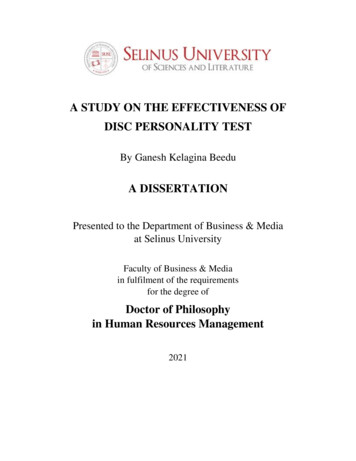
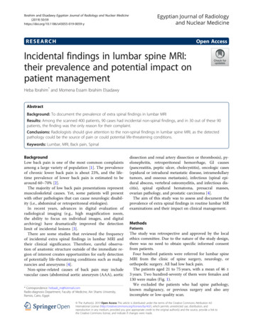
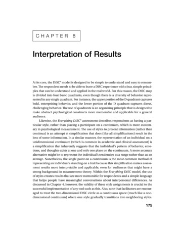
2528D.F. Fardon et al. / The Spine Journal 14 (2014) 2525–2545Fig. 1. Normal lumbar disc. (Top Left) Axial, (Top Right) sagittal, and (Bottom) coronal images demonstrate that the normal disc, composed of central NPand peripheral AF, is wholly within the boundaries of the disc space, as defined, craniad and caudad by the vertebral body end plates and peripherally by theplanes of the outer edges of the vertebral apophyses, exclusive of osteophytes. NP, nucleus pulposus; AF, annulus fibrosus.two terms as almost synonymous. However, in this revision,we regard the term ‘‘tear’’ as nonstandard usage.Degeneration may include any or all of the following:desiccation, fibrosis, narrowing of the disc space, diffuseFig. 2. Fissures of the annulus fibrosus. Fissures of the annulus fibrosus occuras radial (R), transverse (T), and/or concentric (C) separations of fibers of theannulus. The transverse fissure depicted is a fully developed, horizontally oriented radial fissure; the term ‘‘transverse fissure’’ is often applied to a less extensive separation limited to the peripheral annulus and its bony attachments.bulging of the annulus beyond the disc space, fissuring(ie, annular fissures), mucinous degeneration of the annulus, intradiscal gas [28], osteophytes of the vertebral apophyses, defects, inflammatory changes, and sclerosis of theend plates [15,29–34].Herniation is broadly defined as a localized or focal displacement of disc material beyond the limits of the intervertebral disc space. The disc material may be nucleus,cartilage, fragmented apophyseal bone, annular tissue, orany combination thereof. The disc space is defined craniadand caudad by the vertebral body end plates and, peripherally, by the outer edges of the vertebral ring apophyses, exclusive of osteophytes. The term ‘‘localized’’ or ‘‘focal’’refers to the extension of the disc material less than 25%(90 ) of the periphery of the disc as viewed in the axialplane.The presence of disc tissue extending beyond the edgesof the ring apophyses, throughout the circumference of thedisc, is called ‘‘bulging’’ and is not considered a form ofherniation (Fig. 3, Top Right). Asymmetric bulging of disctissue greater than 25% of the disc circumference (Fig. 3,Bottom), often seen as an adaptation to adjacent deformity, is, also, not a form of herniation. In evaluating theshape of the disc for a herniation in an axial plane, the
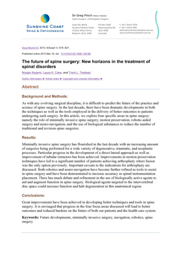
D.F. Fardon et al. / The Spine Journal 14 (2014) 2525–25452529Fig. 3. Bulging disc. (Top Left) Normal disc (for comparison); no disc material extends beyond the periphery of the disc space, depicted here by the brokenline. (Top Right) Symmetric bulging disc; annular tissue extends, usually by less than 3 mm, beyond the edges of the vertebral apophyses symmetricallythroughout the circumference of the disc. (Bottom) Asymmetric bulging disc; annular tissue extends beyond the edges of the vertebral apophysis, asymmetrically greater than 25% of the circumference of the disc.shape of the two adjacent vertebrae must be considered[15, 35].Herniated discs may be classified as protrusion or extrusion, based on the shape of the displaced material.Protrusion is present if the greatest distance between theedges of the disc material presenting outside the disc spaceis less than the distance between the edges of the base ofthat disc material extending outside the disc space. TheFig. 4. Herniated disc: protrusion. (Left) Axial and (Right) sagittal images demonstrate displaced disc material extending beyond less than 25% of the discspace, with the greatest measure, in any plane, of the displaced disc material being less than the measure of the base of displaced disc material at the discspace of origin, measured in the same plane.
2530D.F. Fardon et al. / The Spine Journal 14 (2014) 2525–2545Fig. 5. Herniated disc: extrusion. (Left) Axial and (Right) sagittal images demonstrate that the greatest measure of the displaced disc material is greater thanthe base of the displaced disc material at the disc space of origin, when measured in the same plane.base is defined as the width of disc material at the outermargin of the disc space of origin, where disc material displaced beyond the disc space is continuous with the discmaterial within the disc space (Fig. 4). Extrusion is presentwhen, in at least one plane, any one distance between theedges of the disc material beyond the disc space is greaterthan the distance between the edges of the base of the discmaterial beyond the disc space or when no continuity existsbetween the disc material beyond the disc space and thatwithin the disc space (Fig. 5). The latter form of extrusionis best further specified or subclassified as sequestration ifthe displaced disc material has lost continuity completelywith the parent disc (Fig. 6). The term migration may beused to signify displacement of disc material away fromthe site of extrusion. Herniated discs in the craniocaudad(vertical) direction through a gap in the vertebral bodyend plate are referred to as intravertebral herniations(Schmorl nodes) (Fig. 7).Disc herniations may be further specifically categorizedas contained, if the displaced portion is covered by outerannulus fibers and/or the posterior longitudinal ligament,or uncontained when absent of any such covering. If themargins of the disc protrusion are smooth on axial computed tomography (CT) or magnetic resonance imaging(MRI), then the displaced disc material is likely containedby the posterior longitudinal ligament and perhaps a fewsuperficial posterior annular fibers [21,35–37]. If the posterior margin of the disc protrusion is irregular, the herniationis likely uncontained. Displaced disc tissue is typically described by location, volume, and content, as discussed laterin this document.An alternative scheme of distinguishing protrusion fromextrusion is discussed in the Discussion section.TraumaThe category of trauma includes disruption of the disc associated with physical and/or imaging evidence of violentfracture and/or dislocation and does not include repetitive injury, contribution of less than violent trauma to the degenerative process, fragmentation of the ring apophysis inconjunction with disc herniation, or disc abnormalities inFig. 6. Herniated disc: sequestration. (Left) Axial and (Right) sagittal images show that a sequestrated disc is an extruded disc in which the displaced discmaterial has lost all connection with the disc of origin.
D.F. Fardon et al. / The Spine Journal 14 (2014) 2525–25452531Morphologic variant of unknown significanceInstances in which data suggest abnormal morphologyof the disc, but in which data are not complete enough tosupport a diagnostic categorization can be categorized asa morphologic variant of unknown significance.Discussion of nomenclature in detailFig. 7. Intravertebral herniation (Schmorl node). Disc material is displaced beyond the disc space through the vertebral end plate into the vertebral body, as shown here in sagittal projectionassociation with degenerative subluxations. Whether or not a‘‘less than violent’’ injury has contributed to or been superimposed on a degenerative change is a clinical judgment thatcannot be made based on images alone; therefore, from thestandpoint of description of images, such discs, in the absenceof significant imaging evidence of associated violent injury,should be classified as degeneration rather than trauma.Inflammation/infectionThe category of inflammation/infection includes infection, infection-like inflammatory discitis, and inflammatoryresponse to spondyloarthropathy. It also includes inflammatory spondylitis of the subchondral end plate and bone marrow manifested by Modic Type I MRI changes [29,30, 38]and usually associated with degenerative pathologicchanges in the disc. To simplify the classification scheme,the category is inclusive of disparate conditions; therefore,when data permit, the diagnosis should be subcategorizedfor appropriate specificity.NeoplasiaPrimary or metastatic morphologic changes of disc tissues caused by malignancy are categorized as neoplasia,with subcategorization for appropriate specificity.Miscellaneous paradiscal masses of uncertain originAlthough most intraspinal cysts are of meningeal or synovial origin, a minority arise from the disc and create a paradiscal mass that does not contain nuclear material. Epiduralbleeding and/or edema, unrelated to trauma or other knownorigin may create a paradiscal mass or may increase the sizeof herniated disc material. Such cysts and hematomas maybe seen acutely and unaccompanied by other pathology ormay be a component of chronic disc pathology.This document provides a nomenclature that facilitatesthe description of surgical, endoscopic, or cadaveric findings as well as imaging findings; and also, with the caveatthat it addresses only the morphology of the disc, it facilitates communication for patients, families, employers, insurers, and legal and social authorities and permitsaccumulation of more reliable data for research.Normal discCategorization of a disc as ‘‘normal’’ means the disc isfully and normally developed and free of any changes ofdisease, trauma, or aging. Only the morphology, and notthe clinical context, is considered. Clinically ‘‘normal’’(asymptomatic) people may have a variety of harmlessimaging findings, including congenital or developmentalvariations of discs, minor bulging of the annuli, agerelated desiccation, anterior and lateral marginal vertebralbody osteophytes, prominence of disc material beyondone end plate as a result of luxation of one vertebral bodyrelative to the adjacent vertebral body (especially commonat L5–S1), and so on [39]. By this article’s morphologybased nomenclature and classification, however, such individual discs are not considered ‘‘normal,’’ but rather aredescribed by their morphologic characteristics, independentof their clinical import unless otherwise specified.Disc with fissures of the annulusThere is a general agreement about the various forms ofloss of integrity of the annulus, such as radial, transverse,and concentric fissures. Yu et al. [40] have shown that annular fissures, including radial, concentric, and transversetypes, are present in nearly all degenerated discs [41]. Ifthe disc is dehydrated on an MRI scan, it is likely that thereis at least one or more small fissures in the annulus. Relatively wide, radially directed annular fissures, with stretchof the residual annular margin, at times involving avulsionof an annular fragment, have sometimes been called ‘‘annular gaps,’’ although the term is relatively new and not accepted as a standard [27].The terms ‘‘annular fissure’’ and ‘‘annular tear’’ havebeen applied to the findings on T2-weighted MRI scansof localized high intensity zones (HIZ) within the annulus[30,42–44]. High intensity zones represent fluid and/orgranulation tissue and may enhance with gadolinium.Fissures occur in all degenerative discs but are not all
2532D.F. Fardon et al. / The Spine Journal 14 (2014) 2525–2545visualized as HIZs. Discography reveals some fissures notseen by the MRI, but not all fissures are visualized bydiscography. Description of the imaging findings is mostaccurate when limited to the observation of an HIZ or discographically demonstrated fissure, with the understoodcaveat that there is an incomplete concordance with theHIZs, discogram images, and anatomically observedfissures.As far back as the 1995 NASS document, authors haverecommended that such lesions be termed ‘‘fissures’’ ratherthan ‘‘tears,’’ primarily out of concern that the word ‘‘tear’’could be misconstrued as implying a traumatic etiology[9,30,45,46]. Because of potential misunderstanding ofthe term ‘‘annular tear,’’ and consequent presumption thatthe finding of an annular fissure indicates that there hasbeen an injury, the term ‘‘annular tear’’ should be considered nonstandard and ‘‘annular fissure’’ be the preferredterm. Imaging observation of an annular fissure does notimply an injury or related symptoms, but simply definesthe morphologic change in the annulus.Degenerated discBecause there is a confusion in the differentiation ofchanges of pathologic degenerative processes in the discfrom those of normal aging [17,31,47–49], the classification ‘‘degenerated disc’’ includes all such changes, thusdoes not compel the observer to differentiate the pathologicfrom the normal consequence of aging.Perceptions of what constitutes the normal aging processof the spine have been greatly influenced by postmortemanatomic studies involving a limited number of specimens,harvested from cadavers from different age groups, withunknown past medical histories and the presumption of absence of lumbar symptoms [23,50–57]. With such methods,pathologic change is easily confused with consequences ofnormal aging. Resnick and Niwayama [31] emphasized thedifferentiating features of two degenerative processes involving the intervertebral disc that had been previouslydescribed by Schmorl and Junghanns [58]; ‘‘spondylosisdeformans,’’ which affects essentially the annulus fibrosusand adjacent apophyses (Fig. 8, Left) and ‘‘intervertebralosteochondrosis,’’ which affects mainly the nucleus pulposus and the vertebral body end plates and may include extensive fissuring of the annulus fibrosus that may befollowed by atrophy (Fig. 8, Right). Although Resnickand Niwayama stated that the cause of the two entitieswas unknown, other studies suggest that spondylosis deformans is the consequence of normal aging, whereas intervertebral osteochondrosis, sometimes also called ‘‘deteriorateddisc,’’ results from a clearly pathologic, although not necessarily symptomatic, process [29,31,42,59,60].Degrees of disc degeneration have been graded based ongross morphology of midsagittal sections of the lumbarspine (Thompson scheme) [19]; postdiscography CT ob
PURPOSE: To provide a resource that promotes a clear understanding of lumbar disc terminology amongst clinicians, radiologists, and researchers. All the concerned need standard terms for the nor-mal and pathologic conditions of lumbar discs that can be used accurately and consistently and thus best serve patients with disc disorders.