Transcription
Dr Greg FinchFRACS FAOrthASpine Surgeon Orthopaedic SurgeonSuite 16a, Bld BNucleus Medical Suites23 Elsa Wilson DriveBuderim, QLD, 4556PO Box 5536Maroochydore BCQLD 4558Australiat: 61 7 5444 7579f: 61 7 5444 stspine.com.auSurg Neurol Int. 2013; 4(Suppl 1): S15–S21.Published online 2013 Mar 19. doi: 10.4103/2152-7806.109186The future of spine surgery: New horizons in the treatment ofspinal disordersNoojan Kazemi, Laura K. Crew, and Trent L. Tredway*Author information Article notes Copyright and License information AbstractBackground and Methods:As with any evolving surgical discipline, it is difficult to predict the future of the practice andscience of spine surgery. In the last decade, there have been dramatic developments in boththe techniques as well as the tools employed in the delivery of better outcomes to patientsundergoing such surgery. In this article, we explore four specific areas in spine surgery:namely the role of minimally invasive spine surgery; motion preservation; robotic-aidedsurgery and neuro-navigation; and the use of biological substances to reduce the number oftraditional and revision spine surgeries.Results:Minimally invasive spine surgery has flourished in the last decade with an increasing amountof surgeries being performed for a wide variety of degenerative, traumatic, and neoplasticprocesses. Particular progress in the development of a direct lateral approach as well asimprovement of tubular retractors has been achieved. Improvements in motion preservationtechniques have led to a significant number of patients achieving arthroplasty where fusionwas the only option previously. Important caveats to the indications for arthroplasty arediscussed. Both robotics and neuro-navigation have become further refined as tools to assistin spine surgery and have been demonstrated to increase accuracy in spinal instrumentationplacement. There has much debate and refinement in the use of biologically active agents toaid and augment function in spine surgery. Biological agents targeted to the intervertebraldisc space could increase function and halt degeneration in this anatomical region.Conclusions:Great improvements have been achieved in developing better techniques and tools in spinesurgery. It is envisaged that progress in the four focus areas discussed will lead to betteroutcomes and reduced burdens on the future of both our patients and the health care system.Keywords: Future developments, minimally invasive surgery, navigation, robotics, spinesurgery
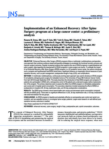
Dr Greg FinchFRACS FAOrthASpine Surgeon Orthopaedic SurgeonSuite 16a, Bld BNucleus Medical Suites23 Elsa Wilson DriveBuderim, QLD, 4556PO Box 5536Maroochydore BCQLD 4558Australiat: 61 7 5444 7579f: 61 7 5444 stspine.com.auINTRODUCTIONAlthough it is difficult to predict the rapidly changing practice of spinal surgery, we focusthis article on four areas of burgeoning interest: Minimally invasive spinal surgery, motionpreservation, robotics and neuro-navigation and the use of biologics. All four of these areaswill most likely see further significant growth in the near, as well as distant future. Minimallyinvasive techniques have been developed and honed for decades; however, recenttechnological advances combined with the development of computer software to aid inrobotic surgery and neuro-navigation will hopefully allow for safer and more efficaciousminimally invasive procedures for our future patients. Moreover, the use of biologicallyactive agents may actually reduce the number of traditional spinal surgeries while providingpatients with reasonable non-operative treatment options. Definitive treatment will mostlikely be preventative in nature, which is in direct contrast to our current surgical treatmentmodalities which mainly focus on treating the “end-result” of spinal pathology. While thisarticle is not intended to be a comprehensive menu of future products, techniques and trendsin spinal neurosurgery, it is our goal to present the possibilities that will be available in ourspinal armamentarium as we approach 2020.Go to:MINIMALLY INVASIVE APPROACHES TO THE SPINEThe introduction of the tubular retractor system (METRx , Medtronic, Memphis, TN, USA)by Foley and Smith in the late 1990s allowed spine surgeons to treat symptomatic herniateddiscs with minimally invasive techniques.[47] These pioneers of minimal access surgerydemonstrated that a routine procedure, the microdiscectomy, could be performed using anovel retractor system combined with an attachable endoscope, allowing for excellentvisualization with minimal obstruction for the passage of instruments through the diminutiveworking channel. This technique has also been employed over recent years to treat a varietyof spinal pathologies including symptomatic spinal stenosis, synovial cysts, intraduralextramedullary tumors, intramedullary tumors, tethered cords, and spinal cordsyrinxes[17,41,42,53,59,60] [Figure 1].Figure 1(a) METRx system with endoscope, (b) View of microendoscopic decompression for lumbarstenosis, (c) Microendoscopic view of filum detethering, (d) Minimally invasive resection ofintradural schwannomaAccordingly, the tubular retractor system has undergone various modifications over the past10 years and has allowed the spine surgeon to expand the role of minimally invasiveapproaches to include lumbar fusion techniques (i.e., minimally invasive transforaminallumbar interbody fusions or MI-TLIFs) for treatment of spondylolisthesis, degenerative disc
Dr Greg FinchFRACS FAOrthASpine Surgeon Orthopaedic SurgeonSuite 16a, Bld BNucleus Medical Suites23 Elsa Wilson DriveBuderim, QLD, 4556PO Box 5536Maroochydore BCQLD 4558Australiat: 61 7 5444 7579f: 61 7 5444 stspine.com.audisease, short segment spinal deformity, as well as traumatic fractures. Excellent outcomescombined with fewer complications, shorter hospital stays, and less blood loss have beenreported with this technique.[27,45] It is quite conceivable that this technique will be the“gold standard” utilized when evaluating emerging techniques for the treatment ofdegenerative disc disease and spondylolisthesis in the future.Another minimally invasive procedure for the thoraco-lumbar spine is the eXtreme lateralinterbody fusion (XLIF , NuVasive, San Diego, CA, USA) or direct lateral interbody fusion(DLIF). This lateral, trans-psoas muscle approach has been popularized by Pimenta andothers for the treatment of degenerative disc disease and has also been utilized to treat avariety of spinal pathologies.[44] The direct lateral approach allows for excellent access tothe thoracolumbar spine from either side. This novel approach also provides a workingcorridor to the disc space as well as the vertebral body and has been utilized to treatdegenerative disease processes, infections, as well as tumors and fractures of the spine. It hasalso been used as a powerful adjuvant in deformity corrective surgery combined withposterior instrumentation.[24,25,57]This direct lateral approach has been utilized for the placement of an intervertebral artificialdisc outside of the United States and is currently in an FDA-Investigational DeviceExemption (FDA-IDE) trial.[48] It is the authors’ opinion that this approach, combined withdisc replacement, may be the primary treatment for symptomatic degenerative disc diseasesince it may reduce the risk of injury to the iliac vessels, neural elements, and genitourinarytract that has been reported with anterior lumbar approaches. With its ever increasingpopularity among spine surgeons, the direct lateral approach will continue to be utilized inthe future treatment of thoraco-lumbar spinal disease.Go to:MOTION PRESERVATIONThe past decade has witnessed a change in philosophy regarding cervical and lumbar fusionsurgery. Traditionally, the anterior cervical discectomy and fusion procedure has providedmany patients with symptomatic relief of radicular, and occasionally, axial neck pain withexcellent clinical outcomes and high fusion rates.[13,66] To a lesser degree, lumbar fusiontechniques including posterior-lateral fusions, anterior lumbar interbody fusions (ALIFs),posterior lumbar interbody fusions (PLIFs), and transforaminal lumbar interbody fusions(TLIFs) have provided some pain relief in carefully chosen patients with correlativesymptoms and imaging studies. Since results have been varied and there has been muchdebate as to patient selection criteria and procedural success, a trend toward motionpreservation in the form of disc arthroplasty has become more popular. The Charite disc(DePuy, Ranham, MA, USA) was the first device approved in the United States for use in thelumbar spine. Since its approval in 2004, there have been many reports documenting thebenefit for relief of axial back pain, as well as some reports suggesting only ,38,69,71] The ProDisc-L (Synthes, Paoli, PA,USA) was released in 2006 and has also demonstrated some encouraging results compared tohistorical fusion procedure success rates.[10,70,71] It is likely that longer follow-up may
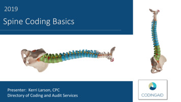
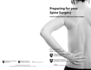
Dr Greg FinchFRACS FAOrthASpine Surgeon Orthopaedic SurgeonSuite 16a, Bld BNucleus Medical Suites23 Elsa Wilson DriveBuderim, QLD, 4556PO Box 5536Maroochydore BCQLD 4558Australiat: 61 7 5444 7579f: 61 7 5444 stspine.com.audefine the efficacy of these products as well as their comparison to previously reportedoutcomes in lumbar fusion surgery.Cervical disc arthroplasty complications are rare; however, lumbar disc arthroplastyapproaches carry well-documented risks to the lumbosacral vessels, neural elements, andgenitourinary structures. This issue may be improved in the future with better preoperativeimaging assessment and possibly through minimal access approaches. Furthermore, the directlateral or extreme lateral approach may reduce these risks and thus become the preferredsurgical approach by both spine and general access surgeons.In regard to the commercially available artificial disc products, two of the FDA-approvedcervical disc arthroplasty devices and both of the FDA-approved lumbar implants arecomposed of materials that do not allow for adequate visualization of the operative disc leveland often obscure adjacent segments when evaluated with MRI. In a study evaluating theability to visualize the spinal canal and adjacent segments after cervical disc arthroplasty,only the Bryan (Medtronic) and Prestige LP (Medtronic) allowed for adequatevisualization.[54] The artifact produced by these implants can be circumvented with the useof non-ferromagnetic biomaterials allowing for postoperative imaging and evaluation in thefuture. Polymers and ceramics may also be employed in the design of future intervertebraldisc prostheses. These products will provide mechanical support, motion sparing, and can beeasily imaged with MRI[43] [Figure 2].Figure 2(a) Intraoperative view of bryan Artificial Disc (Medtronic, Memphis, TN, USA), (b)Postoperative radiograph of Bryan Artificial Disc (Medtronic), (c) Intraoperative view ofProDisc-C Artificial Disc (Synthes, Paoli, PA, USA), (d) Postoperative radiograph .Although disc arthroplasty may be a reasonable option for patients with cervical or lumbardisc disease, many patients do not fit the criteria for arthroplasty due to lumbarspondylolistheses, cervical subluxation, lack of motion related to severe spondylosis, andmore commonly secondary to insurance-related denials. In this population, fusion surgerywill most likely be the mainstay. Traditional titanium-alloy plates and screws are stillpopular; however, we are witnessing a trend toward bio-absorbable plates and screws, as wellas significantly smaller plating systems that purportedly reduce the incidence of iatrogenicadjacent segment disease spondylosis. Bio-absorbable instrumentation has been utilized inplastic surgery and neurosurgery for craniofacial reconstruction for many years, but bioabsorbable anterior cervical plating systems have only been available in the United Statessince 2006[35,51] [Figure 3].Figure 3
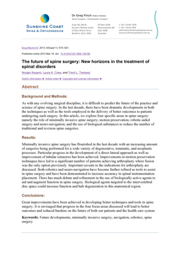
Dr Greg FinchFRACS FAOrthASpine Surgeon Orthopaedic SurgeonSuite 16a, Bld BNucleus Medical Suites23 Elsa Wilson DriveBuderim, QLD, 4556PO Box 5536Maroochydore BCQLD 4558Australiat: 61 7 5444 7579f: 61 7 5444 stspine.com.au(a) Mystique resorbable plate (Medtronic, Memphis, TN, USA) immediate postoperativeradiograph. (b) Mystique resorbable plate (Medtronic) 5 years postoperativeThese products offer an alternative to their metal counterparts which can often be difficult toremove when re-operation is necessary. The absorbable plating system also negates theartifact that is often visualized on postoperative MRIs.Go to:ROBOTICS AND NEURO-NAVIGATIONThere are many potential hazards associated with spine surgery, which include risks to vitalstructures including important vessels, viscera and of course neural elements. In addition, ifinstrumentation is utilized, there is a risk of malpositioning of the hardware leading tocompromise of the anatomy required for spinal stability. Therefore, good control andaccuracy in the operative approach is a necessity in spine surgery. For example, it has beenestimated that a dural breach occurs in 3.5% of primary initial discectomies and around 13%of revision discectomies.[55] This could arise as a result of surgical technique mistakes interms of visualization or instrument control. Faced with these challenges as well as problemswith ergonomics, surgical dexterity and the surgeon-instrument interface, robotic spinesurgery has been conceptualized and developed.Although robotics has been employed for longer than a decade in many surgicalsubspecialties, it has only recently been utilized in the area of spine surgery. The advantageof the current robotic platform is that it allows the surgeon real-time procedural manipulationcombined with heuristic instrument control, real-scale magnification, and elimination oftremor. All of this can now be combined with anatomical spine navigation to allow precisesurgical technique with minimal spinal bony damage and blood loss. However, until recently,robotics has always had limited application in spine surgery due to the challenges ofvisualization, cost, adequate training, and the development of minimally invasive techniquesthat have offered a viable and more widely utilized alternative.Several procedures have been recently tested using the Da Vinci robotic system (IntuitiveSurgical Inc., Sunnyvale, CA, USA) including laminectomy, laminotomy, discectomy, anddural repairs on an in vivo porcine model.[49] In the same model, it has also been used in anALIF procedure[28,68] [Figure 4]. In humans, the Da Vinci robotic model has been utilizedin assisted transoral odontoid resection for decompression of the craniocervical junction.[31]It has also been used laparoscopically in robotic-assisted resection of a thoracolumbarneurofibroma,[40] thoracoscopically for resection of a mediastinal schwannoma, andtransperitoneally in the resection of paravertebral lumbosacral masses.[52,67]
Dr Greg FinchFRACS FAOrthASpine Surgeon Orthopaedic SurgeonSuite 16a, Bld BNucleus Medical Suites23 Elsa Wilson DriveBuderim, QLD, 4556PO Box 5536Maroochydore BCQLD 4558Australiat: 61 7 5444 7579f: 61 7 5444 stspine.com.auFigure 4Da Vinci robotic system console (Intuitive Surgical Inc., Sunnyvale, CA, USA)Renaissance (Mazor Robotics, Inc., Caesarea, Israel) is a miniature robotic guidancesystem which can be either mounted on table or on the patient's spinous process. This systemtogether with its precursor, Spine Assist (Mazor Robotics, Inc.) has now been employed forlonger than 5 years. In its development, it has been cadaverically tested using percutaneousplacement of pedicle and translaminar facet screws, as well as being employed for theplacement of transpedicular screws via open and percutaneous techniques inPLIFs.[3,34,46,56]In a retrospective multi-center trial, using the Spine Assist, screw placement was determinedto be clinically acceptable in 98% of cases based on intraoperative X-ray imaging.Postoperative imaging demonstrated that 98.3% of the screws fell within a safe zone, whereasin 9%, screws breached the pedicle by up to 2 mm. The remainder of the screws breachedbetween 2 and 4 mm, and in only two cases was there a breach greater than 4 mm.Neurological deficit was seen in four patients, but following revision of the instrumentation,all of these deficits resolved.[12]Overall, robotic-assisted surgery provides several advantages to the surgeon. These include asignificant improvement of dexterity with a reduction in physiological tremor, significantreduction in X-ray–induced radiation, visualization in three dimensions, and significantimprovement in ergonomics with significantly less pain, strain, andstiffness.[26,31,36,39,40,49,52,68] Other potential advantages could include the possibilityof minimal muscle dissection, retraction, and bleeding.[28,34,49] The learning curve does notseem to be too steep as has been reported in performing radical nephrectomy using the DaVinci system.[2]There are of course several disadvantages to robotics in spine and these include the lack ofhaptic feedback from tissue manipulation, increased operating time, and the need forincreased training not just for the surgeon but also other operating room staff. There is alsothe issue of significant cost. The estimated cost of acquiring a Da Vinci system, for example,lies in the 1.1- 1.7 million range.[49] This does not include the additional costs of increasedoperating time, increased education, and additional funds needed to transform this particularplatform into that applicable to spine. For example, this could include additional attachmentssuch as bone drills and rongeurs. Despite the disadvantages, it appears clear that the future ofspine surgery will have to include devices and tools that will increase patient safety, improvesurgical outcomes, improve surgeon dexterity while reducing discomfort and do so in aseamless and expedient fashion.
Dr Greg FinchFRACS FAOrthASpine Surgeon Orthopaedic SurgeonSuite 16a, Bld BNucleus Medical Suites23 Elsa Wilson DriveBuderim, QLD, 4556PO Box 5536Maroochydore BCQLD 4558Australiat: 61 7 5444 7579f: 61 7 5444 stspine.com.auIn concert with robotics, spinal and neuro-navigational devices can be utilized. Many largehospitals offering complex spine surgery do not consider navigation a routine component ofcurrent treatment. Often, the reasons for this include the significant investment in cost, space,and the belief that its utility does not improve outcomes significantly over and above nonnavigation techniques.Although intraoperative navigation has been available for the past decade, it has evolvedfrom plain fluoroscopy to 2D and then 3D fluoroscopy acquired intraoperatively and then coregistered with preoperative imaging.[22] Further development in navigation has led to thecreation of an intraoperative CT scanner. The O-arm (Medtronic, Inc., Louisville, CO, USA)is a CT scanner that permits imaging of the screws intraoperatively. In this system, areference pin or anatomical landmark is co-registered with an acquired CT scan and is linkedwith a navigational software program.A recent study compared navigated and non-navigated pedicle screws using the O-armsystem and assessed their relative accuracy. The study found that 4.9% of the non-navigatedscrews needed to be removed intraoperatively compared with 0.6% of the navigated screws.It also reported that a “significant” medial breach, as defined by 50% excursion of the screwdiameter, was almost 8 times more likely to occur without navigation. Image guidance leadsto more accurate placement of pedicle screws.[61]Overall, navigation appears to improve accuracy of surgical instrumentation while alsosignificantly reducing radiation exposure. However, as with robotic-assisted platforms, it canbe associated with increased operative time, risk of exposure to infection during draping andun-draping process, and the required learning for both surgical and non-surgical operatingroom staff. We predict that while it may add little benefit to routine surgical cases wherebony anatomical landmarks are sufficient for placement of screws and instrumentation, itsgreatest application may be observed in cases with significant deformity or dysplasticchanges in the spine. For example, it could be of benefit in scoliosis cases with significantrotational deformity of the spine and perhaps in the thoracic region where accuracy is critical.Go to:SPINAL BIOLOGICSPerhaps the most exciting area in the future treatment of spinal disorders is best witnessed inthe development of biologically active agents used to augment and possibly restore thefunction of the spine. One of the first biologics to be approved in the United States wasrhBMP-2 (Infuse, Medtronic). This powerful osteoinductive agent stimulates the productionof bone and is utilized to augment fusion surgeries of the spine. Further development of thecommercially available product has revolutionized the spine surgeon's ability to obtain afusion mass in patients that may have problems achieving an adequate fusion. Anotherbiologically active product that was approved by the United States FDA was rhBMP-7 orOP-1 (Stryker, Kalamazoo, MI, USA). This product also promotes bone growth and wasmarketed in a putty form compared to the absorbable sponge carrier utilized with Infuse. Thisadjuvant to achieving successful fusion has been documented in preclinical as well as clinicalstudies.[7,11,29,50,62–65] Over the past decade, surgeons have theorized the potential
Dr Greg FinchFRACS FAOrthASpine Surgeon Orthopaedic SurgeonSuite 16a, Bld BNucleus Medical Suites23 Elsa Wilson DriveBuderim, QLD, 4556PO Box 5536Maroochydore BCQLD 4558Australiat: 61 7 5444 7579f: 61 7 5444 stspine.com.aubenefit of these biologics and their impact on the future of spinal surgery.[6,20] Althoughthere are few biologics available for the spine surgeon to date, the future promises a numberof products that may alter the way in which we practice. The concept of placing abiologically active agent in the intervertebral disc to help regenerate or stabilize thedegenerative processes of aging has been theorized for decades.[16,30,58]Studies have demonstrated the feasibility of placing these agents through minimally invasiveprocedures.[33] An and colleagues demonstrated that by placing osteogenic protein-1 vianeedle injection into a New Zealand white rabbit model, changes in the disc degenerationprocess were halted as evidenced by changes observed on MRI.[1] Of course, data gatheredfrom animal models need to be evaluated for safety and efficacy in humans through theclinical trial process. It is the authors’ belief that this type of preventative intervention may infact reduce the number of spinal surgeries performed in the future.As we progress through this decade, we must keep in mind that predicting the future of spinesurgery may also be affected by the economic impact on healthcare that looms in the UnitedStates and throughout the world. It is conceivable that we will be asked to provide care formore individuals while possibly receiving less payment in return. This could greatly impactthe amount of innovation and interest that has allowed our specialty to improve care for ourpatients. It is also important that our leaders in innovation be allowed to work with industryin an open and honest collaboration in order to benefit our future patients.Go to:FootnotesAvailable FREE in open access from: 4/2/15/109186Go to:REFERENCES1. An HS, Takegami K, Kamada H, Nguyen CM, Thonar EJ, Singh K, et al. Intradiscaladministration of osteogenic protein-1 increases intervertebral disc height and proteoglycancontent in the nucleus pulposus in normal adolescent rabbits. Spine (Phila Pa1976) 2005;30:25–31. discussion 31-2. [PubMed]2. Artibani W, Fracalanza S, Cavalleri S, Iafrate M, Aragona M, Novara G, et al. Learningcurve and preliminary experience with da Vinci-assisted laparoscopic radicalprostatectomy. Urol Int. 2008;80:237–44. [PubMed]3. Barzilay Y, Liebergall M, Fridlander A, Knoller N. Miniature robotic guidance for spinesurgery-introduction of a novel system and analysis of challenges encountered during theclinical development phase at two spine centres. Int J Med Robot. 2006;2:146–53. [PubMed]4. Blumenthal S, McAfee PC, Guyer RD, Hochschuler SH, Geisler FH, Holt RT, et al. Aprospective, randomized, multicenter Food and Drug Administration investigational deviceexemptions study of lumbar total disc replacement with the CHARITE artificial disc versuslumbar fusion: Part I: Evaluation of clinical outcomes. Spine (Phila Pa 1976) 2005;30:1565–75. discussion E387-91. [PubMed]5. Blumenthal SL, Ohnmeiss DD, Guyer RD, Hochschuler SH. Prospective study evaluatingtotal disc replacement: Preliminary results. J Spinal Disord Tech. 2003;16:450–4. [PubMed]
Dr Greg FinchFRACS FAOrthASpine Surgeon Orthopaedic SurgeonSuite 16a, Bld BNucleus Medical Suites23 Elsa Wilson DriveBuderim, QLD, 4556PO Box 5536Maroochydore BCQLD 4558Australiat: 61 7 5444 7579f: 61 7 5444 stspine.com.au6. Boden SD. Biology of lumbar spine fusion and use of bone graft substitutes: Present,future, and next generation. Tissue Eng. 2000;6:383–99. [PubMed]7. Brown A, Stock G, Patel AA, Okafor C, Vaccaro A. Osteogenic protein-1: A review of itsutility in spinal applications. BioDrugs. 2006;20:243–51. [PubMed]8. Caspi I, Levinkopf M, Nerubay J. Results of lumbar disk prosthesis after a follow-upperiod of 48 months. Isr Med Assoc J. 2003;5:9–11. [PubMed]9. Cinotti G, David T, Postacchini F. Results of disc prosthesis after a minimum follow-upperiod of 2 years. Spine (Phila Pa 1976) 1996;21:995–1000. [PubMed]10. Delamarter RB, Bae HW, Pradhan BB. Clinical results of ProDisc-II lumbar total discreplacement: Report from the United States clinical trial. Orthop Clin NorthAm. 2005;36:301–13. [PubMed]11. Delawi D, Dhert WJ, Rillardon L, Gay E, Prestamburgo D, Garcia-Fernandez C, et al. Aprospective, randomized, controlled, multicenter study of osteogenic protein-1 ininstrumented posterolateral fusions: Report on safety and feasibility. Spine (Phila Pa1976) 2010;35:1185–91. [PubMed]12. Devito DP, Kaplan L, Dietl R, Pfeiffer M, Horne D, Silberstein B, et al. Clinicalacceptance and accuracy assessment of spinal implants guided with Spine Assist surgicalrobot: Retrospective study. Spine (Phila Pa 1976) 2010;35:2109–15. [PubMed]13. Gaetani P, Tancioni F, Spanu G, Rodriguez y Baena R. Anterior cervical discectomy: Ananalysis on clinical long-term results in 153 cases. J Neurosurg Sci. 1995;39:211–8. [PubMed]14. Geisler FH, Blumenthal SL, Guyer RD, McAfee PC, Regan JJ, Johnson JP, et al.Neurological complications of lumbar artificial disc replacement and comparison of clinicalresults with those related to lumbar arthrodesis in the literature: Results of a multicenter,prospective, randomized investigational device exemption study of Charite intervertebraldisc. Invited submission from the Joint Section Meeting on Disorders of the Spine andPeripheral Nerves, March 2004. J Neurosurg Spine. 2004;1:143–54.[PubMed]15. Geisler FH. The CHARITE Artificial Disc: Design history, FDA IDE study results, andsurgical technique. Clin Neurosurg. 2006;53:223–8. [PubMed]16. Gruber HE, Hanley EN., Jr Recent advances in disc cell biology. Spine (Phila Pa1976) 2003;28:186–93. [PubMed]17. Guiot BH, Khoo LT, Fessler RG. A minimally invasive technique for decompression ofthe lumbar spine. Spine (Phila Pa 1976) 2002;27:432–8. [PubMed]18. Guyer RD, McAfee PC, Banco RJ, Bitan FD, Cappuccino A, Geisler FH, et al.Prospective, randomized, multicenter Food and Drug Administration investigational deviceexemption study of lumbar total disc replacement with the CHARITE artificial disc versuslumbar fusion: Five-year follow-up. Spine J. 2009;9:374–86. [PubMed]19. Guyer RD, McAfee PC, Hochschuler SH, Blumenthal SL, Fedder IL, Ohnmeiss DD, etal. Prospective randomized study of the Charite artificial disc: Data from two investigationalcenters. Spine J. 2004;4:252S–9S. [PubMed]20. Helm GA, Alden TD, Sheehan JP, Kallmes D. Bone morphogenetic proteins and bonemorphogenetic protein gene therapy in neurological surgery: Areview. Neurosurgery. 2000;46:1213–22. [PubMed]
Dr Greg FinchFRACS FAOrthASpine Surgeon Orthopaedic SurgeonSuite 16a, Bld BNucleus Medical Suites23 Elsa Wilson DriveBuderim, QLD, 4556PO Box 5536Maroochydore BCQLD 4558Australiat: 61 7 5444 7579f: 61 7 5444 stspine.com.au21. Hochschuler SH, Ohnmeiss DD, Guyer RD, Blumenthal SL. Artificial disc: Preliminaryresults of a prospective study in the United States. Eur Spine J. 2002;11:S106–10. [PMC freearticle] [PubMed]22. Holly LT, Foley KT. Intraoperative spinal navigation. Spine (Phila Pa1976) 2003;28:S54–61.[PubMed]23. Hopf C, Heeckt H, Beske C. [Disc replacement with the SB Charite endoposthesisexperience, preliminary results and comments after 35 prospectively performed operations] ZOrthop Ihre Grenzgeb. 2002;140:485–91. [PubMed]24. Hsieh PC, Koski TR, Sciubba DM, Moller DJ, O’Shaughnessy BA, Li KW, et al.Maximizing the potential of minimally invasive spine surgery in complex spinaldisorders. Neurosurg Focus. 2008;25:E19.[PubMed]25. Isaacs RE, Hyde J, Goodrich JA, Rodgers WB, Phillips FM. A prospective,nonrandomized, multicenter evaluation of extreme lateral interbody fusion for the treat
Postoperative radiograph of Bryan Artificial Disc (Medtronic), (c) Intraoperative view of ProDisc-C Artificial Disc (Synthes, Paoli, PA, USA), (d) Postoperative radiograph . Although disc arthroplasty may be a reasonable option for patients with cervical or lumbar disc disease, many patients do not fit the criteria for arthroplasty due to lumbar