Transcription
Ibrahim and Elsadawy Egyptian Journal of Radiology and Nuclear Medicine(2019) tian Journal of Radiologyand Nuclear MedicineRESEARCHOpen AccessIncidental findings in lumbar spine MRI:their prevalence and potential impact onpatient managementHeba Ibrahim* and Momena Essam Ibrahim ElsadawyAbstractBackground: To document the prevalence of extra spinal findings in lumbar MRIResults: Among the scanned 400 patients, 90 cases had incidental non-spinal findings, and in 30 out of these 90patients, the finding was the only reason for their complaint.Conclusions: Radiologists should give attention to the non-spinal findings in lumbar spine MRI, as the detectedpathology could be the source of pain or could potential life-threatening conditions.Keywords: Lumbar, MRI, Back pain, SpinalBackgroundLow back pain is one of the most common complaintsamong a large variety of population [1]. The prevalenceof chronic lower back pain is about 23%, and the lifetime prevalence of lower back pain is estimated to bearound 60–70% [2].The majority of low back pain presentations representmusculoskeletal causes. Yet, some patients will presentwith other pathologies that can cause neurologic disability (i.e., abdominal or retroperitoneal etiologies).In recent years, advances in digital evaluation ofradiological imaging (e.g., high magnification zoom,the ability to focus on individual images, and digitalarchiving) have dramatically improved the detectionlimit of incidental lesions [3].There are some studies that reviewed the frequencyof incidental extra spinal findings in lumbar MRI andtheir clinical significance. Therefore, careful observation of anatomic structure outside of the immediate region of interest creates opportunities for early detectionof potentially life-threatening conditions such as malignancies and aneurysms [4].Non-spine-related causes of back pain may includevascular cases (abdominal aortic aneurysm (AAA), aortic* Correspondence: hebaali m@hotmail.comRadio-diagnosis Department, Faculty of Medicine, Ain Shams University,Ramsis, Cairo, Egyptdissection and renal artery dissection or thrombosis), pyelonephritis, retroperitoneal hemorrhage, GI causes(pancreatitis, peptic ulcer, cholecystitis), oncologic cases(epidural or intradural metastatic disease, intramedullarytumors, and osseous metastasis), infectious (spinal epidural abscess, vertebral osteomyelitis, and infectious discitis), spinal epidural hematoma, presacral masses,ovarian pathology, and prostatic carcinoma [4].The aim of this study was to assess and document theprevalence of extra spinal findings in routine lumbar MRexaminations and their impact on clinical management.MethodsPatientsThe study was retrospective and approved by the localethics committee. Due to the nature of the study design,there was no need to obtain specific informed consentfrom patients.Four hundred patients were referred for lumbar spineMRI from the clinic of spine surgery, neurology, ororthopedic surgery. All had low back pain.The patients aged 21 to 75 years, with a mean of 46 3 years. Two hundred-seventy of them were females and130 were males (Fig. 1).We excluded the patients who had spine pathology,known malignancy, or previous surgery and also anyincomplete or low-quality scan. The Author(s). 2019 Open Access This article is distributed under the terms of the Creative Commons Attribution 4.0International License (http://creativecommons.org/licenses/by/4.0/), which permits unrestricted use, distribution, andreproduction in any medium, provided you give appropriate credit to the original author(s) and the source, provide a link tothe Creative Commons license, and indicate if changes were made.
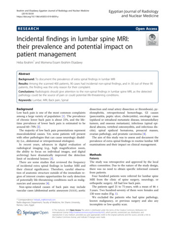
Ibrahim and Elsadawy Egyptian Journal of Radiology and Nuclear Medicine(2019) 50:59Page 2 of 5Fig. 1 47-year-old female with low back pain. Sagittal T2WI of the lumbar spine showing multiple interstitial uterine myomas seen in a (arrow). bshows large posterior wall myomaThe clinician examined the patients for neurologicalfindings but all were referred with a symptom of backpain without any specific sign written in the radiologyreferral sheet.Ten patients had pain referred to the hip joints, socomplementary coverage of the hips was requested.Thirteen patients were recommended to have a contraststudy; only seven came back and received a contrast.Diagnostic workup and MR protocolThe examination was conducted using 0.32 T system,Siemens Magnetom C, Germany, using a surface coil,and scanning was done in supine position with slight hipflexion with a pillow under the knee.The scan included routine MR imaging pulse sequences:sagittal T2-weighted (3190/128/3) [TR/TE/number of excitation] and sagittal T1-weighted (790/17/3) turbo spinecho sequences, axial T1- and T2-weighted, coronal, andaxial gradient 3D thin cuts. The slice thickness is 4 mmwith an intersection gap of 1.4 mm.After the routine MR imaging, short scanning of thehip joints was conducted in 10 patients as per clinicianrequest including sagittal T2 and coronal TIRM (T2 fatsuppression).Based on the radiologist’s recommendation, 13 patients were called back for contrast study, seven of themcame back and received contrast, and post-contrastT1WI was acquired in axial, sagittal, and coronal planesplus DIXON sequence (T1 with fat suppression).Two patients were recommended to do MR spectroscopy for suspected prostate lesion; they referred to highfield magnet to be done.Images interpretationAfter regular interpretation of the lumbar spine MRIusing our facility PACS Hinai ImageNET, special concern was given to the extra spinal findings following adedicated survey list including the presence of any extraspinal finding, the organ involved, the importance ofsuch finding, the reliability of the lumbar MRI to diagnose that pathology, and finally how important andurgent was that finding to report to the referringphysician.Images were interpreted independently by two radiologists, 15 and 14 years of experience as general radiologist, guided by the tailored survey list. Data are collectedand discussed with the referring physician for clinicalcorrelation and further workup.Six months and 1 year of follow up was done either bycontacting the patient, patients coming for rescanning,or through the referring physician.ResultsWe retrospectively included 400 patients with low backpain. Our exclusion criteria included patients with noextra spinal findings, patients with spine pathology,known malignancy or previous surgery, and also any incomplete or low-quality scan.Ninety out of 400 patients (22.5%) had extra spinalfinding including 74 females (82%) and 16 (18 %) males.Their ages ranged between 21 and 75 years, with a meanof 46 3 years.All patients presented with low back pain as per thereferring physician with no other specific sign or symptoms. No previous investigations were done.
(2019) 50:59Ibrahim and Elsadawy Egyptian Journal of Radiology and Nuclear MedicineTwo radiologists interpreted the images and there wasan agreement about detection and localization of thelesion.Both radiologists could detect the extra spinal findingin the 90 included patients (100%); they agreed aboutthe organ of origin, except in suspected cases of metastases (10 cases), accuracy of localization 88.8%, and in twocases of suspected metastases, they detected prostaticmass so they requested dedicated prostatic MRI withspectroscopy.Regarding the organ involved, 40 of the 90 patients(44.5 %) had ovarian findings including simple or complex cyst and dermoid cyst or ovarian mass. Eighteenout of 90 patients (20%) had uterine myomas (Fig. 2).Fifteen out of 90 patients (16.5 %) had renal cysts. Twoout of 90 patients (2.3 %) had paraspinal masses like thelymph node, ten out of 90 (11%) had lumbar and sacralmetastases, seven out of 90 (7.7%) had hip lesions (avascular necrosis (AVN) and insufficiency fracture), andthree out of 90 had aortic aneurysm. Two of the casesshowed paraspinal masses (2.2 %), one of them revealedto be psoas abscess (Pott’s disease), and the other onerevealed to be neurofibroma.There was concordance about the suspected diagnosis (Table 1). The requested further investigation waseither to complete the diagnosis (in twenty-one out of90 cases, 23%) or to better assess the size and relationship as in cases of ovarian and uterine lesionswhere pelvic ultrasound was requested for all of themfor more information.Twenty-one out of 90 patients (23%) needed furthercontrast study, 20 patients (22 %) needed MRI with contrast (5 cases of complex ovarian cyst, 3 cases of ovarianmasses, 2 cases of paraspinal masses, and 2 cases of metastases), and one case needed CT angiography for abdominal aortic aneurysm.Ten patients (11%) submitted to metastatic work up tosearch for the primary and for staging.All cases of ovarian or uterine lesions were recommended to do pelvic ultrasound, not for diagnosis butPage 3 of 5Table 1 Showing the detected extra spinal findingsNumber of findingsExtra spinal findings18Simple ovarian cyst7Complex12Dermoid cyst3Ovarian mass18Uterine myoma15Renal cyst2Paraspinal mass10Metastases7Hip lesions3Aortic aneurysmfor more information about the lesion including its sizeand relationship.In 10 out of 90 patients (11%), the extra spinal findingwas the main cause of low back pain, and symptomssubsided after surgical intervention. Three of them wereposterior wall subserous myoma, four of them wereovarian lesions (Fig. 2), and three out of 90 were paraspinal masses.Regarding the lumbar spine MRI alone, it was enoughand reliable in the diagnosis of the detected lesion insixty-nine out of 90 (75%) patients, the performed MRIwas enough to give a final diagnosis, and no further imaging was needed to diagnose the pathology. However,the recommended imaging (pelvic ultrasound) was tobetter overall assess the condition regarding the size andextension of the lesion.Regarding the urgency of reporting, six out of 90 patients (6.6%) were urgently reported to the referring clinician within 12 h including one case of huge aorticaneurysm, two cases of paraspinal masses, and three casesof hip lesions (Fig. 3) (insufficiency fracture and AVN).DiscussionLow back pain is one of the most common reasons foran outpatient visit. The evaluation for low back painFig. 2 39-year-old female patient with low back pain. a Coronal T2WI showing left adnexal heterogenous mass. b Axial T2WI shows the mass atthe left ovary. Histopathology revealed left ovarian thecoma
Ibrahim and Elsadawy Egyptian Journal of Radiology and Nuclear Medicine(2019) 50:59Page 4 of 5Fig. 3 62-year-old male patient with back pain radiating to the hip joints. a is sagittal T2WI of the left hip joint showing bone marrow edema ofthe acetabulum (arrow), b is TIRM confirming the acetabular edema, and c is coronal T2 TIRM of both hip joints showing bilateral acetabularedema consistent with insufficiency fractureshould include a complete, focused medical history [5].Incidental imaging findings in lumbar MRI are commonand may lead to further tests or investigations [6].General radiologists frequently report lumbar MRI andthey should be aware and familiar with any extra spinalabnormalities either for their clinical significance, medicolegality, or for improving our practice as these findings arenot uncommon. So, it is generally recommended to have asystematic reporting approach to avoid missing anyfinding.We aimed to highlight some of the detected extraspinal pathologies to avoid tunnel view to lumbar MRI;we assessed the prevalence, organ of origin, suspecteddiagnosis, and correlation with patient symptomatologyand level of threat of the detected abnormalities.In our study, the extra spinal findings were detected in22.5% of cases submitted to lumbar MRI for low backpain. Sedat et al. [7] reported a prevalence of 19.8% incidental extra spinal findings in routine lumbar MRI, andthey stated that their rate is less than the values reportedin the literature. Lee et al. [4] reported a rate of 40.5% ofincidental extra spinal findings in lumbar MRI. Thisvariability may be attributed to technical factors, likeFOV, coverage, and using PACS for interpretation.Our study included more females than males (82%), inagreement with the literature. The majority (44.5%) ofour findings were ovarian in origin followed by uterinepathologies, in comparison to Sedat et al. [7] where mostof the findings are renal in origin, and they focused moreabout size criteria of the detected renal cyst. Forty-onepercent of our patients had ovarian cysts, either simple,complicated, or dermoid cysts. The simple cysts represent most of the ovarian lesions; they displayedhomogenous hypointensity on T1WI and hyperintensityon T2WI with no wall thickening. Renal cysts accountfor 17% of our findings.Complex cysts are mostly hemorrhagic; they are brighton T1WI indicating either fat or blood content, on satsuppressed T1WI; and they remain bright, ruling out afatty lesion. If the contrast is given, there will be no enhancement [5].Twenty percent of cases had uterine myomas, beingthe most common are benign gynecologic tumors. Theyprimarily affect women of reproductive age, and the estimated incidence of fibroids is over 70% by 50 years ofage [7]. Although submucosal leiomyomas are the leastcommon, they are most commonly symptomatic, andalso, pedunculated subserous leiomyomas may undergotorsion, which results in infarction accompanied by pain[8].We had 10 cases (10%) of osseous metastasis. Indeed,bone is the third most common organ affected by metastasis, surpassed only by the lungs and liver [9]. Approximately 70% of patients with breast or prostate cancerhave bone metastases [10].Ju Fu et al. [11] focused on the incidental extra spinalmalignancies found on lumbar MRI, the majority of theircases were lymph nodes, they reported 32.1% of theirstudy population as lymphadenopathy either metastasesor lymphoma, and our study included only three casesof lymph nodes suspected of metastases (3% of the totalpopulation).Three out of 90 cases (3%) showed an abdominal aorticaneurysm. Abdominal aortic aneurysms occur most commonly in individuals between 65 and 75 years of age. Theyoften do not cause any symptoms and are found incidentally on physical examination or imaging examinations ofthe abdomen and pelvis [12]. Approximately 90% of all abdominal aortic aneurysms are infrarenal [13].Seven out of 90 cases (8%) found to have hip lesions,namely femoral head avascular necrosis and acetabularfracture. The incidence of AVN has been increasing.The causes include greater use of exogenous steroidsand an increase in trauma [14]. MRI signs include subchondral edema, low signal serpiginous line, and doubleline sign (inner bright line from granulation tissue andouter dark line from sclerotic bone) on T2-weightedimages [15, 16].
(2019) 50:59Ibrahim and Elsadawy Egyptian Journal of Radiology and Nuclear MedicineClinician/radiologist feedback was very crucial as theradiological opinion guided the clinician to further assessment and the radiologists got a short and long feedback from the clinician following the patient.We had some limitations including the inability to doall requested examinations in the same place, and thestudy being retrospective did not facilitate on time complementary ultrasound correlation.We found that MRI of the lumbar spine could conveya lot of information apart from just spine imaging, andlumbar MRI alone could be reliable in some extra spinalpathologies, could raise doubt in some other, and evenwarn against life-threatening or serious pathologies.ConclusionExtraspinal incidental findings are not uncommon andsometimes may explain patient symptomatology.The main recommendation of this study is to avoidthe tunnel vision interpretation of the lumbar spine andto draw the radiologist’s attention towards other scannednon-spinal organs during reading lumbar spine MRI, either this incidental finding was significant or not. Inaddition, encouraging clinician-radiologist feedback, discussion, and further workup of certain cases are nsMRI: Magnetic resonance imagingAcknowledgementsThe authors thank all radiographers for their support.Authors’ contributionsIH suggests the research idea, reviewing the literature, and data collectionand analysis; perform statistical analysis; revise the manuscript; and preparedthe MRI cases. EM contributed to the reviewing of the literature and wrotethe manuscript and final editing. All authors have read and approved thefinal manuscript.FundingThis study had no funding from any resource.Availability of data and materialsThe datasets used and/or analyzed during the current study are availablefrom the corresponding author on reasonable request.Ethics approval and consent to participateThis study was approved by the local Research Ethics Committee. It was aretrospective study, and no consent was taken from the patients.Consent for publicationNot applicableCompeting interestsThe authors declare that they have no competing interests.Received: 11 July 2019 Accepted: 26 September 2019References1. Friedman BW et al (2010) Diagnostic testing and treatment of low backpain in US Emergency Departments. A National Perspective. Spine 35(24):E1406–E14112. Balague F et al (2011) Non-specific low back pain. The Lancet 379(9814):482–491. 2198225616.Page 5 of 5Wagner SC, Morrison WB, Carrino JA, Schweitzer ME, Nothnagel H (2002)Picture archiving and communication system: effect on reporting ofincidental findings. Radiology. 225:500–505Lee SY, Landis MS, Ross IG, Goela A, Leung AE (2012) Extraspinal findings atlumbar spine CT examinations: prevalence and clinical importance.Radiology. 263:502–509Bigos S, Bowyer O, Braen G et al (2012) Acute low back problems in adults.Radiology 95:0642Levine D et al (2010) Management of asymptomatic ovarian and otheradnexal cysts imaged at US: society of radiologists in ultrasound consensusconference statement. Radiology 256:943–954Sedat Alpaslan Tuncel MD, Bekir Çaglı MD, Aslan Tekataş MD, MehmetYadigar Kırıcı MD, Ercüment Ünlü MD, Hakan Gençhellaç MD (2015)Extraspinal incidental findings on routine MRI of lumbar spine: prevalenceand reporting rates in 1278 patients. Koran J of Radiology. 16(4):866–873Spencer JA1, Forstner R, Cunha TM, Kinkel K (2010) ESUR guidelines for MRimaging of the sonographically indeterminate adnexal mass: an algorithmicapproach. Eur Radiol. 20(1):25–35Sue W, Sarah S-B (2009) Radiological appearances of uterine fibroids. IndianJ Radiol Imaging. 19(3):222–231Elizabeth Kagan Arleo1, Peter E. Schwartz2, Pei Hui3 and Shirley McCarthy4(2017) Uterine leiomyoma (Fibroid) imaging, Updated,;34-45.Chen-Ju F, Chen H-W, Wu C-T, Chen L-H, Wong Y-C, Wang L-J, Wan Y-L, YiKang K (2013) Extraspinal malignancies found incidentally on lumbar spineMRI: prevalence and etiologies. J Radiol Sci 38:85–91Cuccurullo V, Cascini GL, Tamburrini O, Rotondo A, Mansi L (2013) Bonemetastases radiopharmaceuticals: an overview. Curr Radiopharm. 6:41–47Choi J, Raghavan M (2012) Diagnostic imaging and image-guided therapyof skeletal metastases. Cancer Control. 19:102–112Keisler B, Carter C (2015) Abdominal aortic aneurysm. Am Fam Physician.91(8):538–543Ajay D. Wadgaonkar, MD James H. Black III, MD Elizabeth K. Weihe, MDStefan L. Zimmerman, MD Elliot K. Fishman, MD Pamela T. Johnson, MD.Abdominal aortic aneurysms revisited: MDCT with multiplanarreconstructions for identifying indicators of instability in the pre- andpostoperative patient, Volume 35, Issue 1, January, 2015; 680-700.Michael R Aiello, MD and Felix S Chew, MD. (2017) Isssmaging in avascularnecrosis of the femoral head, 11,; 120-128.Publisher’s NoteSpringer Nature remains neutral with regard to jurisdictional claims inpublished maps and institutional affiliations.
22.5% of cases submitted to lumbar MRI for low back pain. Sedat et al. [7] reported a prevalence of 19.8% inci-dental extra spinal findings in routine lumbar MRI, and they stated that their rate is less than the values reported in the literature. Lee et al. [4] reported a rate of 40.5% of incidental extra spinal findings in lumbar MRI. This