Transcription
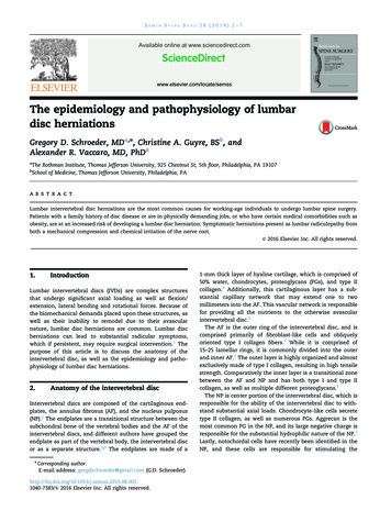
SE M I NSPI N ESUR G28 (2016) 2–7Available online at The epidemiology and pathophysiology of lumbardisc herniationsGregory D. Schroeder, MDa,n, Christine A. Guyre, BSb, andAlexander R. Vaccaro, MD, PhDaaThe Rothman Institute, Thomas Jefferson University, 925 Chestnut St, 5th floor, Philadelphia, PA 19107School of Medicine, Thomas Jefferson University, Philadelphia, PAbabstractLumbar intervertebral disc herniations are the most common causes for working-age individuals to undergo lumbar spine surgery.Patients with a family history of disc disease or are in physically demanding jobs, or who have certain medical comorbidities such asobesity, are at an increased risk of developing a lumbar disc herniation. Symptomatic herniations present as lumbar radiculopathy fromboth a mechanical compression and chemical irritation of the nerve root.& 2016 Elsevier Inc. All rights reserved.1.IntroductionLumbar intervertebral discs (IVDs) are complex structuresthat undergo significant axial loading as well as flexion/extension, lateral bending and rotational forces. Because ofthe biomechanical demands placed upon these structures, aswell as their inability to remodel due to their avascularnature, lumbar disc herniations are common. Lumbar discherniations can lead to substantial radicular symptoms,which if persistent, may require surgical intervention.1 Thepurpose of this article is to discuss the anatomy of theintervertebral disc, as well as the epidemiology and pathophysiology of lumbar disc herniations.2.Anatomy of the intervertebral discIntervertebral discs are composed of the cartilaginous endplates, the annulus fibrosus (AF), and the nucleus pulposus(NP).2 The endplates are a transitional structure between thesubchondral bone of the vertebral bodies and the AF of theintervertebral discs, and different authors have grouped theendplate as part of the vertebral body, the intervertebral discor as a separate structure.2,3 The endplates are made of anCorresponding author.E-mail address: gregdschroeder@gmail.com (G.D. 08.0031040-7383/& 2016 Elsevier Inc. All rights reserved.1-mm thick layer of hyaline cartilage, which is comprised of50% water, chondrocytes, proteoglycans (PGs), and type IIcollagen.4 Additionally, this cartilaginous layer has a substantial capillary network that may extend one to twomillimeters into the AF. This vascular network is responsiblefor providing all the nutrients to the otherwise avascularintervertebral disc.5The AF is the outer ring of the intervertebral disc, and iscomprised primarily of fibroblast-like cells and obliquelyoriented type I collagen fibers.3 While it is comprised of15–25 lamellar rings, it is commonly divided into the outerand inner AF.2 The outer layer is highly organized and almostexclusively made of type I collagen, resulting in high tensilestrength. Comparatively the inner layer is a transitional zonebetween the AF and NP and has both type I and type IIcollagen, as well as multiple different proteoglycans.6The NP is center portion of the intervertebral disc, which isresponsible for the ability of the intervertebral disc to withstand substantial axial loads. Chondrocyte-like cells secretetype II collagen, as well as numerous PGs. Aggrecan is themost common PG in the NP, and its large negative charge isresponsible for the substantial hydrophilic nature of the NP.3Lastly, notochordal cells have recently been identified in theNP, and these cells are responsible for stimulating the
SEM I NSP I N ESUcollagen and PG production, as well as preventing apoptosisof the chondrocyte-like cells.7,83.Defining a lumbar disc herniationPrior to discussing the epidemiology and pathophysiologyof a herniated lumbar disc, it is critical to have a cleardefinition a lumbar disc herniation (Fig. 1). While oftentimes the terms disc herniation, disc protrusion, and discbulge are used interchangeably in the literature, accordingto the combined task forces of the North American SpineSociety, the American Society of Spine Radiology, and theAmerican Society of Neuroradiology, these pathologies arenot the same; they define a disc herniation as “localized orfocal displacement of disc material beyond the limits ofthe intervertebral disc space.”9 This distinction is critical,because it establishes that diffuse annular expansionextending beyond the disc space (a disc bulge) is not a discherniation, but rather a form of disc degeneration. A trueherniated disc is a focal pathology that affects less than 25%of the intervertebral disc.9 Herniated discs can be categorized as protrusions, extrusions, or sequestrations (Fig. 2).Protrusions are wide-based herniations in which the diameter at the base of the herniation is wider than the diameterof the herniation in the canal. Extrusions have a narrowbase, with a large herniation in the canal, and sequestrations are herniations in which there is no continuitybetween the herniation and the remaining intervertebraldisc.94.Epidemiology of lumbar disc herniationsSignificant research into the epidemiology of lumbar discherniations has been performed, and many possible riskfactors have been identified. In an analysis of patientsR G28 (2016) 2–73enrolled in the intervertebral disc arm of the Spine PatientOutcomes Research Trial (SPORT), Cummins et al.10 reportedthat the average age of patients with a herniated disc was 41years, and the diagnosis was slightly more common in malesthan females (57% versus 43%, respectively).An elevated body mass index (BMI) is a risk factor forlumbar disc herniation, and it is thought to be due to theincreased axial load on lumbar spine.11 In a Finnish study,Bostman reported 27% of the patients undergoing surgery fora lumbar disc herniation were obese, whereas the populationprevalence of obesity in Finland at that time was only 16%.Similarly, a recent meta-analysis found that overweightpatients (BMI: 25–30) and obese patients (BMI 4 30) had astatistically significant increase risk of being diagnosed withlumbar radiculopathy than patients with a BMI o 25.12Furthermore, obesity has been linked to an increased risk ofrecurrent disc herniations after a microdiscectomy, as Meredith et al.13 reported obese patients (BMI 4 30) were 12 timesmore likely to have a recurrent herniation, and 30 times morelikely to undergo a revision surgery than non-obese patients.Other medical comorbidities such as diabetes, hyperlipidemia, and smoking have also been reported as possible riskfactors for lumbar disc herniations. Sakellaridis compared acase series of 102 patients requiring surgical intervention fora lumbar disc herniation to 98 patients undergoing electivesurgery for another reason and found a statically significantincrease in the rate of diabetes in patients undergoing alumbar microdiscectomy (32% versus 19%, p ¼ 0.001). Furthermore, Mobbs et al.14 reported that the needed for revisionsurgery for diabetic patients was 7 times higher than nondiabetic patients. Similarly, in a case–control study, Longoet al.15 reported that plasma concentration of triglyceridesand total cholesterol was elevated in patients with a lumbardisc herniation. Lastly, a recent meta-analysis of 49 articlesidentified smoking as an independent risk factor for lumbardisc herniations.16 While the mechanism by which thesecomorbidities increase the rate of lumbar disc herniationsFig. 1 – A 40-year-old female with persistent L5 radiculopathy. Sagittal (A) and axial (B) T2 MRI images, demonstrating a rightpoterolateral L4/5 herniated disc
4SEM I NSPIN ESUR G28 (2016) 2–7Fig. 2 – Schematic drawing representing a protrusion (A), an extrusion (B), and a sequestration (C).has yet to be definitively proven, authors have speculatedthat these comorbidities may either lead to a decrease in themicrocirculation to the intervertebral discs or an alteration inthe cytokine expression. Either of these mechanisms couldthen lead to annular degeneration and an increased risk of alumbar disc herniation.11,15Occupational risk factors for lumbar disc herniations havebeen extensively studied, as this pathology is more commonin working aged individuals than other lumbar spine disorders. Cummins et al.10 reported that 20% of the patients inlumbar disc herniation arm of the SPORT had a workers’compensation claim, compared to only 8% of patients withspinal stenosis and 7% of the patients with a degenerativespondylolisthesis. Furthermore, multiple biomechanicalstudies have demonstrated that the combination of an axialload and twisting mechanism or an axial load and a flexionmechanism can lead to a herniated disc.17,18 In the Copenhagen Male Study, a prospective epidemiologic cohort study ofover 5000 men, strenuous physical activity at work was themost significant risk factor [hazard ratio of 3.90 (1.82–8.38)] fora subject requiring hospitalization for a lumbar herniateddisc.19 Furthermore, in a multi-center, case–control study,Seidler et al.20 found a dose–response relationship betweenthe total work-related lumbar load and the incidence oflumbar disc herniations.While the increase in lumbar disc herniations with heavylifting seems intuitive, studies have shown that it is not justmanual labor that may increase the risk of a lumbar discherniation. In a separate study by Seidler et al.,21 lumbar discherniations were significantly higher in patients who were inhigh stress jobs, specifically patients whose jobs resulted inrepeated time-based deadlines, and patients with lower jobsatisfaction. Similarly, in a case–control study of over 4000Chinese patients, Zhang et al.28 found a statistically significant increase in the rate of lumbar disc herniations inpatients who reported having substantial time pressure intheir job.Additionally patients who spend significant time drivingmay also be at an increased risk for lumbar discherniations.16,22 Two different mechanisms have been proposed for how driving may lead an increased risk of discherniation. The first is that sitting decreases the lumbarlordosis, which increases the load through the posteriorportion of the intervertebral disc. This alteration in loaddistribution then increases the risk of a posterolateral discherniation.23 The second is that the constant vibration fromthe vehicle may lead to a weakening of the posterior annulus.However, while multiple case series have reported lumbardisc herniations in professional drivers,24–26 a recent case–control study of over 1000 patients, as well as a meta-analysisfound no statistical increased risk of lumbar disc herniationsin patients whose occupation involves significant driving.16,22Finally, there is a clear genetic link to lumbar disc degeneration and lumbar disc herniations.27 In a case–control studyof over 4000 patients, Zhang et al.28 reported that a familyhistory of a lumbar disc herniation was the most importantrisk factor in predicting patients who would develop a lumbardisc herniation (odds ratio ¼ 3.6). While the genetics behindlumbar disc herniations are likely multifactorial, a possiblepathway has been described. The collagen IX tryptophanallele (Trp2) has been linked to an increased severity of discdegeneration in patients less than 40 years of age with alumbar disc herniation.295.Pathophysiology of lumbar disc herniationsAs patients age, there is a natural degenerative process of theintervertebral disc that may predispose the discs to injury,and these changes being within the first few years of life. Bythe age of three, there is a significant decrease in the numberof capillaries extending from the endplate to the AF, changesin the cell morphology, and cell density within the NP, andsmall clefts formed in the AF.30,31 While a multitude ofstudies have been designed to characterize every step in thedegenerative process of the intervertebral disc as patientsage, the changes to the AF may be particularly important inthe development of intervertebral disc herniations. As
SEM I NSP I N ESUpatients age, there is a steady increase in number andseverity of annular clefts as well as clustering and apoptosisof fibroblast-like cells.30,32 Furthermore, starting in the seconddecade of life the clear boundary between the AF and NPbegins to slowly disappear.3 Importantly, the outer layer ofR G28 (2016) 2–75the AF is not affected until later in life,3 and the integrity ofthe outer layer of the AF may help prevent disc herniations.While a herniated disc is traditionally thought of as aherniation of the NP through the AF, histologic examination of surgical specimens has shown that a pureFig. 3 – “Histologic sections of rat L5 nerve root exposed to tumor necrosis factor alpha (TNF alpha).45 All the animals weretreated similarly, but tissue was removed at the following times after TNF alpha application: day 1 (A), day 3 (B), day 5 (C), day7 (D), day 28 (E and F). From plastic embedded specimens, 2 lm-thick sections were cut and stained with methylene blueAzure II. All magnification 400 . (A) Exposure to TNF alpha caused Schwann cell activation (evidenced as cytoplasmichypertrophy), edema (evidenced as structureless space), and early changes in myelin integrity (*) within the first 24 h afterTNF alpha exposure. (B) By day 3, Wallerian degeneration of nerve fibers is evident, seen here (*) as dark-staining axons andmyelin blebbing. (C) The degenerative process continues with further blebbing of myelin and hydropic, swollen axoplasm (*).(C) Approximately 7 days after initial exposure to TNF alpha, the pathology is at its most severe level, with extensiveWallerian degeneration and demyelination evident in this illustration. Note numerous dark-staining axons, myelin blebbing,and active phagocytosis of degenerating nerve fibers. This phagocytosis is associated with activated macrophage invasion. Apeculiar but characteristic feature of TNF alpha injury is splitting and expansion of the myelin sheaths (*). This may lead toeither demyelination or Wallerian degeneration of the nerve fiber. (E) There is continuing degeneration of myelin evident even28 days after TNF alpha exposure. (F) Remyelination, seen in this section, is characterized by thinly myelinated nerve fibers (*).Sections (E) and (F) both illustrate an increase in the density of collagen fibers both inside and outside the perineurium.Collagen stains red in these illustrations. This deposition is subsequent to TNF alpha-induced activation of fibroblasts, whichcan be seen to have proliferated in the spaces adjacent to the perineurium.”
6SEM I NSPIN ESUherniation of NP is rare. The annulus makes up a portion ofthe herniation in two-thirds of the cases, and approximately20% of all herniations include a portion of the cartilaginousendplate.3Lumbar disc herniations are most commonly posterolateralherniations that affect the traversing nerve root, and painmay either be from mechanical compression or chemicalirritation of the nerve root. Mechanical compression candeform and stretch the nerve, as well as compress themicrocirculation leading to ischemia and radicular symptoms; additionally, the herniation stimulates a substantialinflammatory cascade that is critical in the resorption of thedisc herniation, but it can also lead to chemical irritation ofthe nerve root and radicular symptoms.33–42The elevation of multiple cytokines including TNF alpha,interleukin-1 (IL1), fibroblast growth factor (FGF), intracellularadhesion molecule-1, lymphocyte function-associated antigen, midkine (MK), monocyte chemotactic protein-3 (MCP-3),monocyte chemotactic protein-4 (MCP-4), RANTES, and interferon gamma-induced protein-10 (IG-10) have all been identified after a lumbar disc herniation33–42; however, the role ofonly a few cytokines on intervertebral disc resorption andradicular symptoms has been fully identified. The concentration of FGF is elevated in surgical specimens of humanlumbar disc herniations, and this cytokine potently attractsmacrophages to the injury site.33 Additionally, larger herniations such as extrusions and sequestrations have a significantly increased level of FGF compared to smallerprotrusions. In a rabbit model, the increase in FGF concentration has been shown to be beneficial, as it leads to anincrease in disc resorption.39 Similarly MK is increased inherniated disc tissue, and in a similar model, MK has beenshown to increase herniated disc resorption.42While the inflammatory cascade is beneficial because itleads to resorption of the herniated discs, it also is partlyresponsible for the symptoms from a herniated disc. Whilethere is little innervation to uninjured intervertebral discs,after being exposed to the inflammatory cascade, up to 80% ofdisc herniations have nerves present after being surgicallyremoved.3 Additionally, there is a significant increase in theconcentration of TNF alpha in lumbar disc herniationscompared to intact intervertebral discs,43 and TNF alphahas been repeatedly demonstrated to stimulate radiculitis.41,44–47 In a rat study, Igarashi et al.45 demonstrated thatthe exogenous application of TNF alpha to lumbar nerve rootsleads to behavioral changes in the rats similar to a lumbarherniated disc, and furthermore, histological changes in thenerve root such as edema in the endoneurial space, alterationto the myelin, and eventual Wallerian degeneration of portions of the nerve were observed (Fig. 3). Similarly, Genevayet al.35 reported an increase in the TNF-alpha concentrationin the epidural fat of patients with a herniated lumbar discand radiculopathy compared to patients undergoing lumbarspine surgery without radicular symptoms.6.ConclusionThe lumbar intervertebral disc is an avascular structure withlimited regenerative capabilities that is required to withstandR G28 (2016) 2–7significant axial load, as well as flexion/extension, lateralbending, and rotational forces. Because of this, lumbar discherniations are common. Multiple risk factors includingfamily history, heavy lifting, and obesity have been identified,and the injury results in both mechanical compression of thenerve root as well as chemical irritation.re fe r en ces1. Lurie JD, Tosteson TD, Tosteson AN, et al. Surgical versusnonoperative treatment for lumbar disc herniation: eightyear results for the spine patient outcomes research trial.Spine. 2014;39(1):3–16.2. Colombier P, Clouet J, Hamel O, Lescaudron L, Guicheux J. Thelumbar intervertebral disc: from embryonic development todegeneration. Joint Bone Spine. 2014;81(2):125–129.3. Roberts S, Evans H, Trivedi J, Menage J. Histology andpathology of the human intervertebral disc. J Bone Joint SurgAm. 2006;88(suppl 2):10–14.4. Roberts S, Menage J, Urban JP. Biochemical and structuralproperties of the cartilage end-plate and its relation to theintervertebral disc. Spine. 1989;14(2):166–174.5. Taylor JR. Growth of human intervertebral discs and vertebralbodies. J Anat. 1975;120(Pt 1):49–68.6. Eyre DR, Muir H. Types I and II collagens in intervertebral disc.Interchanging radial distributions in annulus fibrosus. Biochem J. 1976;157(1):267–270.7. Erwin WM, Ashman K, O’Donnel P, Inman RD. Nucleuspulposus notochord cells secrete connective tissue growthfactor and up-regulate proteoglycan expression by intervertebral disc chondrocytes. Arthritis Rheum. 2006;54(12):3859–3867.8. Erwin WM, Islam D, Inman RD, Fehlings MG, Tsui FW.Notochordal cells protect nucleus pulposus cells from degradation and apoptosis: implications for the mechanisms ofintervertebral disc degeneration. Arthritis Res Ther. 2011;13(6):R215.9. Fardon DF, Williams AL, Dohring EJ, Murtagh FR, GabrielRothman SL, Sze GK. Lumbar disc nomenclature: version2.0: recommendations of the combined task forces of theNorth American Spine Society, the American Society of SpineRadiology and the American Society of Neuroradiology. SpineJ. 2014;14(11):2525–2545.10. Cummins J, Lurie JD, Tosteson TD, et al. Descriptive epidemiology and prior healthcare utilization of patients in the SpinePatient Outcomes Research Trial’s (SPORT) three observational cohorts: disc herniation, spinal stenosis, and degenerativespondylolisthesis. Spine. 2006;31(7):806–814.11. Weiler C, Lopez-Ramos M, Mayer HM, et al. Histologicalanalysis of surgical lumbar intervertebral disc tissue providesevidence for an association between disc degeneration andincreased body mass index. BMC Res Notes. 2011;4(4):497.12. Shiri R, Lallukka T, Karppinen J, Viikari-Juntura E. Obesity as arisk factor for sciatica: a meta-analysis. Am J Epidemiol.2014;179(8):929–937.13. Meredith DS, Huang RC, Nguyen J, Lyman S. Obesity increasesthe risk of recurrent herniated nucleus pulposus after lumbarmicrodiscectomy. Spine J. 2010;10(7):575–580.14. Mobbs RJ, Newcombe RL, Chandran KN. Lumbar discectomyand the diabetic patient: incidence and outcome. J Clin Neurosci. 2001;8(1):10–13.15. Longo UG, Denaro L, Spiezia F, Forriol F, Maffulli N, Denaro V.Symptomatic disc herniation and serum lipid levels. Eur SpineJ. 2011;20(10):1658–1662.16. Jordan J, Konstantinou K, O’Dowd J. Herniated lumbar disc.BMJ Clin Evid. 2009;3(3):1118.
SEM I NSP I N ESU17. Parkinson RJ, Callaghan JP. The role of dynamic flexion inspine injury is altered by increasing dynamic load magnitude.Clin Biomech (Bristol, Avon). 2009;24(2):148–154.18. Marshall LW, McGill SM. The role of axial torque in discherniation. Clin Biomech (Bristol, Avon). 2010;25(1):6–9.19. Sorensen IG, Jacobsen P, Gyntelberg F, Suadicani P. Occupational and other predictors of herniated lumbar disc disease—a 33-year follow-up in the Copenhagen male study. Spine.2011;36(19):1541–1546.20. Seidler A, Bergmann A, Jager M, et al. Cumulative occupational lumbar load and lumbar disc disease—results of aGerman multi-center case–control study (EPILIFT). BMC Musculoskelet Disord. 2009;10(1):48.21. Seidler A, Bolm-Audorff U, Siol T, et al. Occupational riskfactors for symptomatic lumbar disc herniation; a case–control study. Occup Environ Med. 2003;60(11):821–830.22. Palmer KT, Griffin M, Ntani G, et al. Professional driving andprolapsed lumbar intervertebral disc diagnosed by magneticresonance imaging: a case–control study. Scand J Work EnvironHealth. 2012;38(6):577–581.23. Wilder DG, Pope MH, Frymoyer JW. The biomechanics oflumbar disc herniation and the effect of overload and instability. J Spinal Disord. 1988;1(1):16–32.24. Bovenzi M, Betta A. Low-back disorders in agricultural tractordrivers exposed to whole-body vibration and postural stress.Appl Ergon. 1994;25(4):231–241.25. Boshuizen HC, Bongers PM, Hulshof CT. Self-reported backpain in fork-lift truck and freight-container tractor driversexposed to whole-body vibration. Spine. 1992;17(1):59–65.26. Hannerz H, Tuchsen F. Hospital admissions among maledrivers in Denmark. Occup Environ Med. 2001;58(4):253–260.27. Battie MC, Videman T, Kaprio J, et al. The twin spine study:contributions to a changing view of disc degeneration. Spine J.2009;9(1):47–59.28. Zhang YG, Sun Z, Zhang Z, Liu J, Guo X. Risk factors forlumbar intervertebral disc herniation in Chinese population:a case–control study. Spine. 2009;34(25):E918–E922.29. Higashino K, Matsui Y, Yagi S, et al. The alpha2 type IXcollagen tryptophan polymorphism is associated with theseverity of disc degeneration in younger patients with herniated nucleus pulposus of the lumbar spine. Int Orthop. 2007;31(1):107–111.30. Boos N, Weissbach S, Rohrbach H, Weiler C, Spratt KF, NerlichAG. Classification of age-related changes in lumbar intervertebral discs: 2002 Volvo award in basic science. Spine. 2002;27(23):2631–2644.31. Edelson JG, Nathan H. Stages in the natural history of thevertebral end-plates. Spine. 1988;13(1):21–26.32. Johnson WE, Eisenstein SM, Roberts S. Cell cluster formationin degenerate lumbar intervertebral discs is associated withincreased disc cell proliferation. Connect Tissue Res. 2001;42(3):197–207.33. Doita M, Kanatani T, Harada T, Mizuno K. Immunohistologicstudy of the ruptured intervertebral disc of the lumbar spine.Spine. 1996;21(2):235–241.R G28 (2016) 2–7734. Doita M, Kanatani T, Ozaki T, Matsui N, Kurosaka M, YoshiyaS. Influence of macrophage infiltration of herniated disctissue on the production of matrix metalloproteinases leading to disc resorption. Spine. 2001;26(14):1522–1527.35. Genevay S, Finckh A, Payer M, et al. Elevated levels of tumornecrosis factor-alpha in periradicular fat tissue in patients withradiculopathy from herniated disc. Spine. 2008;33(19):2041–2046.36. Hatano E, Fujita T, Ueda Y, et al. Expression of ADAMTS-4(aggrecanase-1) and possible involvement in regression oflumbar disc herniation. Spine. 2006;31(13):1426–1432.37. Kawaguchi S, Yamashita T, Katahira G, Yokozawa H, TorigoeT, Sato N. Chemokine profile of herniated intervertebral discsinfiltrated with monocytes and macrophages. Spine. 2002;27(14):1511–1516.38. Kawaguchi S, Yamashita T, Yokogushi K, Murakami T,Ohwada O, Sato N. Immunophenotypic analysis of theinflammatory infiltrates in herniated intervertebral discs.Spine. 2001;26(11):1209–1214.39. Minamide A, Hashizume H, Yoshida M, Kawakami M, HayashiN, Tamaki T. Effects of basic fibroblast growth factor onspontaneous resorption of herniated intervertebral discs. Anexperimental study in the rabbit. Spine. 1999;24(10):940–945.40. Minamide A, Tamaki T, Hashizume H, Yoshida M, KawakamiM, Hayashi N. Effects of steroid and lipopolysaccharide onspontaneous resorption of herniated intervertebral discs. Anexperimental study in the rabbit. Spine. 1998;23(8):870–876.41. Yamashita M, Ohtori S, Koshi T, et al. Tumor necrosis factoralpha in the nucleus pulposus mediates radicular pain, butnot increase of inflammatory peptide, associated with nervedamage in mice. Spine. 2008;33(17):1836–1842.42. Zhou G, Dai L, Jiang X, et al. Effects of human midkine onspontaneous resorption of herniated intervertebral discs. IntOrthop. 2010;34(1):103–108.43. Roberts S, Evans H, Menage J, et al. TNFalpha-stimulated geneproduct (TSG-6) and its binding protein, IalphaI, in the humanintervertebral disc: new molecules for the disc. Eur Spine J.2005;14(1):36–42.44. Andrade P, Visser-Vandewalle V, Philippens M, et al. Tumornecrosis factor-alpha levels correlate with postoperative painseverity in lumbar disc hernia patients: opposite clinicaleffects between tumor necrosis factor receptor 1 and 2. Pain.2011;152(11):2645–2652.45. Igarashi T, Kikuchi S, Shubayev V, Myers RR. Volvo awardwinner in basic science studies: exogenous tumor necrosisfactor-alpha mimics nucleus pulposus-induced neuropathology. Molecular, histologic, and behavioral comparisons in rats2000. Spine. 2000;25(23):2975–2980.46. Olmarker K, Rydevik B. Selective inhibition of tumor necrosisfactor-alpha prevents nucleus pulposus-induced thrombusformation, intraneural edema, and reduction of nerve conduction velocity: possible implications for future pharmacologictreatment strategies of sciatica. Spine. 2001;26(8):863–869.47. Aoki Y, Rydevik B, Kikuchi S, Olmarker K. Local application ofdisc-related cytokines on spinal nerve roots. Spine. 2002;27(15):1614–1617.
Finally, there is a clear genetic link to lumbar disc degen-eration and lumbar disc herniations.27 In a case-control study of over 4000 patients, Zhang et al.28 reported that a family history of a lumbar disc herniation was the most important risk factor in predicting patients who would develop a lumbar disc herniation (odds ratio ¼ 3.6).