Transcription
SPINE Volume 41, Number 19, pp 1493–1502ß 2016 Wolters Kluwer Health, Inc. All rights reservedCERVICAL SPINECervical Disc Arthroplasty Versus AnteriorCervical Discectomy and Fusion for Incidenceof Symptomatic Adjacent Segment DiseaseA Meta-Analysis of Prospective Randomized Controlled TrialsYuhang Zhu, MD, Boyin Zhang, MD, Haochuan Liu, MD, Yuntao Wu, MD, and Qingsan Zhu, MD, PhDStudy Design. Meta-analysis of randomized controlled trials.Objective. To evaluate the reported rate of adjacent segmentdisease (ASD) of cervical disc arthroplasty (CDA) compared withanterior cervical discectomy and fusion (ACDF).Summary of Background Data. Motion-maintaining technologies such as CDA have developed rapidly because of theconcern of ASD. Till date, however, it still has been underdebate whether CDA is superior to ACDF regarding theincidence of ASD.Methods. We comprehensively searched PubMed, EMBASE,and Cochrane Central Register of Controlled Trails for prospective randomized controlled trials (RCTs) that reported theincidence of ASD between CDA and ACDF. The retrieved resultswere last updated on November 20, 2015 without languagerestrictions. Two independent authors selected qualified studies,assessed methodological quality, and extracted requisite data.Results. Fourteen relevant RCTs involving 3235 individualswith a follow-up period of 2 to 7 years were included in themeta-analysis (1696 in CDA group and 1539 in ACDF group).The outcomes indicated that CDA was superior to ACDFconsidering the lower rate of ASD (risk ratio, 0.57; 95%confidence interval, 0.37 to 0.87; P ¼ 0.009). And comparedwith ACDF, there were significantly fewer adjacent segmentreoperations in the CDA group (risk ratio, 0.47; confidenceinterval, 0.32 to 0.70; P ¼ 0.0002). Subgroup analysis stratifiedby different types of disc prostheses was also performed.From the Department of Orthopedics, China-Japan Union Hospital of JilinUniversity, Changchun, China.Acknowledgment date: December 7, 2015. First revision date: January 12,2016. Acceptance date: February 15, 2016.The article submitted does not contain information about medical device(s)/drug(s).No funds were received in support of this work.No relevant financial activities outside the submitted work.Address correspondence and reprint requests to Qingsan Zhu, MD, PhD,Department of Orthopedics, China-Japan Union Hospital of Jilin University,Changchun, China; E-mail: zhuqingsan1@foxmail.comDOI: 10.1097/BRS.0000000000001537SpineConclusion. CDA was superior to ACDF regarding fewer ASDsand relative reoperations on the basis of available evidence froma meta-analysis of 14 RCTs. CDA may be a better surgicalprocedure to reduce the incidence of ASD for patients withcervical disc disease compared with ACDF. Further welldesigned studies should continue to pay attention to excellentpatients with longer-term follow-up to evaluate the incidence ofASD of these two procedures.Key words: adjacent segment disease, anterior cervicaldiscectomy and fusion, cervical disc arthroplasty, discprostheses, randomized controlled trials.Level of Evidence: 1Spine 2016;41:1493–1502Anterior cervical discectomy and fusion (ACDF) is aconventional and well-accepted surgical procedureas the ‘‘gold standard’’ to treat symptomatic cervical disc disease. Clinical studies have reported high successrates, favorable outcomes, and relief of symptoms afterACDF.1–3 However, there is evidence showing that ACDFmay result in adjacent segment degeneration (radiographicchanges of degeneration at levels adjacent to a spinal fusion)and adjacent segment disease (development of new symptoms correlating with adjacent segment degeneration).4Some biomechanical studies also indicate that the adjacentlevels of fusion may suffer higher articular surface load andkinematic strain leading to mechanical instability anddisc generation,5,6 for which additional operations areoften needed.Cervical disc arthroplasty (CDA) has become a progressively popular surgical procedure to substitute ACDF inrecent years. The purposes of CDA are to accomplish thesame neural decompression as that of conventional fusionsurgery and to restore disc height and maintain the motionof joint. Thus, it can normalize the kinematics of cervicalspine in theory and possibly retard degeneration of adjacentsegments. However, a few clinical studies have specificallyaimed to assess the incidence of adjacent segment disease(ASD) after CDA.www.spinejournal.com1493Copyright 2016 Wolters Kluwer Health, Inc. Unauthorized reproduction of this article is prohibited.
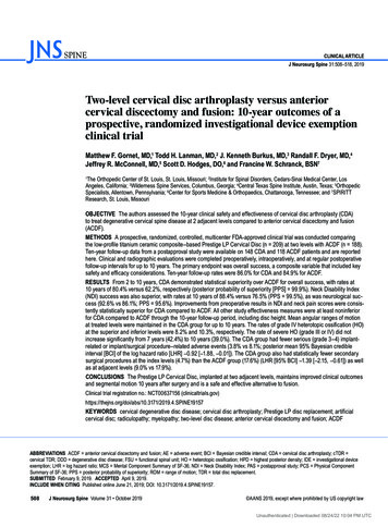
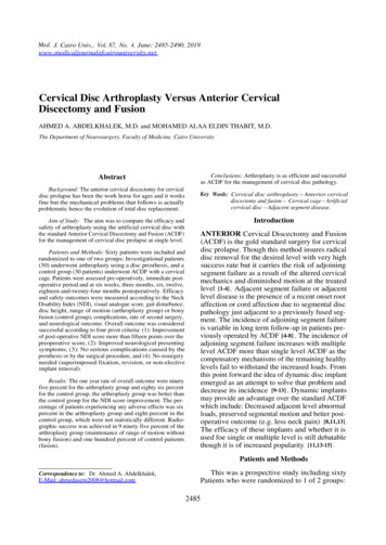
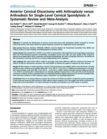
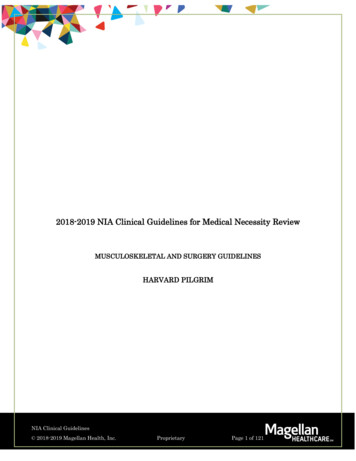
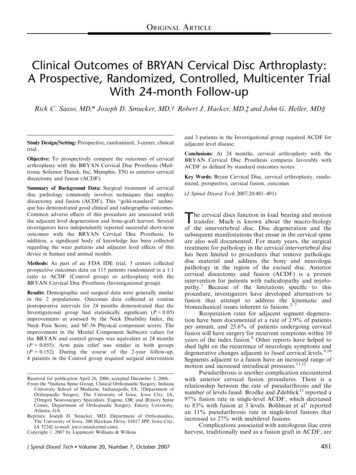
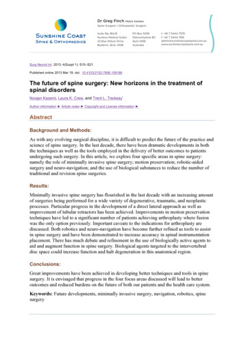
CERVICAL SPINECervical Disc Arthroplasty Versus Anterior Cervical Discectomy and Fusion Zhu et alA few previous meta-analyses38–40 have focused on thisscope, but they have different conclusions about whetherarthroplasty in cervical spine will accomplish its primarypurposes to improve clinical outcomes and reduce ASDs. Toclarify this debate further, we made a search of availablepublished data and performed a meta-analysis to comparethe rate of ASD between CDA and ACDF.MATERIALS AND METHODSSearch StrategyTo make an exhaustive search of all relevant literatures,two independent reviewers (Y.H.Z. and B.Y.Z.) conducteda PRISMA (Preferred Reporting Items for SystematicReviews and Meta-Analyses) compliant search of PubMed,EMBASE, and Cochrane Central Register of ControlledTrails (CENTRAL). The following Mesh and free textsearch terms included: ‘‘anterior cervical decompressionand fusion,’’ ‘‘cervical arthrodesis,’’ ‘‘disc replacement,’’‘‘disc prostheses,’’ and ‘‘cervical arthroplasty’’ with a limitof ‘‘clinical trial.’’ The retrieved results were last updatedon November 20, 2015 without language restrictions.References cited in the relevant literatures were alsoreviewed.Criteria for Selecting TrialsWe included studies that were eligible for the followingcriteria: (i) randomized controlled trial (RCT) of comparingcervical arthroplasty and cervical arthrodesis; (ii) patientswere definitely diagnosed with ASD or suffered adjacentsegment reoperations; (iii) the individuals were older than18 years; (iv) a minimum of 2-year follow-up. The exclusioncriteria were as follows: (i) case reports, reviews, or observational studies; (ii) descriptive or graphic outcomes with nonumerical values; (iii) only evidence of radiographicdegeneration at adjacent segments; (iv) the same data hadbeen published repeatedly. Two reviewers (Y.H.Z. andH.C.L.) independently selected the potentially qualifiedtrials according to the inclusion and exclusion criteria.Any disagreement was resolved by discussion and conformity was reached.Data ExtractionTwo independent reviewers (Y.H.Z. and Y.T.W.) extractedthe data from eligible studies. Any discrepancy was eitherresolved by discussion or by involving a third reviewer(Q.S.Z.) when necessary until a consensus for all itemswas achieved. The indispensable data extracted fromeligible researches included the study design, interventiondetails, sample size, age, sex distribution, missing size, andduration of follow-up. The pooled outcomes included therate of ASD and adjacent segment reoperation. Although therate of reoperation at adjacent segments is not synonymouswith ASD, it indirectly reflects the incidence of ASD and weutilized it also as a secondary assessment standard. Inaddition, we did subgroup analysis stratified by differenttypes of disc prostheses.1494www.spinejournal.comQuality AssessmentTwo reviewers (Y.H.Z. and B.Y.Z.) independently used the12 criteria recommended by the Cochrane Back ReviewGroup to assess the risk of bias of included studies.7 Theevaluation details of 12 criteria were based on a previousmeta-analysis of Wei et al.8 If at least six of the criteria wentthrough without serious flaws; studies were defined asmeeting ‘‘low risk of bias.’’ If not, we defined the studiesas having ‘‘high risk of bias.’’ Moreover, the GRADE(Grades of Recommendation, Assessment, Development,and Evaluation) approach was used to rate the strengthof evidence for all pooled outcomes. According to theassessment of study design, risk of bias, consistency, directness, and precision, the quality of outcomes was categorizedas very low, low, moderate, or high.9Statistical AnalysisThe x2 test and I2 test were used to assess the statisticalheterogeneity. It demonstrated significant heterogeneitywhen a P value of the x2 test was less than 0.10 or I2 valuesexceeded 50%. A random effects model was used whensignificant heterogeneity was observed among the includedstudies. Otherwise, a fixed-effects model was used for nosignificant heterogeneity. Only dichotomous data werepooled in this study, so the risk ratio (RR) and its 95%confidence interval (CI) were calculated. The funnel plotsand Egger tests were used to assess the possibility of publication bias. Sensitivity analyses were executed only formulticenter studies. This meta-analysis was performed byRevMan 5.3 software (Cochrane Collaboration, Copenhagen, Denmark), and the Egger test was accomplished byStata 13.0 (Stata Corporation, College Station, TX). Thestatistically significant level was set at P 0.05.RESULTSSearch ResultsThe process of identifying relevant studies was displayed inFigure 1. A total of 453 relevant researches were initiallyinspected from PubMed (N ¼ 149), EMBASE (N ¼ 151),CENTRAL (N ¼ 141), and reference lists (N ¼ 12). Thenumber of 164 trials was remained after excluding theduplicates. After reviewing the titles and abstracts, 115trials were excluded because they did not reach the standardof inclusion criteria. A full text review was accessed in theretaining 49 studies, and finally, 14 RCTs10–23 with 3235individuals (CDA ¼ 1696, ACDF ¼ 1539) were included inthis meta-analysis. We recorded the extracted data of thecharacteristics of 14 included papers (Table 1), and thesupplementary characteristics and details (Table 2).Quality AssessmentAccording to the 12 criteria of assessing the risk of bias, 11studies with ‘‘low risk of bias’’ and three study with ‘‘highrisk of bias’’ were indicated (Figure 2). Most of the biasesfocused on lacking of adequate allocation concealment,detailed blinding methods and intend to treat analysis.October 2016Copyright 2016 Wolters Kluwer Health, Inc. Unauthorized reproduction of this article is prohibited.
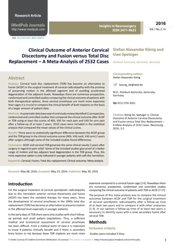
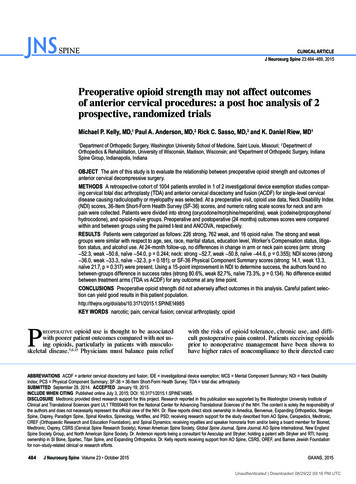
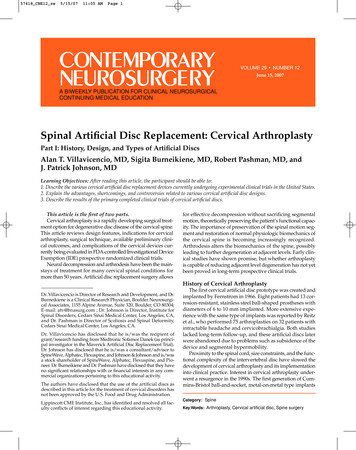
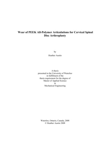
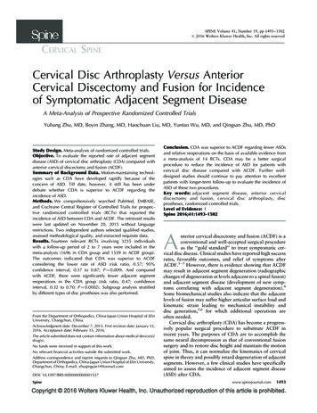
CERVICAL SPINECervical Disc Arthroplasty Versus Anterior Cervical Discectomy and Fusion Zhu et alFigure 1. Flow diagram of selecting relevantstudies.According to the GRADE approach, four pooled outcomesshowed moderate quality evidences and three displayed lowquality evidences (Table 3).Analysis of Pooled OutcomesEight studies with a total of 1346 patients (708 in CDAgroup and 638 in ACDF group) were pooled in the outcomeof ASD. The rate of ASD in the CDA group was significantlylower than that in the ACDF group (RR, 0.57; 95% CI, 0.37to 0.87; P ¼ 0.009) with no obvious heterogeneity (P ¼ 0.66,I2 ¼ 0%) (Figure 3).Ten studies with a total of 2416 patients (1250 in CDAgroup and 1166 in ACDF group) were analyzed in adjacentsegment reoperation. There were fewer adjacent segmentreoperations in the CDA group compared with the ACDFgroup (RR, 0.47; CI, 0.32 to 0.70; P ¼ 0.0002) with lowheterogeneity (P ¼ 0.13, I2 ¼ 35%). After sensitivity analysis,the heterogeneity substantially decreased (P ¼ 0.55, I2 ¼ 0%)and CDA still had fewer adjacent segment reoperations thanACDF (RR, 0.61; CI, 0.40 to 0.94; P ¼ 0.02) (Figure 4).SpineIn addition, subgroup analysis stratified by different typesof prostheses was performed (Figure 5). CDA with Bryandisc presented no significant difference compared withACDF regarding ASD (RR, 0.37; CI, 0.08 to 1.77;P ¼ 0.21) or adjacent segment reoperation (RR, 0.72; CI,0.35 to 1.51; P ¼ 0.39). Similarly, there was no significantdifference in ASD between CDA with ProDisc-C disc andACDF (RR, 0.34; CI, 0.08 to 1.41; P ¼ 0.14). However,CDA with Prestige disc displayed significant lower rate ofASD than ACDF (RR, 0.42; CI, 0.21 to 0.82; P ¼ 0.01)(Table 4).Publication BiasThe funnel plots for ASD indicated no obvious evidence ofpublication bias (Figure 6A), and the Egger test also showedno evidence of publication bias (t ¼ –0.66, P ¼ 0.548).However, the funnel plots for adjacent segment reoperationdisplayed the possibility of publication bias (Figure 6B) withwhich the result of Egger test was consistent (t ¼ –2.81,P ¼ 0.026).www.spinejournal.com1495Copyright 2016 Wolters Kluwer Health, Inc. Unauthorized reproduction of this article is prohibited.
CERVICAL SPINECervical Disc Arthroplasty Versus Anterior Cervical Discectomy and Fusion Zhu et alTABLE 1. Characteristics of the Studies Included in the 1120122012201320142014201520152015Porchet et al10Nabhan et al11Garrido et al12Coric et al13Coric et al14Sasso et al15Zhang et al16Nunley et al17Delamarter et al18Burkus et al19Zhang et al20Skeppholm et al21Phillips et al22Gornet et al23DesignSample Size(CDA/ACDF)Mean Age(CDA/ACDF)Sex(Male/Female)(CDA/ACDF)Missing Size(CDA/ACDF)RCT 4 sitesRCT 1 siteRCT 1 siteRCT 1 siteRCT 21 sitesRCT 30 sitesRCT 3 sitesRCT 2 sitesRCT 13 sitesRCT 31 sitesRCT 11sitesRCT 3 sitesRCT 24 sitesRCT 20 sites55 (27/28)41 (20/21)47 (21/26)98 (57/41)269 (136/133)463 (242/221)120 (60/60)182 (120/62)209 (103/106)541 (276/265)111 (55/56)151 (81/70)403 (218/185)545 77/531/4564/82NR5/955/628/42ACDF indicates anterior cervical discectomy and fusion; CDA, cervical disc arthroplasty; NR, not reported.Sensitivity Analysis by All Multicenter StudiesIn 14 included RCTs, three single-center studies11–13 basedon small sample size were excluded in the sensitivityanalysis. Six multicenter studies reported the incidence ofASD and eight reported the incidence of adjacent segmentreoperation (Figure 7). There were fewer ASDs in the CDAgroup compared with the ACDF group (RR, 0.58; 95% CI,0.38 to 0.91; P ¼ 0.02). Moreover, the rate of adjacentsegment reoperation in the CDA group was significantlylower than that in the ACDF group (RR, 0.48; 95% CI, 0.32to 0.73; P ¼ 0.0006). Sensitivity analyses indicated therobustness of the rates of adjacent segment disease andreoperation in multicenter studies compared with the pooledresults in all included studies.DISCUSSIONAlthough ACDF is considered to be the ‘‘gold standard’’ totreat symptomatic myelopathy and/or radiculopathy withnotable curative effect, ASD emerges gradually as a commoncomplication. Hilibrand et al1 reported the rate of symptomatic ASD as 2.9% per year and 25.6% for 10-year followup after cervical fusion. Similarly, Cho et al24 reported theTABLE 2. Supplementary Characteristics and Clinical Details of Included StudiesNumberof InvolvedLevelsPatientsof ASD(CDA/ACDF)Events ofReoperations(CDA/ACDF)FollowUp(Years)10Porchet et alNabhan et al11Garrido et al12Coric et al131111 or 20/20/11/3NRNRNR1/31/32342Coric et al14Sasso et al15Zhang et al16Nunley et al171111 or 2NRNR1/319/99/710/101/3NR2424Delamarter et al18Burkus et al19Zhang et al20Skeppholm et al21Phillips et al22Gornet et al231111 or esDisc TypePrestige IIProDisc-CBryanBryan, Discover orKineflex CKineflex CBryanBryanKineflex C, Mobi-C orAdventProDisc-CPrestigeMobi-CDiscoverPCMPrestige LPACDF indicates anterior cervical discectomy and fusion; CDA, cervical disc arthroplasty; NR, not reported.1496www.spinejournal.comOctober 2016Copyright 2016 Wolters Kluwer Health, Inc. Unauthorized reproduction of this article is prohibited.
CERVICAL SPINECervical Disc Arthroplasty Versus Anterior Cervical Discectomy and Fusion Zhu et alincidence of ASD as 3% annually and 25% within the first10 years after fusion. Lee et al25 indicated that secondarysurgery at adjacent segments occurred at a rate of 2.4% peryear and predicted that 22.2% of patients would needadjacent segment reoperations by 10 years postoperation.The definite risk factor of causing adjacent segment degeneration is still unclear. Some studies indicate that postoperative disc height, congenital stenosis, and postoperativekyphotic change may be risk factors for developing radiographic degeneration at adjacent segments after ACDF.33–35Additional plate augmentation for ACDF can lower the rateof ASD, and factors increasing the risk of secondary surgeryat adjacent segments were smoking, female sex, and thenumber of arthrodesis segments.36 In a RCT study comparing ACDF with CDA, Nunley et al17 reported that osteopenia and concurrent lumbar degeneration diseasesignificantly increased the risk of ASD after anterior cervicalsurgery. It is also indicated that patients are more likely todevelop ASD and adjacent segment degeneration at thesuperior adjacent level compared with the inferior adjacentlevel.26–28 It remains under debate whether ASD caused byspinal fusion attributes to iatrogenic motion restriction.4,29However, Matsumoto et al30 compared 64 patients with201 asymptomatic volunteers for 10-year follow-up, andfound that ACDF patients had significantly higher incidenceof disc degeneration at adjacent segments than asymptomatic volunteers, which implied that ACDF could acceleratethe nature history of spinal segmental degeneration. Theoriginal design purpose of CDA is to maintain the motion ofjoint, so it can normalize the kinematics of cervical spinetheoretically and possibly retard adjacent segment degeneration. CDA has demonstrated the advantage of keepingnormal motion and reducing intradiscal pressure and facetloads at adjacent segments in some in vitro biomechanicalstudies.5,6,31,32 However, it still lacks of sufficient clinicalFigure 2. Risk of bias of included studies.TABLE 3. Summary of Strength of Evidence With Regard to the OutcomesOutcomesASDASD (Bryan)ASD (Prestige)ASD (ProDisc-C)Adjacent segmentreoperationAdjacent segmentreoperation(Bryan)Adjacent segmentreoperationzStudiesPatients(CDA/ACDF)Risk of Bias InconsistencyIndirectness 1250/1166SeriousSerious Serious Serious Serious LowModerateLowModerate3323/307Serious NoNoSeriousyLow91032/981Serious NoNoNoModerateACDF indicates anterior cervical discectomy and fusion; ASD, adjacent segment disease; CDA, cervical disc arthroplasty. Serious risk of performance bias, detection bias, or attrition bias.yWide confidence intervals are around the estimate of the effect or total population size is less than 400.zAdjacent segment reoperation after the sensitivity analysis.Spinewww.spinejournal.com1497Copyright 2016 Wolters Kluwer Health, Inc. Unauthorized reproduction of this article is prohibited.
CERVICAL SPINECervical Disc Arthroplasty Versus Anterior Cervical Discectomy and Fusion Zhu et alFigure 3. Forest plot comparing the incidence of adjacent segment disease between cervical disc arthroplasty and anterior cervical discectomyand fusion. CI indicates confidence interval; df, degrees of freedom; MH, Mantel-Haenszel.evidences to prove that CDA is superior to ACDF regardinglower rate of ASD.In our meta-analysis, we selected 14 RCTs comparingACDF with CDA that specifically reported the rate ofsymptomatic ASD or adjacent segment reoperation at thefollow-up of two to seven years. And two studies17,18mainly aimed to report the rate of ASD and relativereoperation. Studies with only data of radiographic changesof degeneration at adjacent levels were not included,because a simple observation of ossification at adjacentsegments cannot be directly correlated to the developmentof symptomatic disease.37 Eight different types of discprostheses were involved in the pooled CDA group. Phillipset al22 reported a relatively low rate of adjacent segmentreoperation in the CDA group in a RCT study comparingPCM prosthesis with ACDF. This study resulted in quantitative heterogeneity in the pooled outcome and wasremoved in the sensitivity analysis. In the subgroupanalysis, Bryan and ProDisc-c prostheses were equivalentto ACDF regarding the incidence of ASD, respectively.Only Prestige prosthesis showed fewer ASDs comparedwith cervical fusion, which implied that the differentFigure 4. Forest plot comparing the incidence of adjacent segment reoperation between cervical disc arthroplasty and anterior cervicaldiscectomy and fusion. CI indicates confidence interval; df, degrees of freedom; MH, Mantel-Haenszel.1498www.spinejournal.comOctober 2016Copyright 2016 Wolters Kluwer Health, Inc. Unauthorized reproduction of this article is prohibited.
CERVICAL SPINECervical Disc Arthroplasty Versus Anterior Cervical Discectomy and Fusion Zhu et alFigure 5. Subgroup analyses of the incidences of adjacent segment disease and reoperation by different prosthesis types. CI indicatesconfidence interval; df, degrees of freedom; MH, Mantel-Haenszel.prosthesis types might influence the rate of ASD. However,the validity of subgroup analysis was limited by low qualityevidences in GRADE approach because of risk of bias andimprecisions.There have been a few meta-analyses comparing thesafety and efficacy between ACDF and CDA. Yanget al38 and Verma et al39 respectively performed a metaanalysis involving six RCTs to compare the radiographicadjacent segment degeneration or symptomatic ASDbetween the two surgical procedures. These two studieswere both current up to 2011 and indicated no significantdifference in the incidence of ASD when comparing ACDFwith CDA. However, four new RCTs16,20 –22 reporting therate of ASD have been published in recent 4 years, and threeoriginal RCTs17–19 have updated new outcome in longerfollow-up period. Luo et al40 reported that CDA had fewerASDs than ACDF in a recent meta-analysis. However, itonly included eight RCTs, and the rates of radiographicdegeneration and symptomatic disease at adjacent segmentswere pooled together into the outcome. Moreover, all theprevious meta-analyses ignored the subgroup analysis ofdifferent prosthesis types.There are a few strengths in our study. First, this is thelatest and most comprehensive meta-analysis to evaluate theTABLE 4. Pooled Results in the Subgroup Analysis of Different Prosthesis TypesOutcomesASD (Bryan)ASD (Prestige)ASD (ProDisc-C)Adjacent segmentreoperation ixedRR(95% –1.41)(0.35–1.51)I2 (%) 140.39No differenceCDANo differenceNo differenceASD indicates adjacent segment disease; CDA, cervical disc arthroplasty; CI, confidence interval; RR, risk ratio.Spinewww.spinejournal.com1499Copyright 2016 Wolters Kluwer Health, Inc. Unauthorized reproduction of this article is prohibited.
CERVICAL SPINECervical Disc Arthroplasty Versus Anterior Cervical Discectomy and Fusion Zhu et alFigure 6. (A) Funnel plots for the adjacent segment disease. (B) Funnel plots for the adjacent segment reoperation.incidence of ASD between CDA and ACDF. Second, weused Cochrane risk of bias and GRADE approach to assessthe quality of evidence. Additional strengths contained arigorous search strategy, only RCTs included, no languagelimitations, sensitivity analysis, and subgroup analysisstratified by different types of prostheses to guarantee theconsistency and accuracy. However, the validness of ourstudy is limited by several factors. First, the pooled outcomeof adjacent segment reoperation indicated the possibility ofpublication bias. Second, the results of subgroup analysisshould be cautiously accepted because of low quality evidences. Third, various surgery interventions and surgicaltechnologies at different centers may influence the outcome.Moreover, the follow-up period of six included RCTs is 2years, and this period is too short to assess a comprehensiverate of ASD. So these studies should continue to focus onreporting the incidence of ASD in longer-follow-up period.During our search of relevant studies, ASD was not mentioned in some studies, which were designed as RCTs toevaluate the effectiveness and safety of disc prostheses andonly focused on the improvement of symptoms at thesurgical segment. So this study also aims to highlight theimportance of considering ASD as an outcome in futureprospective studies.In summary, our meta-analysis indicated that CDA wassuperior to ACDF regarding fewer ASDs and adjacentsegmental reoperations. Compared with ACDF, CDAmay be a better surgical procedure to reduce the incidenceof ASD for patients with cervical disc disease. Further welldesigned studies should continue to focus on excellentpatients with longer-term follow-up to evaluate the incidence of ASD of these two procedures.Figure 7. Sensitivity analyses of the incidences of adjacent segment disease and reoperation in all multicenter studies. CI indicates confidenceinterval; df, degrees of freedom; MH, Mantel-Haenszel.1500www.spinejournal.comOctober 2016Copyright 2016 Wolters Kluwer Health, Inc. Unauthorized reproduction of this article is prohibited.
CERVICAL SPINECervical Disc Arthroplasty Versus Anterior Cervical Discectomy and Fusion Zhu et alKey Points15.This meta-analysis of prospective randomizedcontrolled studies provided the latest evidenceof comparing the incidence of ASD between CDAand ACDF.Compared with ACDF, CDA presented fewersymptomatic ASDs and relevant adjacentsegment reoperations.The different types of disc prostheses may affectthe incidence of ASD.More high quality studies with longer follow-upare needed to explore the occurrence of ASD ofthese two surgical procedures.References1. Hilibrand AS, Carlson GD, Palumbo MA, et al. Radiculopathyand myelopathy at segments adjacent to the site of aprevious anterior cervical arthrodesis. J Bone Joint Surg Am1999;81:519–28.2. Emery SE, Bohlman HH, Bolesta MJ, et al. Anterior cervicaldecompression and arthrodesis for the treatment of cervical spondylotic myelopathy. Two to seventeen-year follow-up. J Bone JointSurg Am 1998;80:941–51.3. Goffin J, Geusens E, Vantomme N, et al. Long-term follow-upafter interbody fusion of the cervical spine. J Spinal Disord Tech2004;17:79–85.4. Hilibrand AS, Robbins M. Adjacent segment degeneration andadjacent segment disease: the consequences of spinal fusion? SpineJ 2004;4:190s–4s.5. Eck JC, Humphreys SC, Lim TH, et al. Biomechanical study on theeffect of cervical spine fusion on adjacent-level intradiscal pressureand segmental motion. Spine 2002;27:2431–4.6. Matsunaga S, Kabayama S, Yamamoto T, et al. Strain on intervertebral discs after anterior cervical decompression and fusion.Spine 1999;24:670–5.7. Furlan AD, Pennick V, Bombardier C, et al. 2009 updated methodguidelines for systematic reviews in the Cochrane Back ReviewGroup. Spine 2009;34:1929–41.8. Wei J, Song Y, Sun L, et al. Comparison of artificial total discreplacement versus fusion for lumbar degenerative disc disease: ameta-analysis of randomized controlled trials. Int Orthop2013;37:1315–25.9. Guyatt GH, Oxman AD, Schunemann HJ, et al. GRADE guidelines: a new series of articles in the Journal of Clinical Epidemiology. J Clin Epidemiol 2011;64:380–2.10. Porchet F, Metcalf NH. Clinical outcomes with the Prestige IIcervical disc: preliminary results from a prospective randomizedclinical trial. Neurosurg Focus 2004;17:E6.11. Nabhan A, Steudel WI, Nabhan A, et al. Segmental kinematics andadjacent level degeneration following disc replacement versusfusion: RCT with three years of follow-up. J Long Term EffMed Implants 2007;17:229–36.12. Garrido BJ, Taha TA, Sasso RC. Clinical outcomes of Bryancervical disc arthroplasty a prospective, randomized, controlled,single site trial with 48-month follow-up. J Spinal Disord Tech2010;23:367–71.13. Coric D, Cassis J, Carew JD, et al. Prospective study of cervicalarthroplasty in 98 patients involved in 1 of 3 separate investigational device exemption studies from a single investigational sitewith a minimum 2-year follow-up. Clinical article. J NeurosurgSpine 2010;13:715–21.14. Coric D, Nunley PD, Guyer RD, et al. Prospective, randomized,multicenter study of cervical arthroplasty: 269 patients from theKineflex C artificial disc investigational device exemption 9.30.31.32.with a minimum 2-year follow-up: clinical article. J NeurosurgSpine 2011;15:348–58.Sasso RC, Anderson PA, Riew KD, et al. Results of cervicalarthroplasty compared with anterior discectomy and fusion:four-year clinical outcomes in a prospective, randomized controlled trial. J Bone Joint Surg Am 2011;93:1684–92.Zhang X, Zhang X, Chen C, et al. Randomized, controlled,multicenter, clinical trial comparing BRYAN cervical disc arthroplasty with anterior cervical decompression and fusion in China.Spine 2012;37:433–8.Nunley PD, Jawahar A, Kerr EJ, et al. Factors affecting theincidence of symptomatic adjacent-level disease in cervical spineafter total disc arthroplasty: 2- to 4-year follow-up of 3 prospectiverandomized trials. Spine 2012;37:445–51.Delamarter RB, Zigler J. Five-year reoperation rates, cervical totaldisc replacement versus fusion, results of a prospective randomizedclinical trial. Spine 2013;38:711–7.Burkus JK, Traynelis VC, Haid RW, et al. Clinical and radiographic analysis of an artificial cervical disc: 7-year follow-up fromthe Prestige prospective randomized controlled clinical trial:clinical article. J Neurosurg Spine 2014;21:516–28.Zhang HX, Shao YD, Chen Y, et al. A prospective, randomised,controlled multicentre study comparing cervical disc replacementwith anterior cervical decompression and fusion. Int Orthop2014;38:2533–41.Skeppholm M, Lindgren L, Henriques T, et al. The Discoverartificial disc replacement versus fusion in cervical radiculopathy:a randomized controlled
CERVICAL SPINE Cervical Disc Arthroplasty Versus Anterior Cervical Discectomy and Fusion Zhu et al . CDA with Bryan disc presented no significant difference compared with ACDF regarding ASD (RR, 0.37; CI, 0.08 to 1.77; P¼0.21) or adjacent segment reoperation (RR, 0.72; CI, 0.35 to 1.51; P¼0.39). Similarly, there was no significant