Transcription
Faculty of Life SciencesTowards Structured andStandardized MRI Data Archive Development of a Tool for DICOMFile Arrangement in JavaMaster ThesisSubmitted by:Dimitrij BaalMatriculation No:Degree Program: Biomedical EngineeringSupervisor:Prof. Dr. Udo van StevendaalDr. Carsten LiessDr. Shuo ZhangHamburg, August 27th 2019
AbstractTo be able to communicate with medical equipment manufactured by different vendors, thevast majority of systems today uses the Digital Imaging and Communications in Medicine(DICOM) standard. Although the information of the modality data elements can be made available through specific programs, there are still limitations for the immediate use. Therefore, inthis thesis a tool is introduced which consists of two function groups. The first one creates atext file with all the extracted DICOM header information sorted in subgroups and the secondfunction stores the data separately depending the image series in subfolders for a standardizedenvironment in the archive. Furthermore, two examples of images series of a fetal brain examination are demonstrated to show the result after application of the software. In future work,the tool could be integrated in the scanner for the enhancement of the online to offline operationto make the data processing faster and more efficient.
Table of ContentsList of Abbreviations .1.Introduction . 12.Theoretical Framework. 33.4.2.1Magnetic Resonance Imaging . 32.2DICOM Standard . 72.2.1DICOM Parameter . 82.2.2Archive and Post-Processing . 9Methodology . 123.1Java and Eclipse . 123.2Function Group 1: DICOM Header Extraction . 153.3Function Group 2: Data Restructuring . 16Results . 174.1Execution of the developed Program . 174.2Comparison with other Programs . 204.3Examples of DICOM Image Series. 235.Discussion . 286.Conclusion . 29Bibliography .AAnnex . B
List of AbbreviationsACRAmerican College of RadiologyCTComputed TomographyDICOMDigital Imaging and Communications in MedicineECGElectrocardiogramFIDFree Induction DecayFTFourier TransformJDKJava Development KitJPEGJoint Photographic Experts GroupJREJava Runtime EnvironmentMRIMagnetic Resonance ImagingNEMANational Electrical ManufacturesPACSPicture Archiving and Communication SystemPETPositron Emission TomographyRFRadio FrequencySPECTSingle Photon Emission Computed TomographyTIFFTagged Image File Format
1. IntroductionThis thesis is written in the field of Clinical Science of the company Royal Philips. “Royal Philipsis a leading health technology company focused on improving people's health and enablingbetter outcomes across the health continuum from healthy living and prevention, to diagnosis,treatment and home care. Philips leverages advanced technology and deep clinical and consumer insights to deliver integrated solutions. Headquartered in the Netherlands, the companyis a leader in diagnostic imaging, image-guided therapy, patient monitoring and health informatics, as well as in consumer health and home care. Philips generated 2018 sales of EUR18.1 billion and employs approximately 77,000 employees with sales and services in morethan 100 countries” [1]. Since the introduction of computed tomography as the first digital imaging method in medicine, the importance of digital medical image processing has steadilyincreased. At the latest with the emergence of the idea of digital archiving of images (PACS)and electronic image distribution in hospitals, the need arose to be able to exchange digitalimages between devices from different manufacturers. DICOM was adopted in 1993 and hasbeen continuously expanded ever since. Since 1995 DICOM has also been accepted as aformal standard in Europe (MEDICOM, ENV 12052) [2]. This standard has developed into anindispensable component for the integration of digital image processing systems in medicine.DICOM offers solutions for a variety of communication applications - in the network as well asoffline. A DICOM file consists of a list of data elements that contain a variety of image-relatedinformation: Information about the patient, e.g. name, date of birth and identification number, information on modality and recording, e.g. instrument parameters, calibration, radiation dose and contrast agent administration, and Image information, such as resolution and windowing.The DICOM Standard defines for each modality exactly which data elements are prescribed,which are optionally and which are prescribed under certain conditions (e.g. only with contrastmedium application). All this information is included when exported from the MRI system andcan be made available through specific programs such as the Philips DICOM Viewer. However, this availability is limited and not accessible by many other dedicated programs, especially in the fields of pediatrics or neurology. This leads to a large limitation for the immediateuse of the exported DICOM files in a clinical environment and poses therefor the followingchallenges. When exporting data from the scanner all data are saved almost randomly in onelarge folder. These are arranged neither in chronological order nor in sequential order. Furthermore, the file names do not reflect imaging information like the image series or information1
about the data structure. Another concern is that new subfolders are created as soon as theimage number succeeds 2048, which leads to a disorganized folder structure. In addition, parameters of the imaging techniques like the pulse sequences are not directly available andhave to be displayed by the mentioned tools. In clinical environment, it would be of great benefitto have direct use of the exported DICOM files. It would smooth the “online to offline” researchactivities, thus from experiments on the scanner to processing on data. In consideration of thepresented limitations, the objective for this work is to split the exported DICOM files, so that: the data names reflect the basic structure and imaging information like the image series, the data can be easily accessed (i.e. loaded and processed) by another software.Furthermore, the goal is to extract the corresponding DICOM header information, so that: important parameters are saved in a separate file, basic information with respect to imaging technique and scan protocol are directly available and can be further used for comparison, investigation, publication and citation.Part of the challenge is to create a tool, which can also deal with the compatibility of variousDICOM files of different software release versions (R3, R4).The remainder of this thesis is organized in the following way. Chapter 2 introduces the readerto the functionality of magnetic resonance imaging and to the DICOM standard. It is describedhow the standard is used by hospital equipment to store and exchange digital images andassociated information. Chapter 3 gives an overview about the programing language Java anda workflow chart is used to illustrate the two functionality groups of the created tool. Chapter 4presents the tool and the distinctions to other DICOM processing programs. In addition, thetwo examples diffusion and T2-weighting DICOM image series are shown. The discussion ofthe results and an outlook of future work is subject of chapter 5. Chapter 6 concludes the workof the project presented in this thesis. In the appendix the source code and the entire list of theparameters to be extracted is given.2
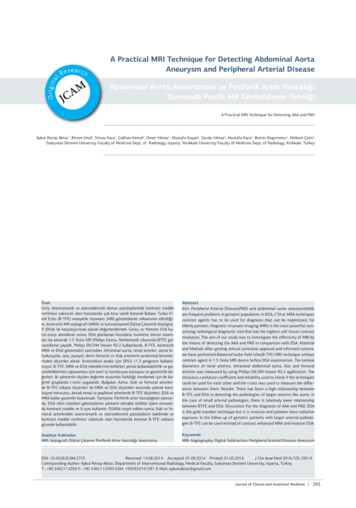
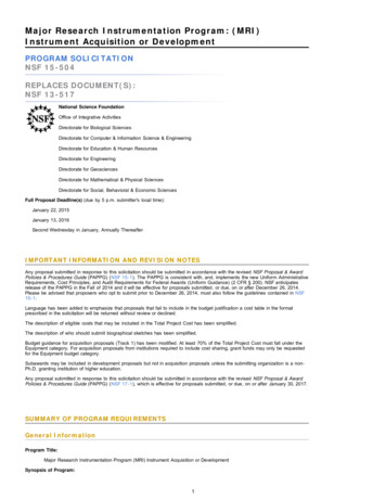
2. Theoretical FrameworkMagnetic Resonance Imaging (MRI) is one of the established imaging methods in clinical routine, which is used primarily in medical diagnostics [3]. The generated data is stored as socalled DICOM files and used for the exchange of information in medical image data management. The physical and medical processes required for the development of these medical images are presented and explained in this chapter.2.1 Magnetic Resonance ImagingMRI is used in medical diagnostics to visualize the structure and function of tissues and organsin the body. Sectional images of the human or animal body are generated which allow anassessment of the organs and many pathological organ changes. No X-rays or other ionizingradiation is generated or used in the system [4]. In general, all atomic nuclei in the body rotatearound their own axis. This angular momentum is also called nuclear spin. Due to their ownrotation, these nuclei generate a minimal magnetic field (Fig. 1). Hydrogen nuclei are particularly important here, as they occur most frequently in the body. The magnetic orientation of thehydrogen nuclei is purely random under natural circumstances (Fig. 2). If, however, a strongmagnetic field is applied to the body from the outside, then these atomic nuclei all arrangethemselves in the same direction, namely in the longitudinal direction of the body (Fig. 3) [5].Fig. 1: Atomic nuclei have a spin and aremagnetic [16].Fig. 2: Atomic nuclei spinning randomly in thetissue [16].By briefly applying an additional high-frequency alternating field in the radio frequency range,this magnetization can be deflected from the direction of the static field, i.e. partially or completely converted into a transverse magnetization. The transverse magnetization begins immediately to precede the field direction of the static magnetic field, i.e. the magnetization direction rotates. This motion is called Larmor precession and can be observed mechanicallyanalogously on a gyroscope, if its axis of rotation is not vertical, but performs a precession3
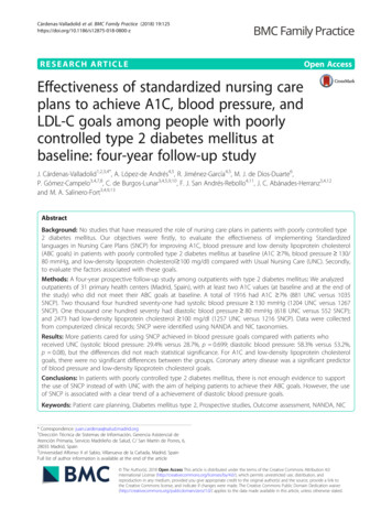
around the vertical (Fig. 4) [3]. Both for excitation and for observation of the signal, a resonancecondition must be fulfilled.Fig. 3: The application of a strong magneticfield makes all the nuclei align in the same direction [16].Fig. 4: Gyroscope performing a precession aroundthe vertical [16].Like the rotation of the magnet in the dynamo in a coil (receiver circuit), this precession movement of the tissue magnetization induces an electrical voltage and can thus be detected. Itsamplitude is proportional to the transverse magnetization [6]. After switching off the high-frequency alternating field, the transverse magnetization decreases again, so that the spins againalign themselves parallel to the static magnetic field. For this so-called relaxation they need acharacteristic decay time. This is dependent on the chemical compound and the molecularenvironment in which the preceding hydrogen nucleus is located. Therefore, the different tissuetypes differ characteristically in their signal, which leads to different signal strengths (brightness) in the resulting image as can for example be seen in Figure 5 and Figure 6 [6].Fig. 5: A typical MR image of the abdomen [6].Fig. 6: A typicial MR image of the head [6].4
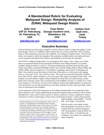
The intensity of magnetic resonance signals is a function of several parameters, including relaxation times 𝑇1 and 𝑇2 , proton density, chemical shift and motion. These parameters are setin the pulse sequence [6]. Such an electromagnetic pulse sequence is a pre-selected set ofradio frequency and gradient pulses that are repeated many times during a scan. In the shorttime interval between the pulses, signals are received and automatically evaluated by a computer (depending on the amplitude and shape of the gradient waves one receive different signals), and images are calculated [6]. Virtually all MR pulse sequences can be separated intotwo functional parts – the spin preparation, which means the manipulation of the MR signalcharacteristics through the use of radio frequency pulses or magnetic field gradients and thesignal production, which describes components necessary to generate the signal and encodethis signal with spatial information (Fig. 7) [6].Signal productionSpin preparationPrepulseTissue presaturationFIDSpin EchoGradient EchoFTIMAGEFig. 7: A pulse sequence separated into two functional elements: signal production and spinpreparation.An MRI system (Fig. 8) includes a combination of a static magnetic field, lo
can be made available through specific programs such as the Philips DICOM Viewer. How-ever, this availability is limited and not accessible by many other dedicated programs, espe-cially in the fields of pediatrics or neurology. This leads to a large limitation for the immediate use of the exported DICOM files in a clinical environment and poses therefor the following challenges. When exporting .