Transcription
19Root Resorption in Orthodontics:An Evidence-Based ApproachLeandro Silva Marques1, Paulo Antônio Martins-Júnior1,Maria Letícia Ramos-Jorge1 and Saul Martins Paiva21FederalUniversity of Vales do Jequitinhonha e Mucuri2Federal University of Minas GeraisBrazil1. IntroductionRoot resorption is a pathological process that causes a shortening of the dental root.Although this condition is generally asymptomatic and missed in diagnosis, it may result intooth mobility and even tooth loss if not diagnosed and treated early (Ahangari et al., 2010).In orthodontics, induced inflammatory root resorption is a form of pathologic rootresorption related to the removal of hyalinized areas of the periodontal ligament followingthe application of orthodontic forces and is considered an undesirable but unavoidableiatrogenic consequence of orthodontic treatment (Brezniak & Wasserstein, 2002a; Brezniak &Wasserstein, 2002b).The root resorption may compromise the continued existence and functional capacity of theaffected tooth, depending on their magnitude (Brezniak & Wasserstein, 1993a, Brezniak &Wasserstein, 1993b), since the root structure (volume and contour) is changed (Consolaro,2002). However, as the process of root resorption during orthodontic treatment is usuallysmooth and ends when the force is removed (Brezniak & Wasserstein, 1993; Levander et al.,1994) some authors have pointed out that the aesthetic and functional improvements justifythe risks (Brezniak & Wasserstein, 1993).1.1 Aims of the chapterThe aims of this chapter are to give a detailed description of root resorption, how it begins,the mechanisms involved in this condition and how the risk factors described in theliterature contribute toward the development of root resorption related to orthodontictreatment. The importance of a thorough patient history and early diagnosis are alsodiscussed. The value of high-quality research, such as longitudinal cohort and prospectivestudies, randomized clinical trials, systematic reviews and meta-analysis, is stressed in lightof the current emphasis on evidence-based dentistry. Care and recommendations, legalimplications and a case description of a patient with root resorption following orthodontictreatment are also presented.www.intechopen.com
430Orthodontics – Basic Aspects and Clinical Considerations2. Etiology of root resorptionDetermining the cause of root resorption requires a thorough history, rescuing the previousdental history, addiction, accidentes, previous treatment, associated diseases and otherdetails relevant to pathogenesis, but not always remembered by patients and identified byorthodontists. Several authors have pointed out that the multifactor etiology of rootresorption is complex, but the condition appears to result from a combination of individualbiologic variability, genetic predisposition and the effect of mechanical factors (Bartley et al.,2011; Weltman et al., 2010; Zahrowski & Jeske, 2011). However, no definitive conclusion hasbeen drawn as to whether sex (Harris et al., 1997; Hendrix et al., 1994; Sameshina & Sinclair,2001), age (Baumrind et al., 1996; Costopoulos & Nanda, 1996; Harris et al., 1997; Harris &Baker, 1990; Owmann-Moll et al., 1995), tooth extractions (Baumrind et al., 1996; Blake et al.,1995; Hendrix et al., 1994; McNab et al., 2000) and duration of active treatment (Baumrind etal., 1996; Beck & Harris, 1994; Harris et al., 1997; Kaley & Phillips, 1991; Kurol et al., 1996;Mirabella & Artun, 1995; Sameshina & Sinclair, 2001) are risk factors for root resorption.Conflicting data are reported on the relationship between root resorption and hypodontia orpartial anodontia (Artun, 2000; Kjaer, 1995, 2000; Lee et al., 1999) and ectopic teeth (Kjaer,2000; Lee et al., 1999).2.1 How root resorption begins?Orthodontic tooth movement is based on force-induced periodontal ligament and alveolarbone remodeling (Abuabara, 2007). So, orthodontic forces represent a physical agent capableof inducing inflammatory reaction in the periodontium (Giannopoulou et al., 2008). When atooth moves, a necrosis of periodontal ligament on the pressure side with formation of acell-free hyaline zone occurs. This event is followed by osteoclast resorption of theneighbouring alveolar bone and bone apposition by osteoblasts on the tension side(Abuabara, 2007). The resorption process of dental hard tissues seems to be triggered by theactivity of some cytokines as well as that of bone. Immune cells migrate out of the capillariesin the periodontal ligament and interact with locally residing cells by elaborating a largearray of signal molecules (Jäger et al., 2005). According Consolaro et al. (2011), the causes ofroot resorption should be related to the loss of root surface cementoblasts.2.2 Orthodontic treatment-related factorsThe ideal force for tooth movement would mimic a physiologic balance between toothmovement and bony adaptation (Paetyangkul et al., 2009). Schwarz (1932) advocated theoptimal force level for tooth movement between 7 and 26 g per square centimeter. He alsostated that, when force exceeded this threshold, root resorption occurs. When pressuredecreases below this limit, root resorption ceases (Owman Moll et al., 1996). This was laterconfirmed by King and Fischlschweiger (1982), who found that light forces producedinsignificant root resorption, whereas intermediate or heavy forces resulted in substantialcrater formation.In this context, several aspects have been related to induce root resorption duringorthodontic treatment. This aspects are as follows: treatment duration (Casa et al., 2001; Fox,2005; Levander & Malmgren, 1988; Otis et al., 2004; Paetyangkul et al., 2011; Sameshima &Sinclair, 2004; Segal et al., 2004), magnitude of the applied forces (Barbagallo et al., 2008;Bartley et al., 2011; Casa et al., 2001; Chan et al., 2005; Harris et al., 2006; Paetyangkul et al.,www.intechopen.com
Root Resorption in Orthodontics: An Evidence-Based Approach4312011), direction of tooth movement (Barbagallo et al., 2008; Han et al., 2005) amount ofapical displacement (Fox, 2005; Segal et al., 2004), force application method (continuous vs.intermittent) (Brezniak & Wasserstein, 2002; Faltin et al., 2001), type of appliance (Brezniak& Wasserstein, 1993; Pandis et al., 2008) and treatment technique (Bartley et al., 2011; Beck &Harris, 1994; Janson et al., 1999; Marques et al., 2010; Pandis et al., 2008; Parker & Harris,1998; Scott et al., 2008).2.2.1 Treatment duration, force application method and magnitude of the appliedforcesIn a study, Acar et al. (1999) compared a 100-g force with elastics in either an interrupted (12hours per day) or a continuous (24 hours per day) application. Group who has teethexperiencing orthodontic movement had significantly more root resorption than the controlgroup. Besides that, continuous force produced significantly more root resorption thandiscontinuous force application.Later, Ballard et al. (2009) conducted a prospective randomized clinical trial to compare rootresorption with two force application patterns (continuous and intermittent) and theyconcluded that the application of intermittent orthodontic forces of 225 cN for 8 weeks (14days of force application, 3 days of rest, then 4 days of force application repeated for 6weeks) caused less root resorption than continuous forces of 225 cN for 8 weeks. Theauthors stated that, although it might not be clinically practical, compared with continuousforces, intermittent forces might be a safer method to prevent significant root resorption.More recently, Paetyangkul et al. (2011) investigated the amounts of root resorptionvolumetrically after the application of controlled light and heavy forces in the buccaldirection for 4, 8, and 12 weeks. They found significant differences in the extent of rootresorption between 4, 8, and 12 weeks of force application (P 0.001), with substantiallymore severe resorption in the longer force duration groups. The light force producedsignificantly less root resorption than did the heavy force. The authors argued that theduration of force application appears to be an important factor in orthodontic rootresorption. Even though the application of light orthodontic forces did not show asignificant difference between 4 and 8 weeks of buccal force application, the amount of rootresorption increased significantly from 8 to 12 weeks of force application. So the duration oforthodontic force application caused more root resorption even when light forces of 25 gwere used. This finding agrees with others studies published by Vardimon et al. (1991) andGonzales et al. (2008). Paetyangkul et al. (2011) affirmed that this might be due to theincreased osteoclastic activity around 8 weeks of force application.In another study, Chan and Darendeliler (2006) found that the mean volume of theresorption craters was 11.59 times greater in the heavy-force group than in the controlgroup. Barbagallo et al. (2008), in a prospective randomized clinical trial compared forcesapplied with removable thermoplastic appliances (TA) and fixed orthodontic appliances.The results showed that teeth experiencing orthodontic movement had significantly moreroot resorption than did the control teeth. They also found that heavy force producedsignificantly more root resorptions (9 times greater than the control) than light force (5 timesgreater than the control).In this context, Harris et al. (2006) conducted a prospective randomized clinical trial toquantify the amount of root resorption when controlled light and heavy intrusive forceswww.intechopen.com
432Orthodontics – Basic Aspects and Clinical Considerationswere applied to human premolars and to establish the sites where root resorption is moreprevalent. They found that the volume of the root resorption craters after intrusion wasdirectly proportional to the magnitude of the intrusive force applied. The findings showedthat the control group had fewer and smaller root resorption craters, the light force grouphad more and larger root resorption craters than the control group, and the heavy forcegroup had the most and the largest root resorption craters of all groups. A trend of linearincrease in the volume of the root resorption craters was observed from control to light toheavy groups, and these differences were statistically significant. The mean volumes of theresorption craters in the light and heavy force groups were 2 and 4 times greater than in thecontrol groups, respectively. The mesial and distal surfaces had the greatest resorptionvolume, with no statistically significant difference between the 2 surfaces.2.2.2 Direction of tooth movementEvaluating the direction of tooth movement (intrusive vs. extrusive force), Han et al. (2005)found that root resorption from extrusive force was not significantly different from thecontrol group. Intrusive force significantly increased the percentage of resorbed root area (4fold). The correlation between intrusion or extrusion and root resorption in the same patientwas r 0.774 (P 0.024).2.2.3 Amount of apical displacementIn orthodontics, total apical displacement might represent a better marker for overalltreatment activation. A tooth that is moved greater distances through bone is subjected tolonger durations of activation. There is no way to move a tooth between two points withfixed appliances, without causing hyalinization. Perhaps, this is why maxillary incisors aremost likely to exhibit severe levels of root resorption (Segal et al., 2004). Segal et al. (2004)conducted a meta-analysis to elucidate possible treatment-related etiological factors - suchas, duration of treatment and apical displacement – for external root resorption and theyfound that mean apical root resorption was strongly correlated with total apicaldisplacement (r 0.822) and treatment duration (r 0.852). In 2005, Fox also found thattreatment-related root resorption is correlated with the distance the apex moves and thelength of time the treatment took.2.2.4 Archwire sequenceMandall et al. (2006) compared 3 orthodontic archwire sequences in terms of: (1) patientdiscomfort, (2) root resorption, and (3) time to working archwire. In that study, all patientswere treated with maxillary and mandibular preadjusted edgewise appliances (0.022-inslot), and all archwires were manufactured by the same manufacturer. The results showedthat there was no statistically significant difference between archwire sequences, formaxillary left central incisor root resorption (F ratio, P 0.58). There was also no statisticallysignificant difference between the proportion of patients with and without root resorptionbetween archwire sequence groups (P 0.8).2.2.5 Type of applianceReukers et al. (1998) compared the prevalence and severity of root resorption after treatmentwith a fully programmed edgewise appliance (FPA) and a partly programmed edgewisewww.intechopen.com
Root Resorption in Orthodontics: An Evidence-Based Approach433appliance (PPA). All FPA patients were treated with 0.022-in slot Roth prescription (‘‘A’’Company, San Diego, Calif), and misplaced brackets were rebonded. All PPA patients weretreated with 0.018-in slot Microloc brackets (GAC, Central Islip, NY), and the archwireswere adjusted for misplaced brackets. They found no statistically significant differences inthe amount of tooth root loss (FPA, 8.2%; PPA, 7.5%) or prevalence of root resorption (FPA,75%; PPA, 55%) between the groups.More recently, Scott et al. (2008) investigated the effect of either Damon3 self-ligatingbrackets or a conventional orthodontic bracket system on mandibular incisor rootresorption. Patients were treated with Damon3 self-ligating or Synthesis (both, Ormco,Glendora, Calif) conventionally ligated brackets with identical archwires and sequencing inall patients. The results showed that mandibular incisor root resorption was not statisticallydifferent (Damon3, 2.26 mm, SD 2.63; Syn-thesis, 1.21 mm, SD 3.39) between systems.2.2.6 Treatment techniqueBrin et al. (2003) examined the effect of 2-phase vs 1-phase Class II treatment on theincidence and severity of root resorption. The results showed that children treated in 2phases with a bionator followed by fixed appliances had the fewest incisors with moderateto severe root resorption, whereas children treated in 1 phase with fixed appliances had themost resorption. However, the difference was not statistically significant. As treatment timeincreased, the odds of root resorption also increased (P 0.04). The odds of a toothexperiencing severe root resorption were greater with a large reduction in overjet duringphase 2.2.3 Patient-related risk factorsPossible patient-related risk factors include a previous history of root resorption (Brezniak &Wasserstein, 1993; Hartsfield et al., 2004; Marques et al., 2010), tooth/root morphology,length and roots with developmental abnormalities (Brin et al., 2003; Fox, 2005; ; Marques etal., 2010; Sameshima & Sinclair, 2001, 2004; Smale et al., 2005), genetic influences (AlQawasmi et al., 2003; Bollen, 2002; Hartsfield et al., 2004; Ngan et al., 2004; Sameshima &Sinclair, 2001), systemic factors (Adachi et al., 1994; Igarashi et al., 1996), including drugs(nabumetone) (Villa et al., 2005), hormone deficiency, hypothyroidism, hypopituitarism(Loberg & Engstrom, 1994; Poumpros et al., 1994), asthma (Brezniak & Wasserstein, 2002;McNab et al., 1999), proximity of root to cortical bone (Horiuchi et al., 1998; Kaley & Phillips,1991; Otis et al., 2004), alveolar bone density (Midgett et al., 1981; Otis et al., 2004), previoustrauma (Brezniak & Wasserstein, 2002; Brin et al., 2003; Hartsfield et al., 2004; Mandall et al.,2006), endodontic treatment (Brezniak & Wasserstein, 2002; Hamilton et al., 1999), severityand type of malocclusion (Brin et al., 2003; Sameshima & Sinclair, 2001; Segal et al., 2004),patient age (Bishara et al., 1999; Fox, 2005; Harris et al., 1993; Levander & Malmgren, 1998;Mavragani et al., 2002) and gender (Chan & Darendeliler, 2006; Fox, 2005; Harris et al., 1997;Sameshima & Sinclair, 2001).2.3.1 Genetic influencesAlthough several studies proved that there is a relationship between orthodontic force androot resorption, individual susceptibility also appears to influence the occurrence of rootwww.intechopen.com
434Orthodontics – Basic Aspects and Clinical Considerationsresorption. Since mechanical forces and other environmental factors do not adequately explainthe variation seen among individual expressions of root resorption, interest has increased ongenetic factors influencing the susceptibility to root resorptions (Hartsfield, 2009). The reactionto orthodontic force, including rate of tooth movement, can differ depending on theindividual’s genetic background (Abass & Hartsfield, 2007; Iwasaki et al., 2008).In this context, pro-inflammatory cytokines like interleukin-1 (IL-1) and tumour necrosisfactor (TNF) are known to induce synthesis of various proteins that, in turn, elicit acute orchronic inflammation. Al-Qawasmi et al. (2003) identified linkage disequilibrium betweenthe IL-1B gene and root resorption in orthodontically treated individuals. Thepolymorphism variation was found to account for 15% of the variation in root resorption inthat sample. Persons in their sample homozygous for the IL-1B allele 1 had a 5.6 fold (95 %CI 1.9–21.2) increased risk of root resorption greater than 2 mm as compared with those whoare not homozygous for the IL-1 beta allele 1. Data indicate that allele 1 at the IL-1B gene,known to decrease the production of IL-1 cytokine in vivo (Pociot et al., 1992), significantlyincreases the risk of root resorption (Al-Qawasmi et al., 2003).2.3.2 Systemic factorsA study conducted by Nishioka et al. (2006) determined whether there is an associationbetween excessive root resorption and immune system factors. The prevalence of rootresorption found was 10.3%. Allergy, abnormalities in root morphology and asthma showedbe high risk factors for the development of excessive root resorption during orthodontictooth movement. The modifying effect of several pharmacological agents on orthodonticroot resorption also has been examined. Among them, L-thyroxine has been shown to havean inhibitory effect and clinical application has been attempted (Shirazi et al., 1999). Studieshave been published describing anti-inflammatory properties of tetracyclines (and theirchemically modified analogues) unrelated to their antimicrobial effect. A significantreduction in the number of mononucleated cells on the root surface was observed. Such cellshave been related to root resorption (Mavragani et al., 2005).Some authors have pointed that bone turnover has an important influence duringorthodontic treatment. High bone turnover, found in patients with hyperthyroidism, canincrease the amount of tooth movement compared with the normal or low bone turnoverstate and adult patients. Low bone turnover, found in patients with hypothyroidism, canresult more root resorption, suggesting that in subjects where a decreased bone turnoverrate is expected, the risk of root resorption could be increased (Verna et al., 2003).Bisphosphonates, potent inhibitors of bone resorption, causes a significant dose-dependentinhibition of root resorption in rats after force application. These results prompt that athorough case history regarding possible pathophysiological conditions influencing bonemetabolism should be performed on an individual patient basis. In subjects where increasedbone turnover rates are expected, the reactivation of the appliance could be performed morefrequently. However, in patients where decreased bone turnover rates are expected, thereactivation should be carried out less frequently and the risk of root resorption should becarefully evaluated (Verna et al., 2003).Most studies agree that patients who have extractions during orthodontic treatment havegreater chances of severe resorption than those treated without extractions (Beck & Harris,www.intechopen.com
Root Resorption in Orthodontics: An Evidence-Based Approach4351994; Harris & Baker, 1990; Hendrix et al., 1994; McNab et al., 2000). One possibleexplanation for this could be the increased movement and retraction of the apex to closeextraction spaces.Another risk factor for severe root resorption is triangular roots (Marques et al., 2010). Thegeometric form of dental roots influences the distribution of forces on the alveolar bone andthe dent al structure itself. Blunt roots and pipette-shaped apices (triangular) tend toconcentrate the forces in a smaller area than roots with a normal shape (Marques et al.,2010). Most studies agree that pointed roots undergo resorption more frequently than thosewith normal shape (Hartsfield et al., 2004; Nigul & Jagomagi, 2006; Ng’ang’a & Ng’ang’a,2003; Sameshima & Sinclair, 2001; Smale et al., 2005; Stenvik & Mjor, 1970).2.4 How root resorption is repaired?The transition of active root resorption into a process of repair is associated with theinvasion of fibroblast-like cells from the circumference of the resorption crater into the activeroot resorption site even with a light force. The formation of new tooth-supportingstructures is seen in the pheriphery of the resorption lacunae, whereas active resorption bymultinucleated odontoclast-like cells took place in the central parts. When orthodontic forceis discontinued, the reparative process is similar to early cementogenesis during toothdevelopment (Brudvik & Rygh, 1995a, Brudvik & Rygh, 1995b). It has been suggested thatthe epithelial cell rests of Malassez might have a significant role in mediating repaircementogenesis (Brice et al., 1991; Hasegawa et al., 2003). The resorptive defects are repairedby the deposition of new cementum and the reestablishment of new periodontal ligament(Andreasen, 1973; Barber & Sims, 1981; Brice et al., 1991; Brudvik & Rygh, 1995b; Langford& Sims, 1982; Reitan, 1974).3. Quality of researchMost of the studies cited in this chapter offer a low amount of scientific evidence andtherefore do not yet allow the precise prediction of the interaction between orthodontictreatment, genetic/systemic factors and root resorption. Part of this insufficient evidencemay be explained by the different methodological criteria employed, different sample sizesand the heterogeneity of the study populations. Thus, the findings have been conflicting,which compromises both the credibility and clinical application of the results. Also, thecurrent state of knowledge does not allow orthodontists to identify which patients arevulnerable. In a recent systematic review, Weltman et al. (2010) stated that ‘‘only 11 trialswere considered appropriate for inclusion in this review, and their protocols were toovariable to proceed with a quantitative synthesis. This reflects the state of the publishedscientific research on this topic.’’Furthermore, although severe root resorption can have drastic consequences to bothtreatment and patient health, there is only one study that specifically addresses the riskfactors for this condition (Marques et al., 2010). The main factors directly involved in severeroot resorption are extraction of first premolars, triangle-shaped roots and root resorptionbefore treatment. In cases of extensive root resorption induced by orthodontic movement,there might be flaws in the predictability, prevention, and early diagnosis of this condition.www.intechopen.com
436Orthodontics – Basic Aspects and Clinical ConsiderationsIt is therefore important to determine the magnitude and prevalence of root resorption invarious populations as well as related risk factors (Marques et al., 2010).However, some challenging situations may appear to the orthodontist during orthodontictreatments. For example, in the study published by Marques et al. (2010), they found anexcessive percentage of patients (6%) that experienced pauses in the mechanical treatment,there was a severe root resorption at the end of the treatment. This finding suggests theinfluence of genetic factors and further increases the responsibility of orthodontists withregard to this issue. If severe root resorption is identified, the treatment plan should bereassessed with the patient. Alternative options might include prosthetic solutions to closespaces, releasing teeth from active archwires if possible, stripping instead of extracting, andearly fixation of resorbed teeth (Brezniak & Wasserstein, 2002).4. Care and recommendationsDetermining the cause of root resorption requires a thorough medical history, including thepast history of the tooth involved as well as vices, accidents, types of sports practiced,previous treatment and associated diseases. Relevant details, such as mild trauma(concussion and subluxation) should be analyzed in detail (Consolaro et al., 2011).As root resorption is often asymptomatic, radiographic images constitute the best way todetect the condition and measure its severity in order to establish an early diagnosis (Erasoet al., 2007), especially control radiographs obtained after six to 12 months of orthodontictreatment (Artun et al., 2009; Weltman et al., 2010). Digital radiography (DR) and digitalsubtraction radiography (DSR) can be used for the detection of apical root resorption assmall as 0.5 mm and lingual resorption of 1 mm or more. In this context, DSR frequentlyperforms better than DR (Ono et al., 2011).When an orthodontist identifies root resorption in a patient, the severity of the condition isdecreased with a pause in active orthodontic movement for two to three months with a useof a passive archwire (Weltman et al., 2010; Zahrowski & Jeske, 2011). However, if theresorption is severe, the orthodontist and patient should reassess the treatment plan(Weltman et al., 2010). Alternative options include prosthetic solutions to close spaces,releasing teeth from active archwires when possible, stripping instead of extracting andearly fixation of resorbed teeth (Brezniak & Wasserstein, 2002). If root resorption isdiagnosed on the final radiographs after treatment, follow-up radiographic examinationsare recommended until the resorption has stabilized (Weltman et al., 2010). However, if itcontinues, sequential root canal therapy with calcium hydroxide may be considered (Pizzoet al., 2007).There is little evidence that previous trauma (with no history of root resorption) andunusual tooth morphology play roles in increasing root resorption (Weltman et al., 2010).Caution should be used when retaining the teeth with fixed appliances, as occlusaltrauma to the fixed teeth or segments may lead to extreme root resorption (Brezniak &Wasserstein, 2002). As the magnitude of force has been documented to be directlycorrelated with the severity of root resorption (Casa et al., 2001; Darendeliler et al., 2004;Faltin et al., 2001; Harris et al., 2006), the ideal force for dental movement would mimic aphysiologic balance between tooth movement and bone adaptation (Paetyangkul et al.,www.intechopen.com
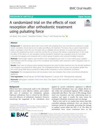
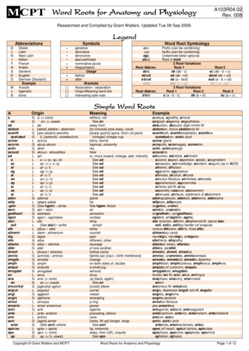
Root Resorption in Orthodontics: An Evidence-Based Approach4372011). It is therefore recommended to employ light forces, especially for intrusivemovements (Weltman et al., 2010).5. Case reportThe case described below illustrates an atypical situation, since with only four months oftreatment using alignment and leveling wires (0.14 and 0.16), a severe root resorption wasdetected. This situation led the orthodontist to stop the orthodontic treatment. Fortunately,the case had low complexity and did not involve extensive tooth movements. In such cases,the orthodontist should be aware of the systematic radiological examinations.Fig. 1. Initial situation of the patient.www.intechopen.com
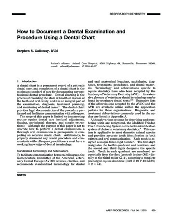
438Fig. 2. Panoramic radiograph.Fig. 3. (a, b). Alignament using wire 0.14.www.intechopen.comOrthodontics – Basic Aspects and Clinical Considerations
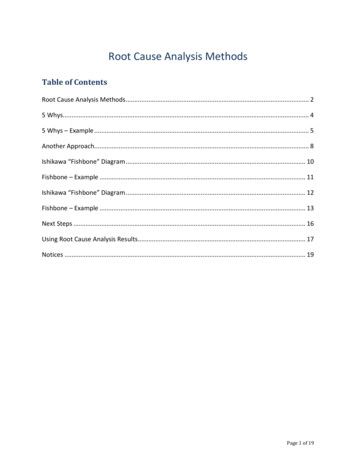
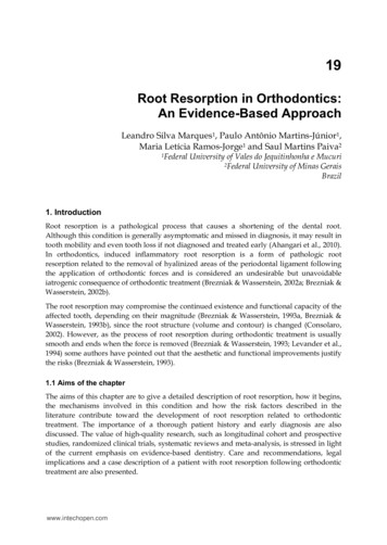
Root Resorption in Orthodontics: An Evidence-Based ApproachFig. 4. (a, b). Periapical radiographs showing root resorption of superior incisors.Fig. 5. (a, b). Final aspect of treatment.www.intechopen.com439
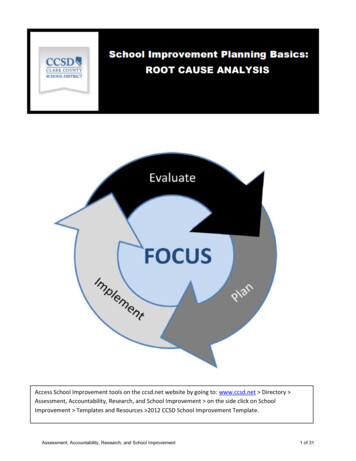
440Orthodontics – Basic Aspects and Clinical ConsiderationsFig. 6. Final panoramic radiograph.6. ConclusionsWhile science provides no consistent evidence for the precise identification of theorthodontic patient that will develop root resorption, orthodontists should keep in mind thevarious indicators known and promote systematic radiographic to monitor their patients.Individualize the diagnosis and treatment plan could meanthe difference between thesuccess and failure of orthodontic treatment.7. AcknowledgmentsThe authors thank the Coordenação de Aperfeiçoamento de Pessoal de Nível Superior(CAPES) for financial support to carry out this research.8. ReferencesAbass, S.K.; Hartsfield, J.K. Jr. (2007) Orthodontics and external apical root resorption. SeminOrthod, 13, 246-56.Abuabara, A. (2007). Biomechanical aspects of external root resorption in orthodontictherapy. Med Oral Patol Oral Cir Bucal, 12, 8, E610-3.Acar, A.; Canyurek, U.; Kocaaga, M.; Erverdi, N. (1999). Continuous vs. dis-continuous forceapplication and root resorption. Angle Orthod, 69, 159-63.Adachi, H.; Igarashi, K.; Mitani, H.; Shinoda, H.; (1994). Effects of topical administration of abisphosphonate (risedronate) on orthodontic tooth movement in rats. J Dent Res, 73,1478-86.Ahangari, Z.; Nasser, M.; Mahdia, M.; Fedorowicz, Z.; Marchesan, M.A. (201). Interventionsfor the management of external root resorption. Cochrane Database Syst Rev, Jun 16,6, CD008003.Al-Qawasmi, R.A.; Hartsfield, J.K. Jr, Everett, E.T. et al. (2003). Genetic predisposition toexternal apical root resorption in orthodontic patients: linkage of chromo-some-18marker. J Dent Res, 82, 356-60.www.intechopen.com
Root Resorption in Orthodontics: An Evidence-Based Approach441Andreasen, J.O. (1973). Cementum repair after apicoectomy in humans. Acta Odontol Scand,31, 211-21.Årtun, J.; Van 't Hullenaar, R.; Doppel, D.; Kuijpers-Jagtman, A.M. (2009) Identificati
19 Root Resorption in Orthodontics: An Evidence-Based Approach Leandro Silva Marques 1, Paulo Antônio Martins-Júnior 1, Maria Letícia Ramos-Jorge 1 and Saul Martins Paiva 2 1Federal University of Vales do Jequitinhonha e Mucuri 2Federal University of Minas Gerais Brazil 1. Introduction Root resorption is a pathological process that causes a shortening of the dental root.