Transcription
Wang et al. BMC Oral Health(2020) SEARCH ARTICLEOpen AccessA randomized trial on the effects of rootresorption after orthodontic treatmentusing pulsating forceJue Wang1, Ejvis Lamani1, Terpsithea Christou1, Peng Li2 and Chung How Kau1*AbstractBackground: An orthodontic device that moves teeth with pulsating force was invented and underwent a singlecenter, controlled, clinical trial to test its safety and efficacy for treatment. The device has a custom-made thermoplastic mouthpiece which fits over the teeth with an inflatable silicone element. A console that measures andcontrols the pulsating force level in real-time controls the air pressure that delivers a pulsating force. In this study,the effect of the device on root resorption during orthodontic treatment was evaluated using 3D cone beamcomputed tomography and compared with a control group of patients who received Invisalign treatment.Methods: Twenty-eight subjects were enrolled in the investigational arm and 15 in the control group. Subjectswere followed until the average score of the mandibular and maxillary teeth achieved a Little’s Irregularity Index of1.5 mm or less.Results: There were no adverse events reported throughout the study for either treatment arm. No clinically significantroot resorption was observed for either group. The investigational device did not cause root resorption greater thanthe control group. Both devices produced a safety profile compared to current orthodontic techniques.Conclusion: The investigational device did not produce more root resorption than similar conventional orthodonticappliances.Trial registration: ClinicalTrials.gov, NCT03421886. Registered 12 January 2018 - Retrospectively registered.Keywords: Radiographic evaluation, Root-crown ratios, Clear aligners, Tooth movement, Cone beam computedtomographyBackgroundEfforts have been continuously made in the orthodonticfield to provide a more effective, esthetic, and comfortable ways of treatment for orthodontic patients.Optimum orthodontic force is critical for the effectiveness and efficiency of orthodontic treatment. However,monitoring the magnitude of the orthodontic force* Correspondence: ckau@uab.edu1Department of Orthodontics, School of Dentistry, Univeristy of Alabama atBirmingham, Room 305, School of Dentistry Building, 1919 7th AvenueSouth, Birmingham, AL 35294, USAFull list of author information is available at the end of the articleapplied by current orthodontic appliances is difficult.Uncontrolled orthodontic force can collapse blood vessels within the periodontal ligament (PDL) leading toundesirable side effects. The cycle of PDL damage (hyalinized area formation and later undermining bone resorption) and repair is usually the main reason forprolonged orthodontic treatment and root damage.Both in vitro and in vivo studies have shown that fullycontrolled pulsating orthodontic force can shorten thetreatment period by potentially preventing the damageto capillary vessels within the PDL [1–4]. The effect ofintermittent force versus continuous force on the The Author(s). 2020 Open Access This article is licensed under a Creative Commons Attribution 4.0 International License,which permits use, sharing, adaptation, distribution and reproduction in any medium or format, as long as you giveappropriate credit to the original author(s) and the source, provide a link to the Creative Commons licence, and indicate ifchanges were made. The images or other third party material in this article are included in the article's Creative Commonslicence, unless indicated otherwise in a credit line to the material. If material is not included in the article's Creative Commonslicence and your intended use is not permitted by statutory regulation or exceeds the permitted use, you will need to obtainpermission directly from the copyright holder. To view a copy of this licence, visit http://creativecommons.org/licenses/by/4.0/.The Creative Commons Public Domain Dedication waiver ) applies to thedata made available in this article, unless otherwise stated in a credit line to the data.
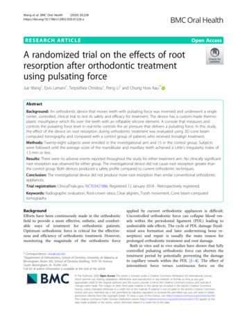
Wang et al. BMC Oral Health(2020) 20:238amount of molar movement was examined in rats [3].The results showed that 70% of the tooth movementwas achieved within 8 h of intermitted force (33.3% relative to continuous). In addition, previous study usingrats showed that an activation of force for 12 h with 12h rest produced similar tooth movement results to thoseachieved when force was applied the whole day [4]. It isalso indicated that there is a diurnal variation regardingthe tooth’s response to orthodontic force, and that theforce applied during the animal’s rest period may bemore effective than that applied during its active period.Notably, when pulsating force was applied to humanmaxillary molars, it is found that both the rate of movement and the total movement of the treated tooth wasgreater than that of the controlled tooth [1].Safety is always the first concern when introducing aninnovation into the medical field. Root resorption hasbeen known as an unavoidable side effect of orthodontictreatment and has degrees of variability. It has been shownthat the resorption in the majority of teeth is less than 2.5mm and differs in the range of 10% for different teeth [5–8]. In addition, the Malmgren index has classified severeroot resorption as more than 4 mm and 1–5% of the rootlength [9]. The severity of root resorption during orthodontic treatment varies largely, and is closely related tomultiple factors. Genetics, ethnicity, individual biologicalvariables, and mechanical factors are common factorsfound relevant [10–13]. Moreover, root morphology,tooth abnormalities, trauma, and severity of malocclusionalso play a part [14]. Notably, mechanical factors, such astype of appliances used, and magnitude and direction ofthe force applied, are associated with the severity of rootresorption during orthodontic treatment [15–17]. Thus,evaluating tooth root resorption is a critical part of assessing the safety profile of an orthodontic appliance.Due to its advantages of comfort and esthetics, clearaligners have become increasingly popular among patientsseeking orthodontic treatment [18, 19]. It also presents asa safe orthodontic appliance as it is found that the prevalence and severity of external root resorption in patientswith clear aligners were less than those in patients withfixed appliances [20, 21]. Thus, we designed the study toassess the external root resorption in patients treated withthe innovative device which delivers pulsating orthodonticforce through inflatable silicone element and compared itwith that in patients treated with clear aligner. Threedimensional cone beam computed tomography (CBCT)was used in the present study as a reliable way to measuretooth crown and root length [22, 23].MethodsPage 2 of 7Number F160418007). The study was designed as anopen label, two-arm study with concurrent, randomizedtreatment and control groups. The study was conductedat a single site, the Department of Orthodontics, University of Alabama at Birmingham.Subjects were derived from incoming patients seekingorthodontic treatment using clear aligners. When such apatient arrived, they were offered the opportunity to beenrolled in the trial. The protocol specified a sample sizeof 45 subjects with allocation ratio of 2:1 between thetreatments (30 active and 15 control). The investigational group received the orthodontic device that delivers pulsating force (Aerodentis System, DrorOrthodesign, Jerusalem, Isreal) while the control groupreceived clear aligners (Invisalign, Align Technology,California, USA). Subjects were followed for a total of upto 15 months or until achieving a Little’s index of 1.5,whichever came first.Subjects will be assigned to one ofthe treatment arms according to randomization list,which was generated using a computerized algorithm.Randomization codes were sealed in individual envelopes per patient. The envelopes were opened only whena single randomization occurred, in order to avoid bias.Patient recruitmentPrior to enrolling subjects in the study, the investigatororthodontist determined through screening if the subjectqualifies for the study and if the investigational device isan appropriate treatment option for the subject usingthe following enrollment criteria: 1) permanent dentition; 2) class l malocclusion with crowding of 6 mm orMild class II, class II subdivision; 3) good oral hygiene.The exclusion criteria for the study were as follows: 1)any medical or dental condition that could negativelyaffect study results during the expected length of thestudy; 2) subject is currently using any investigationaldrug or any other investigational device; 3) subject plansto relocate or move during the treatment period; 4) allergic to acetaminophen (use of aspirin or non-steroidalanti-inflammatory drugs is excluded for subjects whileon the study); 5) use of bisphosphonates (osteoporosisdrugs) during the study; 6) pregnant females; 7) subjectsthat are likely unwilling to be compliant with device use,as determined by the questionnaire for compliance. Allsubjects enrolled in the study met all the enrollmentcriteria.Investigational deviceThe investigational device [24] was recently approved byFood and Drug Administration of United States (FDA #:K192069). It consists of two main components (Fig. 1):Study designThe study was approved by the University of Alabama atBirmingham Institutional Review Board (IRB Protocol1. A thermo-plastic mouthpiece with an integrated inflatable silicone element. The mouthpiece is
Wang et al. BMC Oral Health(2020) 20:238Page 3 of 7Fig. 1 A thermo-plastic mouthpiece with an integrated inflatable silicone element and the consolespecifically designed for the subject and producedfrom orthodontic sheets by vacuum forming technology using CAD/CAM (computer-assisted design/computer assisted manufacturing) and 3Dimaging.2. A small easy-to-operate console with a user-friendlyinterface. It houses the electronics, air pump systemand pressure sensor that measures and controls theelectronic pulsating force level in real-time. An easyto use smart card monitors subject’s compliance intreatment.The inflatable element replaces the wires and bracketsof traditional metal braces. Movement is achieved by applying pneumatic force on the buccal or and lingual surfaces of the teeth being treated.Measuring root resorption by CBCTPrevious studies have shown that the CBCTs are goodsources to measure root resorption [25, 26]. AllCBCT images were taken using the Kodak 9300CBCT (Carestream Dental LLC, USA) machine. TheCBCT scan of a patient was taken with a voxel resolution of 0.3 mm. Each scan was saved in a Dicom fileformat. Two CBCT scans were taken (one at eachtime point) before orthodontic treatment (T1) and atthe completion of tooth alignment (T2). As the pulsating force of the investigational device only appliesto the maxillary and mandibular anterior teeth, measurements were performed on 12 teeth (maxillary andmandibular canines and incisors). All CBCT scanswere approved by IRB and consented by theparticipants.Crown and root lengths were measured and analyzedby using a method developed by Lind [27] and modifiedby Holtta et al. [28]. Midpoint was visually determinedalong a line bisecting the buccal and lingual cementoenamel junctions (CEJ) (Fig. 2). All the teeth measuredwere single-rooted teeth as only anterior teeth were included. Thus, each root was measured from the apex tothe corresponding midpoint. All crown heights weremeasured from the CEJ midpoints perpendicular to theincisal/occlusal reference line (formed tangent to incisaledge or buccal cusps). Root resorption for an individualtooth was assessed using the following formula:Crown f Root fCrown0EARREARR Ratio ¼Root 0EARR ¼ Root 0Where EARR is external apical root resorption, f isthe final measurement, and 0 is the baselinemeasurement.Teeth movement assessment (LII)Teeth movement was assessed by measuring Little’sIndex at 7 weeks and every 2 months thereafter. Toothmisalignment was measured in millimeters by means ofa caliper and Little’s Index was calculated.Fig. 2 Measurement of tooth crown length (blue line) and rootlength (red line) on CBCT
Wang et al. BMC Oral Health(2020) 20:238Page 4 of 7StatisticsThe sample size was calculated based on previous rootresorption studies with similar study designs [20, 29, 30].When the sample sizes in the groups are 15 and 30, atwo-group large-sample normal approximation test ofproportions with a one-sided 0.05 significance level willhave 80% power to reject the null hypothesis that thetest and the standard are not equivalent.The patients’ demographic and clinical characteristicswere summarized as mean and standard deviation (SD)for continuous variables or frequency and proportion forcategorical variables. EARR and EARR ratio were summarized at both tooth and patient level. The group comparison of EARR or EARR ratio at tooth level wasconducted using Wilcoxon test while patient level comparison was conducted with a random effect model totake the clustering effect (multiple teeth within one patient) into account. The false discovery rate (FDR) wasused for the multiple comparison correction. The correlation between LII and patient level EARR or EARR ratiowas evaluated using Spearman correlation coefficient.P 0.05 or FDR 0.05 given multiple comparison wasconsidered as statistically significant. All the analysis wasconducted using SAS 9.4 (Cary, NC).Root morphology measurements at T2 compared to T1The safety endpoint was the incidence of adverse events,whether or not treatment related, and the amount ofroot resorption, as measured by comparing x-rays beforeand at the end of tooth alignment. There were no adverse events reported for either the investigational orcontrol devices.The EARR for the investigation subjects (averagedacross all teeth) was 0.16 0.172 mm and 0.071 0.045mm for control subjects with resorption ranging from 0to 0.84 mm for the investigational group and 0.0–0.2mm for the control group (Table 2). After multiple testing correction, no significant difference was observed forall teeth. The analysis for EARR Ratio yielded similar results, with the root resorption ratio (averaged across allteeth) 1.238% ( 1.326%) in the investigational groupand 0.511% ( 0.359%) in the control group (Table 3). Inany event, the extent of root resorption is minor and notexpected to be clinically relevant as it is in the range ofnormal root resorption [26, 31] and less than 1 mm,which is the generally accepted limit.Correlation between LII and root resorptionResultsSubjectsA total of 36 investigational and 15 control subjects wereenrolled into the trial from August 2016 through June2018. However, 8 of the investigational subjects were excluded after consent as they chose not to participate further or did not conform to the compliance regime. Ofthe remaining 28 investigational and 15 control subjects,5 investigational subjects and 2 control subjects did notcomplete the trial. For the investigational subjects, failure to complete the treatment were due to balloon leakage issues, technical issues with the console, andmouthpiece retention issue. The subjects withdrew fromthe control group stated that they preferred fixed appliances. All subjects that remained in the trial successfullyachieved the primary endpoint. No subjects withdrew asa result of adverse events.Demographics of the participants were shown inTable 1. Subject age ranged from 12 to 65, with theTable 1 Baseline characteristics of participants in the twogroupsAerodentis (N 28)Control (N 15)30.6 12.032.3 16.4Male4 (14.3%)5 (33.3%)Female24 (85.7%)10 (66.7%)Age (Years) (Mean SD)average age being 31. The majority of subjects were female (86%), consistent with most aesthetic devices.Sex (N, %)LII was calculated for both maxilla and mandible for allthe patients; for the experiment group, this ranged from0.5–14.45 mm and, for the control group, 0.7–13.5 mm,which illustrates the degree of crowding. Positive correlation was found between LII and the severity of root resorption but not statistically significant, possibly due tosmall sample size (Table 4).DiscussionNo adverse events were reported throughout the studyfor either study arm, suggesting that the device was safefor clinical purpose. Although the mean EARR for theinvestigation subjects was larger than that for the controlgroup, the difference was not statistically significant. Asa result, the investigational device has a comparablesafety profile to the predicate from the perspective ofroot resorption.Both panoramic radiographs and CBCT were used inthe earlier studies to assess tooth root length [20, 21, 26,30]. Currently, CBCT has been widely used in orthodontic treatment as an important tool for determining impacted tooth location [32], facial asymmetry [33],supernumerary teeth, temporomandibular joint (TMJ)pathology [34], airway construction, as well as for surgical planning [35, 36]. Previous study has shown that thetooth length and root length measured by CBCT has nosignificant difference from the actual tooth length,
Wang et al. BMC Oral Health(2020) 20:238Page 5 of 7Table 2 Root resorption measurement: EARR change from baseline in the two groupsEARR (mean SD, mm)AerodentisControlP-valueFDRMaxillary right canine0.21 0.220.06 0.040.01770.1011Maxillary right lateral incisor0.12 0.110.08 0.030.2030.2436Maxillary right central incisor0.12 0.100.09 0.040.30940.3094Maxillary left central incisor0.11 0.160.04 0.020.11290.1694Maxillary left lateral incisor0.20 0.170.09 0.050.02920.1011Maxillary left canine0.13 0.130.09 0.060.26140.2852Mandibular right canine0.21 0.220.09 0.060.05130.1025Mandibular right lateral incisor0.14 0.160.07 0.040.13030.1737Mandibular right central incisor0.17 0.190.06 0.050.05610.1025Mandibular left central incisor0.13 0.090.07 0.040.03170.1011Mandibular left lateral incisor0.15 0.160.06 0.030.05980.1025Mandibular left canine0.23 0.260.07 0.060.03370.1011SD Standard deviations, FDR False discovery rateindicating that CBCT scan provides enough accuracy fortooth crown and root length measurement [22].Tooth root resorption related to orthodontic treatment mainly occurs in the anterior teeth [37, 38]. Sincethe inflatable silicone element in the mouthpiece of theinvestigational device extends only in the anterior region, root measurements were made to the area of anatomy. Previous studies on tooth root resorption alsofocused on the anterior teeth for practical purpose, aswell as for better accuracy of measurement [37, 39].In the present study, both tooth root length and crownheight were measured and the formula for EARR andEARR ratio were used. This method of measurementwas used according to previous studies [39, 40] andchosen specifically to avoid any magnification errorwhen analyzing CBCT scans. The formula was used onthe basis that tooth crown height remains the same before and after the treatment. As a result, the differenceof root length between the two time points representedthe true root resorption amount.From previous studies, it is generally considered thatEARR of 1 mm or more during any 12-month period ofactive treatment is clinically significant [41, 42]. In thepresent study, the mean EARR for the investigation subjects was 0.16 0.17 mm and was 0.07 0.05 mm forcontrol subjects. Thus, the extent of root resorption forboth groups were not clinically significant. Previousstudy has reported that the degree of EARR for fixed appliances is about 1.67 0.64 mm for maxillary central incisors [40]. Another recent study has shown an averageof 1.12 1.34 mm root resorption for fixed appliance[20]. Thus, our data is in accordance with the previousstudy that the severity of EARR in patients using clearaligners wa
to use smart card monitors subject’s compliance in treatment. The inflatable element replaces the wires and brackets of traditional metal braces. Movement is achieved by ap-plying pneumatic force on the buccal or and lingual sur-faces of the teeth being treated. Measuring root resorption by