Transcription
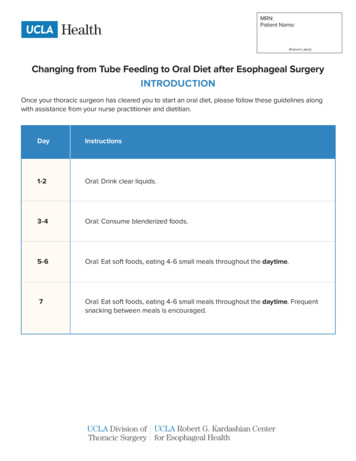
MOH NURSING CLINICAL PRACTICE GUIDELINES 1/2010Nursing Management ofNasogastric Tube Feedingin Adult PatientsJuly 2010
STATEMENT OF INTENTThis set of guidelines serves as a guide for caregivers of adults withnasogastric tube feeding.The recommendations are based on the available research findings andexisting evidence-based guidelines. However there are some aspects inwhich there are insufficient published researches and therefore, consensusof expert in the field has been utilized to provide guidelines specific toconventional practice.As practitioners, each individual must exercise clinical judgement in thenursing management of patients with nasogastric tube feeding. Therefore,these guidelines should be implemented according to individual patient’scondition, overall treatment goal, resource availability, institutional policiesand treatment options available.Copyright 2010 by Ministry of Health, Singapore.i
FOREWORD“Every careful observer of the sick will agree in this that thousands of patientsare annually starved in the midst of plenty, from want of attention to the wayswhich alone make it possible for them to take food.”Florence Nightingale, 1859Nurses have recognised the importance of meeting patients’ nutritional needssince the beginning of our profession. Enteral nutrition is the preferredfeeding method for those patients with normal functioning gastrointestinaltracts but with inability to ingest adequate nutrients solely by mouth.Significant advances and changes have since evolved in nutritional care ofhospitalised patients. Enteral nutrition as compared to parenteral feeding, isconsidered to be safer and less expensive.Nurses have always assumed the role of providing nutritional therapy topatients. They would find the publication of this clinical practice guidelinestimely and useful to guide them in enteral nutrition therapy. Adhering closelyto these guidelines would significantly reduce and, in many instances,prevent complications commonly associated with enteral feeding.PAULINE TAN CJCHIEF NURSING OFFICERii
CONTENTS1 2 INTRODUCTION1 1.1 Background1 1.2 Definition of Nasogastric Tube Feeding1 1.3 Highlights of Patient Management2 1.4 Scope of the Guidelines2 DEVELOPMENT OF GUIDELINES3 2.1 Training and Guidelines3 2.2 Strategy and Literature Review3 2.3 Evaluation of Evidence and Grading of Recommendations3 2.3.1 Individual Study Validity Rating4 2.3.2 Levels of Evidence5 2.3.3 Grade of Recommendation6 2.3.4 Interpretation of the D/4 Grading6 2.4 Guideline Review and Revision7 2.5 Limitations7 3 ALGORITHM FOR MANAGEMENT OF NASOGASTRIC TUBEFEEDING8 4 SELECTION OF NASOGASTRIC TUBES9 4.1 9 5 Feeding Tube SelectionNASOGASTRIC TUBES PLACEMENT AND CARE10 5.1 Tube Insertion and Stabilization10 5.2 pH Testing10 iii
6 7 8 9 5.3 Radiological Determination of Feeding Tube Placement12 5.4 Auscultatory Method13 5.5 Frequency in Checking Placement14 5.6 Tube Clogging14 5.6.1 Maintaining Tube Patency14 ADMINISTRATION OF NASOGASTRIC TUBE FEEDING16 6.1 Preparation of Formula Feeds and Delivery System16 6.2 Feeding Position17 6.3 Bolus Feeding17 6.4 Continuous Feeding18 6.5 Prevention of Diarrhoea19 ADMINISTRATION OF MEDICATIONS21 7.1 Administration of Medications21 MONITORING AND MANAGEMENT23 8.1 23 Monitoring and Management of Gastro-intestinal ToleranceCOMPLICATIONS25 9.1 Management of Feeding Intolerance25 9.2 Declogging25 9.3 Diarrhoea26 9.3.1 Assessment / Monitoring26 9.3.2 Treatment of Diarrhoea26 9.4 Aspiration Pneumonia27 9.5 Feeding Devices28iv
10 QUALITY ASSURANCE29 10.1 Parameters for Evaluation29 10.1.1 Incidence of misplaced tubes29 10.1.2 Incidence of aspiration as a result of nasogastric tube feeding 29 10.2 Sentinel Events29 10.3 Management Role30 11 IMPLEMENTATION OF GUIDELINES31 12 REFERENCES32 13 GLOSSARY36 14 WORKGROUP MEMBERS39 APPENDIX 1NASOGASTRIC TUBE ANCHORING TECHNIQUE41 APPENDIX 2 ALGORITHM FOR ASSESSING PLACEMENT OF THENASOGASTRIC TUBE42 APPENDIX 3SELF ASSESSMENTv43
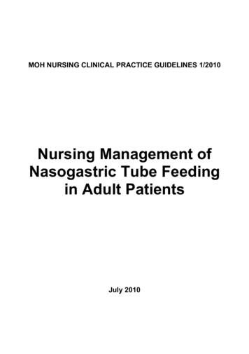
1INTRODUCTIONFigure 1. Schematic picture of nasogastric tube1.1BackgroundPatients in the hospital, as well as home care settings, often requirenutritional supplementation with enteral feeding. Enteral feeding canbe administered via nasogastric, nasoduodenal and nasojejunalmeans. The focus of this clinical practice guideline is on the nursingmanagement of nasogastric tube feeding.Nasogastric tube feeding may be accompanied by complications.Thus, it is important for the practitioner to be aware of how to preventthese complications so that nasogastric tube feeding can beadministered successfully and safely.1.2Definition of Nasogastric Tube FeedingNasogastric tube feeding is defined as the delivery of nutrients fromthe nasal route into the stomach via a feeding tube.1
1.3Highlights of Patient ManagementPatients and their caregivers are important team players in theeffective management of nasogastric tube feeding. The practitionershould: encourage patients and their caregivers to be active participantsin their care develop an effective plan of care that is consistent with thepatient’s goalsThe recommendations focus on: implementation of nasogastric tube feeding monitoring and management of nasogastric tube feeding management of complications1.4Scope of the GuidelinesThis set of guidelines is intended as a simple and readable referencefor caregivers of adults with nasogastric tube feeding.These clinical practice guidelines are tools for assisting clinicaldecision making with regard to the nursing care given to a patientwith a nasogastric tube. They are the result of systematicidentification and synthesis of published research findings and expertopinions. However, they should be adapted locally to suit a particularsituation and patient. It is the intention of the workgroup not to beencyclopaedic in coverage but to produce a concise, readable andpractical format that addresses key topics which can be used as areference and training tool.2
22.1DEVELOPMENT OF GUIDELINESTraining and GuidelinesMembers of the workgroup attended a two-day interactive trainingworkshop to learn about and discuss the theory and practical issuesof developing evidence-based guidelines under the guidance of DrEdwin Chan and Dr Miny Samuel of the then Clinical Trials andEpidemiology Research Unit. The practical training revolved aroundtopic selection and the development of “mock” evidence-basedguidelines which developed into this present set of guidelines.2.2Strategy and Literature ReviewThree highly regarded evidence-based guidelines were reviewed:nd The A.S.P.E.N. Nutrition Support Practice Manual (2 Edition),The American Society for Parenteral and Enteral Nutrition. 2005[ASPEN, 2005] Insertion and management of Nasogastric Tubes for Adults. TheJoanna Briggs Institute: Systematic Reviews, 2006 [JBI, 2006] A.S.P.E.N. Enteral Nutrition Practice Recommendations. 2009[ASPEN, 2009]The members felt that an updated literature search on the specifictopics addressed would suffice. The electronic databases (MEDLINE,EMBASE, Cochrane Library, SPRINGNET and CINAHL) and hardcopies of relevant journals (Journal of Parenteral and EnteralNutrition, Hospital Pharmacy, Critical Care Nurse, American Journalof Critical Care, Medical Surgical Nursing, Journal of Clinical Nursing,Consultant Pharmacist, Gastroenterology Nursing, NursingResearch, Heart and Lung) were searched.2.3Evaluation of Evidence and Grading of RecommendationsThe revised Scottish Intercollegiate Guidelines Network (SIGN)system was adopted as a guide to evaluate the design of individualstudies and assign a grade to each study’s level of evidence. Therecommendation was given a grade after taking into account theexternal validity, result consistency, local constraints and expert3
opinion.The extensive reliance on the ASPEN and JBI guidelines areacknowledged and treated as our main published expert opinion. Forareas where available evidence is inconsistent or inconclusive,recommendations were made based on the clinical experience andjudgement of the workgroup or expert committee reports.2.3.1Individual Study Validity RatingAll primary studies and reviews addressing a particular topic wereappraised using a SIGN checklist appropriate to the study's design.These were individually rated for internal validity using the systembelow:RatingDescription All or most of the criteria have been fulfilled. Where theyhave not been fulfilled the conclusions of the study orreview are thought very unlikely to alter. Some of the criteria have been fulfilled. Those criteria thathave not been fulfilled or not adequately described arethought unlikely to alter the conclusions.–Few or no criteria fulfilled. The conclusions of the studyare thought likely or very likely to alter.4
2.3.2Levels of EvidenceEach study is assigned a level of evidence by combining the designdesignation and its validity rating using the system below:LevelType of Evidence1 High quality meta-analyses, systematic reviews of RCTs, orRCTs with a very low risk of bias.1 Well-conducted meta-analyses, systematic reviews, orRCTs with a low risk of bias.1-Meta-analyses, systematic reviews, or RCTs with a highrisk of bias.2 High quality systematic reviews of case-control or cohort orstudies.High quality case-control or cohort studies with a very lowrisk of confounding or bias and a high probability that therelationship is causal.2 Well-conducted case-control or cohort studies with a lowrisk of confounding or bias and a moderate probability thatthe relationship is causal.2-Case-control or cohort studies with a high risk ofconfounding or bias and a significant risk that therelationship is not causal.3Non-analytic studies e.g. case reports, case series.4Expert opinion.5
2.3.3Grade of RecommendationThe detailed results of each study and mitigating local circumstanceswere considered in formulation of each recommendation which wasthen graded using the system below:GradeRecommendationAAt least one meta-analysis, systematic review, or RCT ,rated as 1 , and directly applicable to the targetpopulation; orA body of evidence, consisting principally of studies rated as 1 , directly applicable to the target population,and demonstrating overall consistency of results.BA body of evidence, including studies rated as 2 ,directly applicable to the target population, anddemonstrating overall consistency of results; or Extrapolated evidence from studies rated as 1 or 1 . A body of evidence including studies rated as 2 ,directly applicable to the target population anddemonstrating overall consistency or results; or Extrapolated evidence from studies rated as 2 .CD2.3.4 Evidence level 3 or 4 ; or Extrapolated evidence from studies rated as 2 .Interpretation of the D/4 GradingThe grading system emphasises the quality of the experimentalsupport underpinning each recommendation. The grading D/4 wasassigned in cases where it would be unreasonable to conduct a RCT because the correctpractice is logically obvious; recommendations derived from existing high quality evidencebased guidelines. We alert the user to this special status byappending the initials of their source e.g. D/4 - ASPEN, 2005.6
2.4Guideline Review and RevisionDrafts of the guidelines were circulated to healthcare institutions forpeer review on validity, reliability and practicality of therecommendations.These guidelines will be reviewed and revised periodically toincorporate the latest relevant evidence and expert clinical opinion.2.5LimitationsThese guidelines offer recommendations that are based on currentscientific evidence and professional judgement. They are notintended as the legal standard of care.Users of these guidelines should determine the appropriate and safepatient care practices, based on assessment of the circumstances ofthe particular patient, their own clinical experiences and theknowledge of the most recent research findings.7
3ALGORITHM FOR MANAGEMENT OF NASOGASTRIC TUBEFEEDING8
44.1SELECTION OF NASOGASTRIC TUBESFeeding Tube SelectionSelect the feeding tubes based on the tube’s composition, intendeduse, estimated length of time required, cost-effectiveness and tubefeatures. Soft, flexible, small diameter tube (8 Fr to 12 Fr) is recommendedfor nasogastric feeding.(D/4 - ASPEN, 2005 & JBI, 2006) Use Polyurethane or silicone tubes for anticipated long termfeeding rather than polyvinylchloride tubes.(D/4 - ASPEN, 2005) Polyvinylchloride (PVC) tubes should be used for a short periodof time usually for gastric drainage, decompression, lavage ordiagnostic procedures.(D/4 - ASPEN, 2005)Rationale:Smaller size feeding tube improves patient comfort. Commoncomplications associated with the use of larger and stiffer tubesinclude nasopharyngeal erosions / necrosis, sinusitis and otitismedia.(Kirby and Opilla, 2005 as cited in ASPEN, 2005)Polyurethane or silicone tubes are better for long-term ( 4-8 weeks)use because they are more flexible and less irritating to tissues.(Kirby and Opilla, 2005 as cited in ASPEN, 2005)PVC feeding tubes are used for short term duration ( 3 weeks) andthey tend to harden and become brittle with time and may causetissue irritation or necrosis.(Kirby and Opilla, 2005 as cited in ASPEN, 2005)For short-term usage, PVC feeding tubes have adequate efficacyand are more cost effective.(Kirby and Opilla, 2005 as cited in ASPEN, 2005)9
55.1NASOGASTRIC TUBES PLACEMENT AND CARETube Insertion and Stabilization Approximate the length of the tube to be inserted by measuringfrom the tip of nose to the earlobe, and then from the earlobe tothe xiphoid process.(D/4 - ASPEN, 2005) Secure the nasogastric tubes (NGT) to the nose with a trousertape. (See Appendix 1 for ‘Nasogastric Tube AnchoringTechnique’.)(D/4)Rationale:Inserting the tube of length measuring from the tip of nose to theearlobe and to the xiphoid process will ensure that the distal endreaches the stomach.(Kirby and Opilla, 2005 as cited in ASPEN, 2005)Stabilizing a tube can reduce the risk of tube displacement (ASPEN,2005). This method increases tape adherence and decreases skinirritation by avoiding excess pressure on the nostril.(Rovinski and Zastocki, 1989)5.2pH Testing A combination of aspirate appearance and pH testing can beused to help make correct predictions about tube placement inthe stomach.(B/1 )10
pH value of aspirates from feeding tubes should be used todifferentiate between gastric and respiratory placement. The pHvalues and their corresponding indications and action are asfollows:ValuepH less than 5IndicationsGastricActionProceed to feedpH 5 – 6Check visualcharacteristics ofaspiratesIf visual characteristicsindicate gastricaspirates, proceed tofeed. Otherwise, docheck x-ray to confirmtube placementpH more than 6Intestinal orRespiratoryDo check x-ray toconfirm tube placement(D/4) Visual characteristics of feeding tube aspirates:GastricMay be grassy greenwith sediment, brown(if blood is presentand has been actedon by gastric acid)May also appearclear and colourless(often with shreds ofoff-white to tanmucus or sediment)IntestinalGenerally moretransparent thangastric aspirates andmay appear bilestained, ranging incolour from light todark golden yellowor brownish-greenRespiratoryTracheobronchealsecretion mayconsist of off whiteto tan sediment(D/4)11
Rationale:Combination of pH and visual characteristics can be helpful indistinguishing between respiratory and gastrointestinal tube position.(Metheny, Reed, Berglund and Wehrle 1994;Metheny and Titler, 2001; Metheny and Stewart, 2002)The pH value can offer correct predication of gastric versusrespiratory placement.(Metheny, Wehrle and Wiersema, 1998)Gastric pH values are lower than that of intestinal or respiratory pH.(Metheny and Titler, 2001; Metheny and Stewart, 2002)pH less than 5 indicates gastric placement, whereas a pH more than5 indicates intestinal or respiratory placement.(Metheny, 1993; Metheny and Stewart, 2002)Patient receiving H2 receptor antagonist or with recent alkaline refluxfrom the intestine may have elevated gastric pH. Proton PumpInhibitors and Histamine receptor blocking agents, such asOmeprazole and Famotidine, fail to decrease gastric pH to below 6.5but tends to elevate gastric pH.(Metheny, Reed, Wiersema, Mcsweeney, Wehrle and Clark, 1993)5.3Radiological Determination of Feeding Tube PlacementRadiological confirmation of tube positioning is recommended underthe following conditions: Aspirate pH 5-6 and visual characteristics not indicative of gastricor intestinal aspirate Aspirate pH 6 No aspirate despite instituting other measures(See Appendix 2 for algorithm for assessing placement of thenasogastric tube to confirm initial tube placement)(D/4 - JBI, 2006)12
Rationale:Radiography determination of tube location is the most accuratemethod of checking tube placement.(Grifffiths et al, 2006 as cited in JBI, 2006)X-ray can visualise the entire course of the tube.(Metheny and Titler, 2001)5.4Auscultatory Method Auscultatory method (also known as air insufflations test) shouldnot be relied on as the sole method to determine the location ofthe feeding tube.(D/4 - ASPEN, 2005 & JBI, 2006)Rationale:The auscultatory method is not effective in distinguishing betweenrespiratory and gastrointestinal placement of feeding tube.“Pseudoconfirmatory gurgling” sound can be heard when feedingtubes were positioned in the tracheobronchial tree or pleural spaces.A tube that is inadvertently placed in the respiratory tract oroesophagus can transmit a sound similar to that of air entry in thestomach.(Metheny, 1993; Metheny and Titler, 2001; Griffiths, Thompson,Chau and Fernandez, 2006 as cited in JBI, 2006)A positive air insufflations test combined with aspiration of gastriccontents is a fairly reliable predictor of successful placement.(Kirby and Opilla, 2005 as cited in ASPEN, 2005;Griffiths et al, 2006 as cited in JBI, 2006)13
5.5Frequency in Checking PlacementMark the intersection where the nasogastric tube enters the nostril,use this marking to check the tube placement: after initial insertion, before each intermittent feeding, and atevery 8-hourly during continuous feedings.(D/4) if patients complain of discomfort, coughing, retching orvomiting and show sudden signs of respiratory difficulties.(D/4) if the visible part of the tube changes in the length.(D/4)Rationale:Regular assessment of feeding tube placement is important becausea tube can be partially pulled out during movement or when tugged atby a confused patient. Tube malposition may be caused by faultyinitial placement or upward dislocation after bouts of coughing orvomiting.(Metheny and Stewart, 2002)5.6Tube Clogging5.6.1Maintaining Tube Patency Flush feeding tubes with 30 ml of water before and afterintermittent feeding, every 4-hourly during continuous feedingand after checking for gastric residuals. More frequent flushingmight be ordered according to patient’s condition.(B/1 ) Flush feeding tube before and after administration of eachmedicine and after checking for residual.(C/2 )14
Rationale:Flushing enteral tubes with water have been found to preventclogging and restore the patency of partially obstructed tubes. Waterwas found to be the most effective flushing solution in maintainingtube patency. However, the patient’s medical condition needs to beconsidered before deciding on the volume of water and frequency forirrigation.(ASPEN, 2009; Mateo, 1994)Compatibility of enteral formula and medications affect drugconcentration and causes clog formation. Irrigating tubes before andafter medication administration resulted in fewer clogged tubes.(Mateo, 1994)Residual, containing gastric aspirate with an average pH of 4.6 orless interacts with protein formula to form clots. Formula residue thatadheres to the tube lumen may cause tube clogging. Thus flushingthe tube with water will maintain the patency by eliminating acidprecipitation of formula in the feeding tube.(Mateo, 1994)15
66.1ADMINISTRATION OF NASOGASTRIC TUBE FEEDINGPreparation of Formula Feeds and Delivery System Wash hands before preparing and handling delivery sets. Use mask if handler has a cold, sore throat or upper respiratorytract infection.(D/4) Use clean technique when handling the feeding system. Limit thehang time of the formula to: 4 hours for powdered, reconstituted formula and enteralnutrition formula with additives 8 hours for sterile, decanted formula in open system 24–48 hours per manufacturer’s guidelines for closed-systementeral nutrition formula(D/4 – ASPEN, 2009) Use pre-prepared feeds for enteral feeding. Initiate all formulas at full strength.(D/4)(D/4)(A/1)Rationale:Handwashing reduces the risk of contamination and cross infection.(Kennedy, 1997)The use of mask has shown to reduce the risk of cross-infection.(Kennedy, 1997)Bacterial contamination is most often due to contamination of aproduct. It has been found that colonization of distal administrationset by enteric bacteria present in the enteral tube hub leads to aneasily accessible reservoir of gastrointestinal organisms.(Eisenberg, 2002)16
Pre-prepared feeds reduce the risk of cross - contamination andcross infection.(Kennedy, 1997)As new formulas and technology develop, it is recognized that theosmolality of formula does not cause diarrhoea and the dilution of theformula to or strength is not necessary.(Eisenberg, 2002)6.2Feeding Position The patient should be placed at a semi-recumbent position oroelevated to an angle of at least 30 during and after feeding for atleast one hour.(D/4 - ASPEN, 2005)Rationale:0Elevation of the head of bed at least 30 during tube feedingdecreases the risk of aspiration of gastric content.(Lord and Harrington, 2005 as cited in ASPEN, 2005)Gravity reduces the likelihood of regurgitation of gastric contentsfrom the distended stomach. There is evidence that a sustainedsupine position (with the head of the bed flat) increasesgastroesophageal reflux (GER) and the probability for aspiration.(Metheny and Stewart, 2002)6.3Bolus Feeding The total volume of feeds administered should not exceed 400mlduring each bolus feed.(D/4)17
Rationale:Both the speed and volume with which formula is delivered has animpact on intragastric pressure and the probability of gastrooesophageal reflux (GER).(Metheny, 2002)Bolus feeding of more than 400 ml and rapid infusion may result inabdominal distension and discomfort.(Marian and Allen, 1998)6.4Continuous Feeding Use feeding pump for administration of continuous feeds. Startwith an initial slow rate of 10 to 40 ml/hour. Advance to the goalrate by increasing the rate by 10 to 20 ml/hour every 8-12 hoursas tolerated. (D/4 - ASPEN, 2005) Continuous feeding is recommended for patients who arecritically ill, receiving jejunal feedings; or patients who are unableto tolerate intermittent feedings. Pump-assisted delivery ofenteral feeds is highly recommended for small-bowel feeding.(D/4 - ASPEN, 2005)Rationale:Continuous feeding minimizes the amount of formula in the stomachat any given time. Less abdominal discomfort occurred duringcontinuous feedings compared with intermittent feedings.(Lord and Harrington, 2005 as cited in ASPEN, 2005)There is evidence that aspiration is less likely with continuousfeedings because this method minimizes the amount of formula inthe stomach at any given time.(Metheny, 2002)18
Pump-assisted continuous feeding helps to prevent gastrointestinalcomplications associated with rapid infusion. The pump usuallydelivers the feed much more accurately than the gravity method andis likely to reduce gastric intolerance(Lord and Harrington, 2005 as cited in ASPEN,2005;Metheny, 2002)6.5Prevention of Diarrhoea Administration sets and containers should be discarded every 24hours.(D/4) Feeds should not be decanted before use. Limit the hang time of the formula to: 4 hours for powdered, reconstituted formula and enteralnutrition formula with additives 8 hours for sterile, decanted formula in open system 24–48 hours per manufacturer’s guidelines for closed-systementeral nutrition formula(D/4 – ASPEN, 2009)(D/4)Rationale:Continuous infusion raises gastric pH and promotes bacteriaovergrowth. Bacteria can spread up the giving set from gastric orenteral sources and thus increase the risk of contamination.(Stroud, Duncan and Nightingale, 2003)Enteral feed is an ideal culture medium for bacteria growth and oncecontaminated, bacteria will multiply rapidly.(Stroud et al, 2003)19
Limiting the hang-time of formula helps to reduce the potential forbacterial contamination of formula feeds.(Bankhead, Boullata, Brantley, Corkins, Guenter, Krenitsky,Lyman, Methany, Mueller, Robbins, Wessel, and theA.S.P.E.N. Board of Directors, 2009 in ASPEN 2009)20
77.1ADMINISTRATION OF MEDICATIONSAdministration of Medications Consult pharmacist to evaluate each patient’s medication profilebefore medications are given via enteral tube.(D/4) Enteric-coated and sustained release medications should not becrushed.(D/4 - ASPEN, 2005) Medications administered through nasogastric tube shouldpreferably be in liquid form.(D/3 - ASPEN, 2005) Flush feeding tube with water prior to medication administration.(B/2 ) Do not administer sublingual and buccal medications via feedingtube.(D/4) Medications should not be added directly to the enteral formulaor into the enteral feeding bag.(D/4)Rationale:Every dosage form (tablet, capsules, immediate release, extendedrelease, enteric coated, elixirs, syrups, granules) has the potential tocause incompatibilities, complications, or intolerance.(Lord and Harrington, 2005 as cited in ASPEN, 2005)21
Pharmaceutical incompatibilities occur when there is an alteration inthe drug form which interferes with drug efficacy, potency, ortolerance. Crushing of enteric–coated medications can inducepharmaceutical incompatibilities.(Guenter, 1999)Medications should be evaluated by the pharmacist to determine ifthe form of medication needs to be changed. Liquid medicationsdecrease the risk of tube clogging and increase absorption of themedication.(Pancorbo-Hidalgo, Garcia-Fernandez and Ramirez-Perez, 2001;Guenter, 1999)Hyperosmolar liquid medications have the potential to causediarrhoea, cramping, and increased gastric residuals.(Guenter, 1999)Flushing the feeding tube prior to drug administration removes anyenteral feeds that remain in the tube to prevent drug nutrientinteraction.(Maka and Murphy, 2000)Sublingual and buckle medications are designed to be absorbed intothe systemic circulation by placement under the patient’s tongue or inthe cheek pouch. Thus, these should not be administered via thefeeding tube.(Lord and Harrington, 2005 as cited in ASPEN, 2005)Adding medications into formula feeds may result in tube cloggingand cause undesirable effects or incompatibilities.(Lord and Harrington, 2005 as cited in ASPEN, 2005)22
88.1MONITORING AND MANAGEMENTMonitoring and Management of Gastro-intestinal Tolerance Gastric residual volume (GRV) should be checked 4-8 hourly incontinuously fed patients and before each intermittent feeding.(B/1) GRV greater than 200 ml should prompt careful bedsideevaluation and initiation of appropriate feeding method andfeeding volume. GRV reading should be evaluated in conjunctionwith physical examination for abdominal distension, absence ofbowel sounds, and presence of nausea and vomiting.(D/4) A trend in GRV may be more important than an isolated highlevel of GRV.(D/3) There is no significant difference between returning and notreturning gastric residuals.(D/3)Rationale:Gastric residuals are more likely to be high during the early phase oftube feedings; therefore it is necessary to monitor residuals moreclosely during that period.(Metheny, 1993)GRV greater than 200 ml was associated with physical findings ofgastrointestinal dysmotility. Bedside evaluation and careful initiationof feeding method may reduce aspiration risk.(Metheny, Schallom and Edwards, 2004)23
GRV volume during intra-gastric feedings is determined by thebalance between the amount of infused formula feeds plus theendogenous secretions of saliva and gastric juice, less the amount offluid emptied from the stomach. This amount varies betweenindividuals. Thus, it is important to note for changes in the trend.(Metheny et al, 2004)Discarding gastric residual may contribute to loss of gastric juicesand subsequently may lead to electrolyte imbalance, howeverreturning the gastric residual may cause potential contamination andtube clogging.(Booker, Niedringhaus, Eden and Arnold, 2000)24
99.1COMPLICATIONSManagement of Feeding IntoleranceWhen patient shows signs of feeding intolerance such as nausea,vomiting, abdominal distension and pain: Perform a physical examination of the abdomen includingassessment for presence of abdominal pain and bowel sounds.(D/3) Feeding should only be stopped abruptly for those patients whodemonstrate overt regurgitation or aspiration.(D/3)Rationale:Presence of nausea, vomiting, abdominal pain, distension, flatus,stool and bowel sounds, and abnormal abdominal X-ray may indicatetube-feeding intolerance.(McClave, Snider, Lowen, McLaughlin, Greene, McCombs,Rodgers, Wright, Roy, Schumer and Pfeifer, 1992 and McClave &Snider, 2003)Inappropriate cessation of feeding may contribute to inadequatecaloric intake and may not be physiologically sound.(McClave et al, 1992)9.2Declogging Use warm water to declog obstructed feeding tubes. Ifunsuccessful, pancreatic enzyme with sodium bicarbonate maybe used.(D4 – ASPEN, 2005)25
Rationale:Warm water is the most effective irrigant in declogging blockedfeeding tubes. However, a solution of digestive enzymes(pancrealipase) mixed with sodium bicarbonate (to activate theenzyme ) has been shown to be fairly effective in dissolving formulaocclusions.(Lord and Harrington, 2005 as cited in ASPEN, 2005; Reising andNeal, 2005; Wilson and Haynes-Johnson, 1987)9.3Diarrhoea9.3.1Assessment / Monitoring Patients on antibiotics should be monitore
iv 5.3 Radiological Determination of Feeding Tube Placement 12 5.4 Auscultatory Method 13 5.5 Frequency in Checking Placement 14 5.6 Tube Clogging 14 5.6.1 Maintaining Tube Patency 14 6 ADMINISTRATION OF NASOGASTRIC TUBE FEEDING 16 6.1 Preparation of Formula Feeds and Delivery System 16 6.2 Feeding Position 17 6.3 Bolus Feeding 17 6.4 Continuous Feeding 18