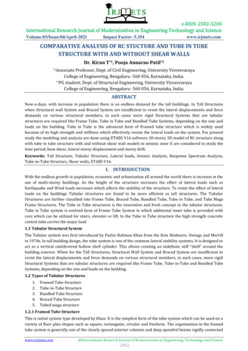
Transcription
Best Practices:Chest Tube ManagementChest tube care: The more you know, the easier it getsCaring for patients with chest tubes can be daunting. This article helps make it less intimidating.By Mark Bauman, MSN, RN, CCRN, and Claudia Handley, MS, RN, MBAMany nurses find chest tube care intimidating—but it doesn’t have to be. Once you understand the basics, you can be confident when caring for patientswho have chest tubes.The practice of using a cannula to drain air or fluid from the pleural space dates back to antiquity. It’s one element in the trinity of life-saving medicalprocedures. (The others are endotracheal intubation and venous cannulation.) Hippocrates and Celsus recorded using hollow tubes to drain loculatedempyemas. By the 1800s, catheters frequently were used to drain and irrigate empyematous cavities.It’s all about negativityA brief review of pulmonary anatomy and physiology helps you understand where chest tubes are placed and how they work. Chest tubes aren’t placed inthe lungs but in the pleural space—a potential rather than actual space between the parietal and visceral pleurae. The parietal (outer) pleura covers the chestwall and diaphragm. It contains a small amount (about 50 mL) of serous fluid that coats the opposing surfaces, allowing the visceral and parietal pleuraeto glide over each other without friction while enabling the pleural surfaces to adhere to each other. Think of two glass plates with a thin coating of water;when you place the second piece of glass atop the first, the two plates slide smoothly. But when you try to separate them, they stick together.
The ability to adhere creates negative pressure within the pleural space, which becomes more negative as the visceral and parietal pleurae are pulled inopposite directions during inspiration. (Picture those two glass plates.) The negative intrapleural (and thus intrapulmonary) pressure generated causes airto flow from positive (atmospheric) pressure into the lungs. Expiration increases intrapleural and intrapulmonary pressures to the point where they exceedatmospheric pressure, creating an opposite pressure differential and causing air to flow out of the lungs into the surrounding atmosphere.A breach in pleural integrity creates a separation between the parietal and visceral pleurae, allowing air or fluid to fill this potential space. (Using the glassplate analogy, the two plates have become separated). The visceral pleura collapses inward along with the lungs, while the parietal pleura recoils outwardalong with the chest wall.Indications for chest tubesChest tubes are used to treat conditions that disrupt the pleural space. The body can absorb small volumes of fluid or air over time. But larger volumes limitlung expansion, causing respiratory distress. In extreme cases, a tension pneumothorax may develop. This condition occurs when injured tissue forms aone-way valve or flap, enabling air to enter the pleural space and preventing it from escaping naturally. Seen mainly with thoracic trauma and line placement, this condition rapidly progresses to respiratory insufficiency, cardiovascular collapse, and ultimately death if unrecognized and untreated. It requiresimmediate live-saving treatment by inserting a needle to relieve pressure (needle thoracentesis), followed by chest-tube insertion. (See Conditions thatdisrupt the pleural space.)Chest tubes also may be used to prevent or mitigate postoperative complications. For example, after cardiac surgery or chest trauma, one or more chesttubes may be inserted in the mediastinum to drain blood and prevent cardiac tamponade. In addition, for proper chest tube care, chest tubes can be usedto instill fluids into the pleural space, such as chemotherapy drugs or sclerosing agents to treat recurrent pleural effusions (a procedure called pleurodesis).Also, blood collected from chest tubes may be used for autotransfusion.(See Autotransfusion: Risks, benefits, and nursing care)Managing pleural-space disruptionsThe overall goal of chest-tube therapy (chest tube care) is to promote lung reexpansion, restore adequate oxygenation and ventilation, and prevent complications. For treatment of pleural-space disruptions, chest-tube therapy should focus on three primary objectives: removing air and fluid as prompt ly as possible1
preventing drained air and fluid from returning to the pleural spacely cut. Usually, bleeding is minor and resolves on its own, but bleeding into restoring negative pressure within the pleural space to reexpand the lung. or around the lung may warrant surgical intervention.Preparing for chest-tube insertionInfection risk increases with duration of tube placement. Regular dressingDepending on the urgency of the situation, the nurse practitioner may insert changes done according to facility policy can help identify and prevent sitea chest tube at the bedside, in the operating room, or in an interventionalinfections. Note changes in drainage amount and character, which may indiradiology suite. When-ever possible, informed consent should be obtained;caregivers should reinforce the benefits of the procedure (for instance,easier breathing with lung expansion).The practitioner administers a local anesthetic, although use of a sedative/amnesic and analgesic agent or moderate sedation should be consideredfor patients without artificial airways. Provide supplemental oxygen andmonitor the patient as you would during any invasive procedure. After chesttube insertion, the patient may lose several hundred milliliters of blood ortransudate, potentially leading to hypotension. So make sure emergencyairway equipment and patent vascular access are available.Equipment to gatherObtain a thoracotomy tray and one or more chest tubes (sometimes calledthoracic catheters) of the appropriate size. Available in sizes ranging frominfant to adult, chest tubes use the French sizing system—the larger the size,the larger the tube. Generally, larger tubes are used to drain blood and transudate, while smaller tubes are for air removal. Adults commonly requiretube sizes between 24 and 40 French. Chest tubes also come in differentconfigurations (curved or straight) and different materials (PVC or silicone)and are available with a heparin coating to reduce friction on insertion.Set up the chest drainage unit (CDU) according to manufacturer’s instructions. (See Understanding chest drainage units)Patient positioningPatient positioning depends on the insertion site, whether air or fluid will bedrained, and the patient’s clinical status. Generally, the patient is positionedflat, with a small wedge or bolster (several folded towels or a blanket) placedunder the shoulder blades to elevate the body and give the practitionereasier access. The arm on the procedural side must be kept out of the way;usually, it’s brought over the patient’s head and secured. Pendulous breastsor excessive adipose tissue may need to be secured out of the way as well.The specific insertion site may vary with the condition being treated.Commonly, a chest tube is inserted at the midaxillary line between thefourth and fifth ribs on a line lateral to the nipple. (See A view of chest-tubeinsertion)Potential complicationsChest-tube insertion may cause bleeding, especially if a vessel is accidental-2
level decreases during inspiration and increases during expiration. If tidaling doesn’t occur, suspect the tubing is kinked or clamped, or a dependenttubing section has become filled with fluid. Also, don’t expect tidaling withcomplete lung expansion or with mediastinal tubes, because respirationsdon’t affect tubes outside the pleural space.Intermittent bubbling, corresponding to respirations in the water-sealchamber, indicates an air leak from the pleural space; it should resolve asthe lung reexpands. If bubbling in the water-seal chamber is continuous,suspect a leak in the system. To locate the leak’s source, such as a looseconnection or from around the site, assess the system from the insertionsite back to the CDU. When searching for the source of an air leak, use rubber-tipped or padded clamps to momentarily clamp the tubing at variouspoints; bubbling stops when you clamp between the air leak and waterseal. If you’ve clamped along the tube’s entire length and still can’t find thesource, the CDU might be faulty; replacement should be considered.cate increased bleeding or new-onset infection.Subcutaneous emphysema may arise as pleural-space air leaks into subcutaneous tissue. When this happens, tissues of the neck, face, and chestswell and you may note crepitus on palpation. Notify the physician if yoususpect subcutaneous emphysema; tube placement and suction level mustbe evaluated.Nursing care: From patient to systemAt least every 2 hours, document a comprehensive pulmonary assessment,including respiratory rate, work of breathing, breath sounds, and arterialoxyhemoglobin saturation measured by pulse oximetry (SpO2). Inspectthe dressing and note any drainage. Assess the insertion site for subcutaneous emphysema and tube migration. Keep all tubing free of kinks andocclusions; for instance, check for tubing beneath the patient or pinchedbetween bed rails. Take steps to prevent fluid-filled dependent loops, whichcan impede drainage.3Assess drainageAssess the color of drainage in the drainage tubing and collection chamber.Know that old drainage in the collection chamber may inaccurately reflectcurrent drainage as shown in the tubing. At regular intervals (at least every8 hours), document the amount of drainage and its characteristics on theclinical flow sheet. Report sudden fluctuations or changes in chest-tubeoutput (especially a sudden increase from previous drainage) or changes incharacter (especially bright red blood or free-flowing red drainage, whichcould indicate hemorrhage). Frequent position changes, coughing, anddeep breathing help reexpand the lung and promote fluid drainage.Don’t milk, strip, or clamp the tubeAvoid aggressive chest-tube manipulation, including stripping or milking,because this can generate extreme negative pressures in the chest tube anddoes little to maintain chest-tube patency. If you see visible clots, squeezehand-over-hand along the tubing and release the tubing between squeezesto help move the clots into the CDU.To promote drainage, keep the CDU below the level of the patient’s chest.Monitor water levels in the water-seal and suction-control chambers. Waterin both chambers evaporates, so be sure to add water periodically to maintain the water-seal and suction levels.As a rule, avoid clamping a chest tube. Clamping prevents the escape of airor fluid, increasing the risk of tension pneumothorax. You can clamp thetube momentarily to replace the CDU if you need to locate the source of anair leak, but never clamp it when transporting the patient or for an extendedperiod, unless ordered by the physician (such as for a trial before chest-tuberemoval).Be aware that tidaling—fluctuations in the water-seal chamber withrespiratory effort—is normal. The water level increases during spontaneousinspiration and decreases with expiration. However, with positive-pressuremechanical ventilation, tidaling fluctuations are the opposite: the waterIn the event of chest-tube disconnection with contamination, you maysubmerge the tube 1” to 2” (2 to 4 cm) below the surface of a 250-mL bottleof sterile water or saline solution until a new CDU is set up. This establishesa water seal, allows air to escape, and prevents air reentry.
Chest-tube removalIndications for chest-tube removal include: improved respiratory status symmetrical rise and fall of the chest bilateral breath sounds decreased chest-tube drainage absence of bubbling in the water-seal chamberduring expiration improved chest X-ray findings.Chest Tube Care: Before starting chest-tube removal,inform the patient that the chest tube will be removed,and briefly describe the steps involved. Make sure thepatient is premedicated to relieve pain and ease anxiety. Teach the patient how to do the Valsalva maneuver,which he or she must perform before tube removal to prevent air from reentering the pleural space.Gather the supplies you’ll need, including sterile gloves, goggles, gown, mask, dressing supplies, sterile suture-removal kit, rubber-tipped hemostats, andwide occlusive tape. Place the patient in the semi-Fowler’s position and put a pad underneath the chest-tube site to catch any drainage.After the dressing is removed and the sutures are cut, the practitioner clamps the chest tube with hemostats. Instruct the patient to perform the Valsalvamaneuver as the practitioner quickly removes the tube at maximum inspiration. Immediately after tube removal, apply an occlusive dressing to the site andsecure it with tape. Another chest X-ray should be taken several hours later to ensure that the lung is still fully inflated.Nursing care after chest-tube removal includes: ongoing respiratory assessment vital-sign documentation monitoring the site for drainage assessing the patient’s comfort level.De-stress over chest tubesBy understanding the indications for chest tubes and providing appropriate nursing care, from chest-tube insertion to removal and beyond, you’ll findchest-tube care less stressful while helping your patient breathe easier and recuperate without complications.Selected referencesAmerican College of Surgeons. ATLS: Advanced Trauma Life Support Program for Doctors. 8th ed. Chicago, IL: American College of Surgeons; 2008.Coughlin AM, Parchinsky C. Go with the flow of chest tube therapy. Nursing. 2006; 36(3):36-41.Morton PG, Fontaine D, eds. Critical Care Nursing: A Holistic Approach. 9th ed. Philadelphia, PA: Lippincott Williams & Wilkins; 2008.Parrillo JE, Dellinger RP, eds. Critical Care Medicine: Principles of Diagnosis and Management in the Adult. 3rd ed. St. Louis, MO: Mosby; 2008.Protocol for autotransfusion: information from the Atrium 2450 self-filling ATS blood bag instruction
Chest Tube Care: Before starting chest-tube removal, inform the patient that the chest tube will be removed, and briefly describe the steps involved. Make sure the patient is premedicated to relieve pain and ease anxi-ety. Teach the patient how to do the Valsalva maneuver, which he or she must perform before tube removal to prevent air from reentering the pleural space. Gather the supplies you .