Transcription
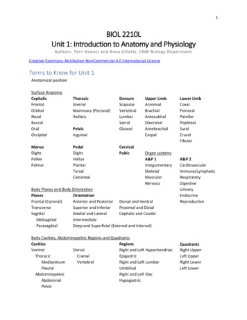
1BIOL 2210LUnit 1: Introduction to Anatomy and PhysiologyAuthors: Terri Koontz and Anna Gilletly, CNM Biology DepartmentCreative Commons Attribution-NonCommercial 4.0 International LicenseTerms to Know for Unit 1Anatomical positionSurface talManusDigitsPollexPalmarThoracicSternalMammary SacralGlutealCervicalPubicUpper alCarpalOrgan systemsA&P 1IntegumentarySkeletalMuscularNervousBody Planes and Body OrientationPlanesOrientationFrontal (Coronal)Anterior and Posterior Dorsal and VentralTransverseSuperior and InferiorProximal and DistalSagittalMedial and LateralCephalic and CaudalMidsagittalIntermediateParasagittalDeep and Superficial (External and Internal)Body Cavities, Abdominopelvic Regions and QuadrantsCavitiesRegionsVentralDorsalRight and Left ertebralRight and Left LumbarPleuralUmbilicalAbdominopelvicRight and Left IliacAbdominalHypogastricPelvicLower rA&P eUrinaryEndocrineReproductiveQuadrantsRight UpperLeft UpperRight LowerLeft Lower
2Learning Objectives (modified from HAPS learning outcomes)1. Anatomical positiona. Describe a person in anatomical position.b. Describe how to use the terms right and left in anatomical reference.2. Body planes & sectionsa. Identify the various planes in which a body might be dissected.b. Describe the appearance of a body presented along various planes.3. Body cavities & regionsa. Describe the location of the body cavities and identify the major organs found in eachcavity.b. List and describe the location of the major anatomical regions of the body.c. Describe either the location of the four abdominopelvic quadrants or the nineabdominopelvic regions and list the major organs located in either each of thequadrants or regions.4. Directional termsa. List and define the major directional terms used in anatomy.b. Describe the location of body structures, using appropriate directional terminology.5. Basic terminologya. Define the terms anatomy and physiology.b. Describe the location of structures of the body, using basic regional and systemicterminology.6. Survey of body systemsa. List the organ systems of the human body and their major components.b. Describe the major functions of each organ system.Explanation of AnatomyFor this first lab of A&P, we’ll begin by exploring surface anatomy, which will be repeated throughoutyour careers as A&P students. In addition, we’ll learn how the body can be divided within body planesand how body orientation terms describe how body areas and regional locations are related to oneanother. The body contains cavities that house internal organs like the brain, heart and stomach. We’lllearn the names of those different body cavities while also looking at how organs are distributed withinabdominopelvic regions, quadrants and organ systems. We’ll finish by learning the general functions ofall the organ systems of the body.Think about how an MRI image looks. It is an image of internal body organs seen as a plane through thebody. Knowing what body cavity the image came from, how the body is divided through the differentbody planes, understanding regional terms, and what organs are within the different organs systems willhelp you better understand an MRI image. This first A&P lab is the first step at gaining a foundationaltool, A&P terminology, that you’ll use again and again as healthcare professionals.Surface AnatomyThe foundation of Anatomy and Physiology (A&P) is learning surface anatomy (see Image 1). Surfaceanatomy are regional names given to external areas of the body that can be used as landmarks to helpclinicians during physical examinations. This terminology is repeated in subsequent chapters and is oftenused for nearby or related anatomical structures. For example, femoral means thigh and the bone thatis the thigh bone is called the femur, the group of muscles that make up the thigh are called the
3quadriceps femoris and the blood vessel that supplies those muscles branches from the femoral artery.It is important to take the time to learn surface anatomy for later study of A&P.Surface anatomy is divided into main body regions and within those main body regions specific areas arenamed. The main body regions are the cephalic (head), cervical (neck), thoracic (upper front torso),dorsum (back), abdominal and pelvic (lower front torso), upper limb, lower limb, manus (hand), pedal(foot), and pubic. Notice that many of the terms are synonyms of words you already know from theEnglish language.Image 1: Surface anatomy - anterior (a) and posterior (b) view of human figure in anatomical positionModified Creative Commons Attribution 4.0 International Openstax URL: Surface anatomySurface anatomy can be used as a noun or an adjective. The noun form can end in “um”, “is”, or “a” likebrachium, oris, axilla, and patella. The adjective form can end in “al”, “ry”, and “ar” like brachial, oral,
4axillary and patellar. When using surface anatomy as an adjective you are describing a noun. A region isa noun so surface anatomy in adjective form is written “brachial region”, “oral region”, “axillary region”,and “patellar region”.Below is a list of the other surface anatomy terms that are within the main body regions: Cephalic region contains the frontal (forehead), orbital (eye), buccal (cheek), nasal (nose), oral(mouth), and occipital (back of the head) regions.Thoracic region contains mammary or pectoral (chest), sternal, and axillary (armpit) regions.Dorsal region contains scapular, vertebral, lumbar, sacral, and gluteal (butt) regions.Abdominal region contains the umbilical region and pelvis contains the inguinal region.Upper limb region can be subdivided into the acromial (shoulder), brachial (upper arm),antecubital (front of elbow), olecranal (elbow), antebrachial (forearm), and carpal (wrist)regions.Lower limb region can be subdivided into the coxal (hip), femoral (thigh), patellar (knee),popliteal (back of the knee), sural (calf or back of lower leg), crural (leg), and fibular (outside,lateral, of lower leg) regions.The manus contains the digits (fingers), pollex (thumb), and palmar (palm) regions.The pedal region contains the digits (toes), hallux (big toe), plantar (bottom of the foot), tarsal(ankle), and the calcaneal (heel) regions.The cervical and pubic regions do not contain further regions/areas.As you are probably discovering, A&P involves a lot of memorization. This requires a daily study oflearning new words. There are many ways to approach the study of A&P, but a key strategy is repetition.Some ways to study include: saying the words and their meaning out loud,writing the words on paper and saying their meaning out loud,drawing what the words mean,using models and labeling models with tape where you’ve written what an area is named,coming up with mnemonics to remember groups of structures,relating the new information to information you already know,forming study groups and quizzing each other, and the list goes on.Find out early what the best ways for you to start committing to memory this new language of the body.Remember, you’ll see these terms not only later in this course, but also in your healthcare career.Mini Activity: Ways to study, top three listWrite out the top three active strategies you will use to start studying A&P terminology. Recommend“read the chapter” is NOT on the list.
5Body Planes and Body OrientationIn anatomy, body planes and body orientation are used when the body is in anatomical position.Anatomical position is standing erect with arms along the side of the body and head, palms of the handand the toes of the feet are facing forward (anteriorly) (see Images 1, 2, and 3).There are three body planes that separate the body into two opposing parts (see Image 2). A frontalplane, also known as the coronal plane, separates the body into anterior (front) and posterior (back)parts; a transverse plane separates the body into superior (upper) and inferior (lower) parts; and asagittal plane separates the body into right and left parts. A midsagittal plane (median) separates rightand left sections of the body equally where a parasagittal plane separates right and left parts unequally.Not only are body orientation terms used when describing how a dissection through a body planeseparates the body, but also to describe the relationship between two different body structures (seeImage 3). Other body orientation terms, in addition to anterior, posterior, superior, and inferior, aremedial (towards the midline), lateral (away from the midline), intermediate (between and in themiddle), proximal (towards the torso or, with tubular organs, towards the beginning of the structure),distal (farther away from the torso or, with tubular organs, towards the end of the structure), deep(internal), and superficial (external). Also, ventral (belly or front), dorsal (back), cephalic (head) andcaudal (tail) are body orientation terms commonly used for quadrupeds. Still, they can be used whendescribing a human and are preferentially used for certain body parts. For example, the ventral anddorsal body cavities, the cephalic surface anatomy term, and the cauda equina, which is an inferiornerve structure in the body.Image 2: Body planes dividing the bodyCreative Commons Attribution 4.0 International Openstax URL:Body planes dividing the body
6Remember that orientation terms are used when the body is in anatomical position. You’ll notice thatthe body orientation terms are paired and opposing one another. When describing the relationshipbetween structures you can use multiple orientation terms.Mini Activity: Examples of how to practice body orientation terms1. What is the relationship between sternal and scapular?You could say that the sternal region is anterior and medial when compared to the scapularregion.2. What is the relationship between acromial and carpal regions on the right arm?In this example, you’ll use either proximal or distal, since these body orientation terms are usedwhen describing structures that are within the same limb. The acromial region is proximal to thecarpal region.So, to understand the relationship of the shoulder as it pertains to the wrist, place your right handon your left wrist. Move your right hand along your upper limb towards your left shoulder. As youmove your hand towards the torso within the upper limb, your right hand is moving closertowards the torso, (namely the axial skeleton) in this relationship. Think of proximal asapproximating, getting closer, to the torso. Distal is the opposite, where the relationship of onestructure to another is farther, a greater distance, from the torso. Once you’ve learned all thesurface anatomy, start to compare the body regions to each other using body orientation terms.Continue practicing terminology in the next Mini-ActivityMini Activity: Superior/inferior versus proximal/distalLet’s get some practice with superior, inferior, proximal, and distal. In the examples below you’ll insertone of those four terms in the blank to compare the two body regions to one another.1. The acromial region is to the brachial region.2. The pedal region is to the femoral region.3. The pelvic region is to the pectoral region.4. The antebrachial region is to the patellar region.
7Image 3: Body orientation terms shown on bodyCreative Commons Attribution 4.0 International Openstax URL:Body orientation terms shown on bodyBody Cavities and Abdominopelvic RegionsSo far we’ve only been looking at the surface regions of the body, but there are cavities that containmore body structures (see Image 4). Just like surface anatomy, body cavities also have a hierarchy ofdivisions where there are main body cavities that are further divided into smaller body cavities. The twomain body cavities are the ventral and dorsal body cavities.The ventral body cavity is the cavity that contains the “guts” of the human body. It is further dividedinto the thoracic body cavity and abdominopelvic body cavity. The diaphragm is a wide muscle thatwhen contracted (tightened) pulls the lungs downward in order to draw air in; we call this breathing. It isthe diaphragm, which runs through a transverse body plane, that separates the thoracic andabdominopelvic body cavities of the ventral body cavity. The thoracic body cavity contains the heart andlungs and the abdominopelvic body cavity mainly contains digestive, urinary, and reproductive organs.The thoracic body cavity is yet further divided where the heart is within a cavity called the mediastinumand the right and left lungs are within each of their own pleural body cavities. The abdominopelvic bodycavity is also further divided into the abdominal body cavity and the pelvic body cavity. The pelvic bodycavity is distinguished from the abdominal body cavity in that its exterior walls are bony, making it moreprotected.The dorsal body cavity is the posterior cavity that contains the central nervous system: brain and spinalcord. This body cavity is further divided into the cranial body cavity whose bony wall protects the brainand the vertebral body cavity that contains the spinal cord. Body cavities typically have at least one partof their wall made of bone. In the case of the dorsal body cavity, the skull forms the bony wall of the
8cranial body cavity that protects the brain and the vertebrae make up the bony wall of the vertebralbody cavity, which protects the spinal cord.Image 4: Body cavitiesCreative Commons Attribution 4.0 International Openstax URL:Body cavitiesThe heart and lungs are important internal organs that also require a substantial amount of bonyprotection. The thoracic body cavity’s bony wall consists of vertebrae and ribs on the posterior side andthe sternum and ribs on the anterior side. The abdominal body cavity is the most vulnerable section ofthe ventral body cavity since its anterior wall is muscle rather than bone. The pelvic body cavity, whichcontains the urinary bladder, has a bony wall that is made up of the sacrum and coccyx (inferior parts ofthe vertebrae) posteriorly along with sections of the coxal (hip) bones laterally and anteriorly.The abdominopelvic body cavity also has named regions. These regions can be used when diagnosingillness and when describing the location of internal body organs. The abdominal body cavity is dividedinto a tic-tac-toe grid (see Image 5) or into four quadrants (see Image 6).In Image 5, the superior row of boxes in the grid are made up of left and right hypochondriac regionslaterally; those two regions are covered by lower rib bones that are connected to the sternum throughcartilage. Hypo- means below and -chondriac means cartilage, making these two regions, and theorgans within them, below the rib cartilage.The box in the midline of the superior row is named the epigastric region. Epi- means above; this regionis above where many of the gastric (stomach) processes occur.To separate the superior and middle rows from each other use the large intestines that travel across(transverse) the body as a line. The middle row of boxes has left and right lumbar (lower back) regionsand the center-most box in the grid is the umbilical region. You may find it confusing that the anteriorabdominal area has two regions called “left and right lumbar” since typically lumbar is referring to the
9posterior (back) of the body. Sometimes, in A&P, historical usage of terms creates conventions that arestrange and those you must simply memorize.The most inferior row in the grid has a left and right iliac region. These are named this because the tworegions are nestled within the shallow depressions of the iliac bones on either side of the body. A&Poften has a lot of synonyms for terms. In some places, the iliac regions are called the inguinal region.You do not need to memorize this but you may come across it in your lecture course.The center box of this inferior row is the hypogastric region; hypo- means below and it is the region thatcontains organs that are below where most gastric processes have already occurred.To separate the inferior row from the middle row, find the crest of the iliac bone, which is the mostsuperior part of the coxal bone. Place your hands on the bony upper part of your hips off to the sides ofyour body. You are resting your hands on your iliac crests! For the two vertical lines in the grid visualizeeach starting just medial to each of the nipples.Mini Activity: Visualize a tic-tac toe grid on a torso modelUse the painter’s tape1 to visualize the grid on a torso model or a full human figure model in the lab.Now that you can see where the lines of the grid are located, take the time to list out what organsbelong in each region (box).Image 5: Abdominopelvic regionsModified Creative Commons Attribution 3.0 Unported Openstax URL: Abdominopelvic regions1Our CNM anatomy lab provides “painter’s tape” for students use because the tape is less sticky than others andwill not damage the models or strip paint from the models. Please do not use any other type of tape on themodels.
10Your instructor might also want you to divide the abdominopelvic body cavity into the four quadrants:right upper quadrant, left upper quadrant, right lower quadrant, and left lower quadrant. Notice inImage 6 that the transverse colon is a good landmark to use to separate the upper and lower quadrants.A cut through the midsagittal plane separates the right and left quadrants from each other.Image 6: Abdominopelvic quadrantsModified Creative Commons Attribution 3.0 Unported Openstax URL: Abdominopelvic quadrantsHierarchy of the Human Body and Organ SystemsYou’ve already learned in your general biology class that there is a hierarchy of life where the cell is thesmallest level in that hierarchy that is alive. As we go up through the levels of life, a group of cellsworking together is a tissue, a structure that has two or more tissues is an organ, organs workingtogether to perform specific tasks are organ systems, and an organism is made up of organ systemsworking together to maintain the organism, in this case the human body. In the A&P I lab course you willstudy in detail four organ systems: Integumentary, Skeletal, Muscular, and Nervous Systems. In theA&P II lab course you will study in detail the remaining seven organ systems: Cardiovascular,Immune/Lymphatic, Respiratory, Digestive, Urinary, Endocrine, and Reproductive Systems. While youwon’t be learning in detail all the organ systems this term, you are expected to know major organswithin each of the organ systems along with the main function of each of the organ systems. See thefollowing Images 7 and 8 for this information.
11Image 7: Organ systems for A&P I labModified Creative Commons Attribution 4.0 International Openstax URL:Organ systems for A&P Ilab
12Image 8: Organ systems for A&P II labModified Creative Commons Attribution 4.0 International Openstax URL:Organ systems for A&P II lab
13Activity 1: Surface Anatomy and Body OrientationPart 1 - Label (write in) all surface anatomy terms on the anterior and posterior view of the humanfigure seen below; take note of what are the major regions and the smaller regions that are within thosemajor regions. You should write the correct term by EVERY leader line and bracket.
14Part 2 - Fill in the blanks below with the appropriate body orientation term while viewing yourcompleted labeling activity of the human figure in Activity 1 Part 1. In some cases, you can combine theorientation terms to be even more specific.Head/Torso/Surface Terms: superior, inferior, anterior, posterior, medial, lateral, superficial, deep1. The sternal region is to the vertebral region.2. The nasal region is to the buccal region.3. The cephalic region is to the thoracic region.4. The umbilical region is to the coxal region.5. The axillary region is to the mammary region.6. The skin is to skeletal muscles.7. Bones are to skeletal muscles.Upper and Lower Limb Terms: proximal, distal, anterior, posterior, ventral, dorsal, medial, lateral,superficial, deep8. The popliteal region is to the patellar region.9. The plantar region is to the dorsum of the pedal region.10. The antebrachial region is to the brachial region.11. The 5th hand digit (pinky finger) is to the pollex.12. The femoral region is to the tarsal region.
15Activity 2: Body PlanesPart 1 - Dissect a banana along the three different body planes. Below is an image of a banana in“anatomical position.” Draw in the designated boxes what the banana looks like when dissected in eachof the different body planes.Sagittal planeCoronal planeTransverseplanePart 2 – Your instructor has laid out models that have the body dissected through a body plane. Fill inthe table below with a description of the model and the body plane that the dissection is travelingthrough within the model.Description (or drawing) of ModelBody Plane of Dissection
16Activity 3: Gunshots GalorePart 1 - You or your instructor, will randomly choose three points to mark a torso model or torso image with an “X”, using painter’s tape in thelab. The “X”s indicate where gunshot wounds (GSWs) are located on a recently deceased person (ie., cadaver) that has come into the NewMexico Office of Medical Investigator (OMI). You will be acting as an employee of the OMI and it is your job to document the location of thebullet wounds using surface anatomy terms where the bullet first entered and/or exited the body, body cavity location that the bullet passedthrough and might still be located within, organs that have been damaged by the bullet along with the organ system that the damaged organsbelong to, and, if the bullet is within an abdominopelvic region, which of the nine regions (tic-tac-toe board) is impacted.Surface Anatomy ofEntry and Exit ofBulletGSW 1GSW 2GSW 3Main BodyCavity (Dorsal orVentral)Specific BodyCavityAbdominopelvicRegion (ifapplicable)Organs PossiblyDamagedOrgan System ofDamaged Organs
17Part 2 - In a court of law, the medical pathologist from OMI needs to be able to specifically describe the location of each GSW using theaccepted orientation/directional terms. In a complete sentence, describe the location of each GSW in relationship to another body region usingthe orientation terms: proximal, distal, anterior, posterior, superior, inferior, medial, lateral. For example, “GSW1 is located lateral to the leftnipple and inferior to the clavicle (collar bone).GSW 1:GSW 2:GSW 3:
5. Basic terminology a. Define the terms anatomy and physiology. b. Describe the location of structures of the body, using basic regional and systemic terminology. 6. Survey of body systems a. List the organ systems of the human body and their major components. b. Describe the major functions of each organ system. Explanation of Anatomy
