Transcription
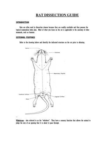
Rat External AnatomyProcedure: Obtained your rat and observe the general characteristics. Key terms are highlighted in grey.The rat's body is divided into six anatomical regions:cranial region - headcervical region - neckpectoral region - area where front legs attachthoracic region - chest areaabdomen - bellypelvic region - area where the back legs attach1. Note the hairy coat that covers the rat and the sensory hairs (whiskers) locatedon the rat's face, called vibrissae.2. The mouth has a large cleft in the upper lip which exposes large front incisors.Rats are gnawing mammals, and these incisors will continue to grow for as longas the rat lives.3. Note the eyes with the large pupil and the nictitating membrane found at theinside corner of the eye. This membrane can be drawn across the eye forprotection. The eyelids are similar to those found in humans.4. The ears are composed of the external part, called the pinna, and the auditorymeatus, the ear canal.5. Locate the teats on the ventral surface of the rat. Check a rat of another sexand determine whether both sexes have teats.6. Examine the tail, the tails of rats do not have hair. Though some rodents, like gerbils, have hair on their tails.7. Locate the anus, which is ventral to the base of the tale.8. Determine whether your rat is male or female by looking near the tail for the male or female genital organs.The Muscular System of the RatCheckpoint—have Your teacher initial lab before continuing.Procedure: Skinning the RatYou will carefully remove the skin of the rat to expose the muscles below. This task is best accomplished bymaking a small incision with your scalpel and the using your probe to separate the connective tissues thatconnects the skin to the first layer of muscles. Do not cut into the muscles! You can start at the incision pointwhere the latex was injected and continue toward the tail. Use the lines on the diagram to cut a similar pattern,avoiding the genital area. Gently peel the skin from the muscles, using scissors and a probe to tease awaymuscles that stick to the skin.Identify the following muscles:1. Biceps brachii - located on the anterior surface of the humerus (arm). Action: flexes lower arm2. Biceps femoris - located on the side of the thigh, in two bundles. Action: flexes the lower leg3. External Oblique - located on the sides of the abdomen. Action: flexes body wall.4. Pectoralis Major/Minor - located in chest area. Action: adducts arm (draws it forward)Rat Dissection John R. Sowash May 2009 Permission to redistribute granted3 Page
The Skeletal System of the RatCheckpoint—have your teacher initial lab before continuing.Procedure: Exposing the bones of the leg.Carefully tease away the biceps femoris and gastrocnemius on one leg to expose the 3 leg bones: Tibia, Fibula,and Femur and the small patella (kneecap). You can also see the ligaments around the knee that attach thebones of the lower leg to the femur and the achilles tendon which attaches the gastrocnemius to the ankle.Remove the muscles from one arm to reveal the ulna, radius, and humerus. Note the size of the radius.The Digestive System of the RatCheckpoint—have Your teacher initial lab before continuing.Procedure: Use scissors to cut through the abdominal wall of the rat following the incision marks in the picture onpg. 2. Be careful not to cut too deeply and keep the tip of your scalpel pointed upwards. Do not damage theunderlying structures.1. Locate the diaphragm, which is a thin layer of muscle that separates the thoracic cavity from the abdominalcavity. The diaphragm is a helpful directional marker.2. DO NOT REMOVE OR CUT THE HEART! The heart is centrally located in the thoracic cavity. The two darkcolored chambers at the top are the atria (single: atrium), and the bottom chambers are the ventricles. The heartis covered by a thin membrane called the pericardium. (We will come back to the heart later.)Rat Dissection John R. Sowash May 2009 Permission to redistribute granted4 Page
The Abdominal Organs1. The coelom is the body cavity withinwhich the viscera (internal organs) arelocated. The cavity is coverd by a membranecalled the peritoneum.2. Locate the liver, which is a large, darkcolored organ suspended just under thediaphragm. The liver has many functions,one of which is to produce bile which aids indigesting fat. The liver also stores glycogenand transforms wastes into less harmfulsubstances. Rats do not have a gall bladderwhich is used for storing bile in other animals.3. The esophagus runs through thediaphragm and moves food from the mouthto the stomach. It is distinguished from thetrachea by its lack of cartilage rings.Liver(many erior4. Locate the stomach on the right sideVena Cava(usually) just under the liver. The functions ofthe stomach include food storage, physicalbreakdown of food, and the digestion ofprotein. The opening between theesophagus and the stomach is called thecardiac sphincter. The outer margin of theSecumcurved stomach is called the greatercurvature, the inner margin is called thelesser curvature.5. The spleen is about the same color as theliver and is attached to the greater curvatureof the stomach. It is shaped like a bananaand is associated with the circulatory systemand functions in the destruction of blood cellsand blood storage. A person can live withouta spleen, but they're more likely to get sickas it helps the immune system function.SmallIntestineSmallIntestineMesentery6. The pancreas is not a clearly identifiable organ but a thin membrane that overlays the stomach and spleen.The pancreas produces digestive enzymes that are sent to the intestine via small ducts (the pancreatic duct). Thepancreas also secretes insulin which is important in the regulation of glucose metabolism. Find the pancreas bylooking for a thin, membrane looking structure that has the consistency of cottage cheese.7. The small intestine is a slender coiled tube that receives partially digested food from the stomach (via thepyloric sphincter). The term “small” refers to its diameter, not its length. It consists of three sections: duodenum,ileum, and jejunum. The small intestine leads to the cecum.8. The cecum is a pouch that connects the large and small intestines. Food is temporarily stored in the cecumwhile helpful bacteria digest the cellulose found in plant cells. Most herbivores such as the rat have a large cecum.Humans and other omnivores and carnivores have a much smaller cecum which is referred to as the appendix.Rat Dissection John R. Sowash May 2009 Permission to redistribute granted5 Page
9. Use your scissors to cut the mesentery (connective tissue and network of blood vessels that connects the smallintestine) of the small intestine, but do not remove the small intestine from its attachment to the stomach andrectum. If you are careful you will be able to stretch it out and untangle it so that you can see the relative lengthsof the large and the small intestine.10. Locate the large intestine, which is the large greenish tube that extends from the small intestine and leads tothe anus. The large intestine is also known as the colon. This is where the finals stages of digestion and waterabsorption occurs and it contains a variety of bacteria to aid in digestion.11. Locate the rectum - the short, terminal section of the colon between the descending colon and the anus. Therectum temporarily stores feces before they are expelled from the body.The Excretory System of the RatCheckpoint—have your teacher initial lab before continuing.The excretory and reproductive systems of vertebrates areclosely integrated and are usually studied together as theurogenital system. However, they do have different functions:the excretory system removes wastes and the reproductivesystem produces gametes (sperm & eggs). The reproductivesystem also provides an environment for the developingembryo and regulates hormones related to sexual development.The primary organs of the excretory system are the kidneys. Theseorgans are large bean shaped structures located toward the back ofthe abdominal cavity on either side of the spine. Renal arteries andveins supply the kidneys with blood.1. Locate the Kidneys. Note the veins and arteries that connect withthe kidneys.2. Remove one of the kidneys and cut it lengthwise. Notice the veryfine veins and arteries within. Blood is filtered through the kidneysapproximately once every 45 minutes.3. The small yellowish glands embedded in the fat atop the kidneysare the adrenal glands which secrete adrenaline into the bloodduring times of crisis.The Excretory and Reproductive System of the RatCheckpoint—have your teacher initial lab before continuing.Male Reproductive Organs (refer to diagram below)1. The major reproductive organs of the male rat are the testes (singular: testis) which are located in the scrotalsac. Cut through the sac carefully to reveal the testis. On the surface of the testis is a coiled tube called theepididymus, which collects and stores sperm cells. The tubular vas deferens moves sperm from the epididymusto the urethra, which carries sperm though the penis and out the body.2. The lumpy brown glands located to the left and right of the urinary bladder are the seminal vesicles. The glandbelow the bladder is the prostate gland and it is partially wrapped around the penis. The seminal vesicles andthe prostate gland secrete materials that form the seminal fluid (semen).Rat Dissection John R. Sowash May 2009 Permission to redistribute granted6 Page
Female Reproductive Organs (refer to diagram below)1. The short gray tube lying dorsal to the urinary bladder is the vagina. The vagina divides into two uterine hornsthat extend toward the kidneys. This duplex uterus is common in some animals and will accommodate multipleembryos (a litter). In contrast, a simple uterus, like the kind found in humans has a single chamber for thedevelopment of a single embryo.2. At the tips of the uterine horns are small lumpy glands called ovaries, which are connected to the uterine hornsvia oviducts. Oviducts are extremely tiny and may be difficult to find without a dissecting scope.Please be sure to observe the reproductive features of both sexes.This will require looking at the rat of another group.The Circulatory System of the RatCheckpoint—have your teacher initial lab before continuing.The general structure of the circulatory system of the rat is almost identical to that of humans. Pulmonarycirculation carries blood through the lungs for oxygenation and then back to the heart. Systemic circulation movesblood through the body after it has left the heart. Use the diagrams on pg. 9, to locate the veins and arteries listedon page 2.Veins (see diagram page 9)Your rat specimen has been double injected with latex to help you identify veins and arteries. Veins carry usedblood (blue) back to the heart and lungs. The lungs re-oxygenate the blood and the heart pumps it back to therest of the body. In the human body, these veins are not the same bright blue that you see in your rat. However, ifyou look at your arm, you can see some bluish veins very close to the skin.Look in your rat specimen for the veins listed on page 2.Rat Dissection John R. Sowash May 2009 Permission to redistribute granted7 Page
Arteries (see diagram page 9)Arteries carry oxygenated blood to the muscles and organs that need it. Blood is essential for life. Bloodcarries nutrients to the body, helps repair cells and tissues, fights against disease, and assists in cleansing toxins.Without blood, we would all be dead.The arteries in your rat are stained red for easyidentification. Find the arteries listed on page 2.HeartAfter completing the procedures above dealing with veinsand arteries, remove the heart from the pericardial sack.You will need to sever the arteries and veins connectingthe heart to the circulatory system. Do this slowly andcarefully so that you do not cut more than isnecessary. Leave as much of the veins and arteriesattached to the heart as possible.Identify the aorta, left and right atrium, and left and rightventricle. Carefully insert your probe into these openingand work it into the center of the heart.Finally, make an incision between the left and rightventricles with your scalpel. Try to locate the bicuspidand semilunar valves which open and close theventricles.Final CheckCan you confidently identify all of the structures listed on pg. 2? These are the structures that you will be testedon. As there are many variations amongst living organisms, it is strongly suggested that you spend some timelooking at the rat of other groups. Color, location, and size of organs will vary from one rat to another.Once you feel confident in your ability to identify the parts on pg. 2, ask your teacher to complete the final checkpoint.Checkpoint—have Your teacher initial lab before continuing.Rat Dissection John R. Sowash May 2009 Permission to redistribute granted8 Page
Rat Dissection John R. Sowash May 2009 Permission to redistribute granted9 Page
Rat External Anatomy Procedure: Obtained your rat and observe the general characteristics. Key terms are highlighted in grey. The rat's body is divided into six anatomical regions: cranial region - head cervical region - neck pectoral region - area where front legs attach thoracic region - chest area abdomen - belly