Transcription
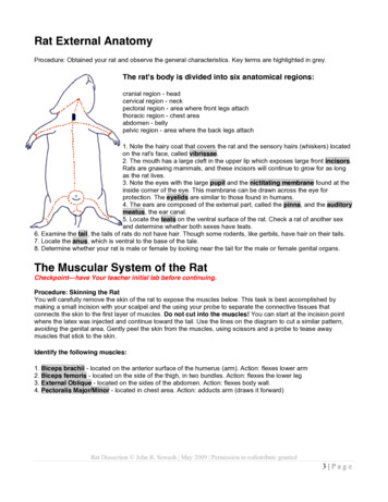
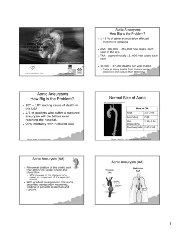
RAT DISSECTION GUIDEINTRODUCTIONRats are often used in dissection classes because they are readily available and they possess thetypical mammalian body plan. Most of what you learn on the rat is applicable to the anatomy of othermammals, such as humans.EXTERNAL FEATURESRefer to the drawing below and identify the indicated structure on the rat prior to skinning.Vibrissae - also referred to as the "whiskers". They have a sensory function that allows the animal tojudge the size of an opening that it is about to pass through.
Nares - the nares (plural) or naris (singular) are the external openings into the nasal cavity.Female urogenital structuresUrethral orifice - is the opening into the urethra (part of the urinary system).Vaginal orifice - is the opening into the vagina (part of the reproductive system).Male urogenital structuresPenis - is hidden on the male rat beneath a fold of skin (the foreskin or prepuce).Scrotum - is a pouch that contains the testes (see the drawing below)
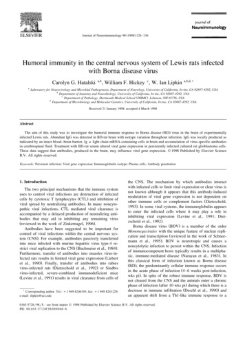
Skinning the ratMake a midventral incision as illustrated on the drawing above. Use your probe, scissors and finger tofree the skin from the underlying tissue. Follow the basic cut pattern illustrated on the first page of thishandout.You should encounter two brownish muscles attached to the skin (the cutaneous maximus in thetrunk area, and the platysma in the neck area). You will probably have to cut these muscles close to theskin before the skin can be removed.MUSCULAR SYSTEMThe diagram below illustrates the muscles of the ventral surface of the rat. Be able to identify thoselisted. Use the photographs on the following pages and your lab atlas to assist you.
Head and Throat musclesDigastric - this V shaped muscle follows the lower jaw line. It functions to open the mouth.Mylohyoid - runs at right angles to the longitudinal axis. You may have to gently raise up the edgeof the digastric muscle on each side of the jaw in order to see this muscle. Itfunctions to raise the floor of the mouth.Sternohyoid - runs parallel to the longitudinal axis on either side of the midline. It functions to pullthe hyoid towards the sternum.Sternomastoid - runs diagonally from the sternum to a point behind the ear. It functions to rotatethe head to one side (when one contracts), or rotates the head down (when bothcontract).Masseter - this is the "cheek" muscle. It functions in mastication (chewing).Chest and Front leg (medial) musclesPectoralis Major - the large triangular muscle covering the upper thorax. It functions to pullthe arm towards the chest.Pectoralis Minor - part of this muscle runs beneath the Pectoralis Major. It function is the sameas the Pectoralis Major.Biceps Brachii - the large muscle located on inside of the upper arm. It functions to flex (bend)the lower arm.Epitrochlearis - this is a flat, thin muscle on the medial surface of the upper arm. It functions toextend the lower arm.Flexors - the collection of small muscles on the medial surface of the lower arm. They function toflex the wrist and hand.Shoulder and Lateral (outside) Muscles of the Front LegBe able to identify the muscles illustrated on the drawing on the next page.Clavotrapezius - one of a group of three muscles that help to stabilize the scapula. Its functionis to pull the clavicle forward.Acromiotrapezius - pulls the scapula forward.Spinotrapezius - pulls the scapula downward.Latissimus dorsi - pulls the arm downward.Serratus ventralis - is made up of fingerlike projections of muscle running between the armpit andthe ribs. Its function is to depress the scapula.
Cleidobrachialis - runs between the clavicle and the humerus. Its function is to pull the armforward.Acromiodeltoid - Runs between the outer clavicle and the scapula. Pulls the upper arm away fromthe ribs.Spinodeltoid - runs between the scapular spine and the upper arm. Pulls the scapula away fromthe ribs.Triceps brachii - this is a muscle with three heads. It runs between the scapula and humerus andthe ulna. Its function is to extend the lower arm.Brachialis - runs next to the Biceps Brachii. Its function is to flex the lower arm.Extensors - a group of small muscles on the lateral side of the lower arm. They extend the wristand hand.Deep Muscles of the ShoulderIdentify the deep muscles illustrated on the drawing above.Rhomboideus - runs between the dorsal border of the scapula and vertebral column. Its functionis to pull the scapula in and forward.
Supraspinatus - covers the lateral side of the scapula, above the scapular spine and attaches to theupper arm. Its function is to pull the upper arm forward.Infraspinatus - covers the lateral side of the scapula, below the scapular spine and attaches to theupper arm. Its functions is the rotate the upper arm outwardly.Teres Major - runs along the outer edge of the scapula to attach to the upper arm. Its function isto rotate the humerus laterally.Hip and Lateral Muscles of the Hind LegIdentify the muscles illustrated on the drawing below.Gluteus Superficialis - covers a large portion of the anterior hip. Its function is to pull thethigh outward.Biceps Femoris - located posterior to the Gluteus. Its function is to pull the upper legoutward and flex the lower leg.Semitendinosus - it is located on the posterior margin of the leg and is partly hidden by theBiceps Femoris. Its function is to flex the lower leg.Gastrocnemius - the major "calf" muscle on the posterior surface of the lower hind leg. Itsfunction is to extend the foot.Soleus - located under the Gastrocnemius. Its function is to extend the foot.Tibialis Anterior - located on the anterior surface of the lower hind leg. Its functions isto flex the foot.
Hip and Medial Muscles of the Hind LegBe able to identify the following muscles illustrated on the drawing on page 3 of this handout.Gracilis - a flat muscle on the caudal (near the tail) portion of the inner thigh. Its functionis to pull the thigh inward.Rectus femoris - located on the anterior (front) surface of the thigh. Its function is toextend the lower hind leg.Vastus medialis - runs parallel to the Rectus femoris on the caudal side. Its function isto extend the lower hind leg.Adductor Magnus and Brevis - runs parallel to and beneath the Gracilis. Its function isto pull the thigh towards the body.Adductor Longus - runs parallel to and beneath the Vastus Medialis. Its function is topull the thigh towards the body.Pectineus - located next to the femoral vein. Its function is to pull the thigh towards the body.Abdominal MusclesBe able to identify the following muscles illustrated on the drawing on page 3 of this handout.External Oblique - covers most of the ventral and lateral abdomen. Its function to compressand hold the internal organs in place.Rectus Abdominis - runs parallel to the longitudinal axis of the body on either side of thelinea alba. Its function is to compress and hold the internal organs.Throat and Oral CavityBe able to identify the structures illustrated on the drawing below. The structures indicated arecomponents of a variety of organ system.Parotid Gland - the major salivary gland. Its function is to secrete saliva containing starchdigesting enzymes.Parotid duct - empties saliva from the Parotid Gland into the oral cavity.Mandibular gland - a salivary gland that secretes a thick mucous.Sublingual gland - a small salivary gland that empties into the oral cavity behind the lowerincisors.Lymph Nodes - part of the immune system.
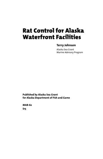
Lacrimal Gland - secretes lacrimal fluid (tears) to lubricate the eye.The Abdominal Cavity and the Digestive SystemBe able to identify the structures illustrated on the drawing below.
Dissection - use scissors to make a midventral cut up the entire length of the abdominal cavity.When you reach the diaphragm (a broad sheet of muscle that separates the thorax fromthe abdomen) make lateral cuts along the lower border of the rib cage.Parietal Peritoneum - a very thin and shiny membrane that lines the inside of the abdominalwall.Visceral Peritoneum - a very thin and shiny membrane that covers the internal organs of theabdominal cavity.Mesenteries - folds of the Visceral Peritoneum that attach the small intestine and colon to theposterior abdominal wall.Liver - is the large brown organ taking up most of the anterior portion of the cavity. I consistsof four lobes. Its function is to manufacture bile, selectively remove and reintroducenutrients into the blood, remove toxins, and manufacture needed proteins and carbohydrates.Gall Bladder - the rat does not have a gall bladder.Stomach - a curved bag-like organ right below the diaphragm. Its function is to mechanicallygrind up the food and start the digestion process.Esophagus - muscular tube that passes through the diaphragm to empty food into the stomach.Small Intestine - composed of three major parts. The stomach empties its contents into the firstsection of the intestine called the Duodemum. The Ileum is the terminalsection of the small intestine that connects with the cecum. The middle section(between the duodenum and ileum) is the Jejunum. The small intestine iswhere most digestion and ingestion occurs.Large Intestine (Colon) - is shorter and wider than the small intestine. The ileum of the smallintestine empties into the first section of the colon, the Cecum. TheVermiform Appendix is a blind-ended sac attached to the cecum. Theappendix functions as a lymphatic organ in immunity and often becomesinfected in humans.Rectum - the terminal portion of the colon leading to the anus.Spleen - is located to the left of the stomach at the end of the pancreas. It functions in immunity.Pancreas - has a glandular lobular appearance and is attached to the duodenum. It has both adigestive function (it secretes enzymes) and a hormonal function.
Respiratory SystemBe able to identify the structures illustrated on the drawing below.Trachea - air tube with conspicuous rings. Gently move the contents of the thorax around until itcan be found.Bronchus - the trachea divides into a left and right bronchus. Gently move the contents of thethorax around until they can be found. Like the trachea they have conspicuous rings.Lungs - appear spongy and on either side of the heart. The right lung on the rat has four lobesand the left lung has three. Each lung is covered by a thin layer of tissue called theVisceral Pleura. The inside of the thoracic wall is covered by a similar layercalled the Parietal Pleura.Diaphragm - is a thin sheet of muscle separating the abdomen from the thorax. When contractedit draws air into the lungs.The Circulatory SystemBe able to identify any of the blood vessels illustrated on the drawing above and below that you canfind without cutting any internal organs out of your specimen.Heart - sits in the space between the left and right lungs. Portions of the right and left atria can beseen as dark flaps on top of the heart. The two anterior vessels and one posterior vesselentering the right atrium are the Vena Cavae that return unoxygenated blood to the heart.
Pulmonary Veins can be seen leaving each lung and entering the Left Atrium.The Ventricles (right and left) can not be differentiated without opening the heart. APulmonary Artery can be seen exiting the right ventricle and passing under the Aorta.Pericardium - is the tough fibrous sack surrounding the heart for protection.MAJOR ARTERIES
Aorta - is the major artery shown exiting the top of the heart and forming an archthat bends to the left. It empties the left ventricle and supplies oxygenatedblood to the entire body.Brachiocephalic Artery - is the first artery to branch off of the Aorta. It suppliesblood to the head and right arm. This artery splits to from the Right SubclavianArtery that takes blood to the right arm, and the Right Carotid Artery thattakes blood to the head.Left Common Carotid Artery - is the second artery to branch off of the Aorta. It suppliesblood to the head.Left Subclavian Artery - is the third artery to branch off of the Aorta. It supplies blood tothe left arm.Common Iliac Artery - divides to form the Internal Iliac Artery (supplying various organs inthe pelvic area) and the External Iliac Artery (passing to the leg).Femoral Artery - is an extension of the External Iliac Artery located on the medial surface of thethigh.MAJOR VEINS
Subclavian Vein - as it returns blood from the arm in connects with the Lateral ThoracicVein, External Iliac Vein, and, Internal Iliac Vein to form theAnterior Vena Cava.Anterior Vena Cava - enters the right atrium on the dorsal side of the heart.External Iliac Vein - runs parallel to the External Iliac Artery.Internal Iliac Vein - runs parallel to the Internal Iliac Artery.Posterior Vena Cava - formed when the right and left Common Iliac Veins unite. It emptiesinto the right Atrium. The Renal Artery and Iliolumbar Arterydrains into it.Hepatic Portal Vein - takes blood from the digestive organs to the liver. Should be injected withyellow latex.FEMALE URINARY AND REPRODUCTIVE STRUCTURESBe able to identify the structures illustrated on the drawing of the Female Urogenital System below.Kidney - can be found close to the dorsal wall of the abdomen surrounded by fat.Ureter - is a whitish tube extending from each kidney and running to the Urinary Bladderwhere urine is stored.Ovary - is a small nodular gland often found buried in fat near each kidney.
Uterus - in female rats the uterus is split into two Uterine Horns. The distal end of eachhorn is located close to each ovary.MALE URINARY AND REPRODUCTIVE STRUCTURESBe able to identify the structures illustrated on the drawing below.Testis - open the scrotum and gently extract a testis. Be careful to leave it attached to theDuctus Deferens.Epididymis - is a highly coiled tube attached to the surface of the testis. It store spermuntil it has time to mature.Ductus Deferens (Vas Deferens) - is a tube that passes inside the Spermatic Cord throughthe Inguinal Canal into the pelvic cavity where it connects to the Urethra.Seminal Vesicles - a pair of glands that secrete an alkaline fluid rich in sugar to nourish thesperm while they search for an egg to fertilize.Prostate Gland - paired accessory glands near the base of the seminal vesicle.Penis - can be observed by opening the urogenital opening and cutting through what is theForeskin (Prepuce)
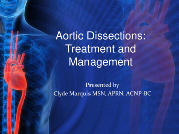
The Mammal SkeletonThe drawing below illustrates the bones of the rat skeleton. In lab we will learn these same bones ona CAT and a MONKEY skeleton. Be able to identify the bones indicated below on these skeletons.
Nares - the nares (plural) or naris (singular) are the external openings into the nasal cavity. Female urogenital structures Urethral orifice - is the opening into the urethra (part of the urinary system). Vaginal orifice - is the opening into the vagina (part of the reproductive system). Male urogenital structures Penis - is hidden on the male rat beneath a fold of skin (the foreskin or prepuce).