Transcription
BIO 113 LAB 1. Anatomical Terminology, Positions, Planes, andSections and moreObjectives Describe the anatomical position verbally or by demonstrating it Demonstrate ability to use anatomical terms describing body landmarks,directions, planes, and surfaces. Name the body cavities, and indicate important organs in each cavity. Understand serial sections and anatomical reconstructionMost of us are naturally curious about our bodies. This curiosity is apparent even ininfants, when they gaze in fascination at their own waving hands or their mother's nose.Unlike the infant, however, an anatomy student must learn to identify body structuresformally.This exercise presents some of the most important anatomical terms you will be usingto describe the body and introduces you to gross anatomy, the study of body structuresyou can see with your naked eye. As you become familiar with this anatomicalterminology, you will have a chance to examine the three-dimensional relationships ofbody structures using illustrations and models.Proper Anatomical PositionWhen doctors refer to specific areas of the human body, they do so relative to astandard position called the anatomical position. In the anatomical position, the humanbody is erect, with head and toes pointed forward and arms hanging at the sides withpalms facing forward (see Figure 3).ACTIVITY 1Demonstrating the Anatomical PositionStand, and assume the anatomical position. Notice that it is not particularly comfortable,because you must hold your hands unnaturally forward instead of allowing them to hangpartially cupped toward your thighs.Surface AnatomyBody surfaces provide a number of visible landmarks that can be used to study thebody. Several of these are described on the following pages.Locating Body LandmarksAnterior Body LandmarksIdentify and use anatomical terms to correctly label the following regions on Figure 1:BIO 113 Fall 2011 LAB 1Page 1
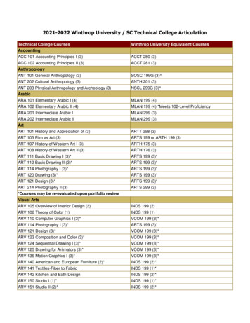
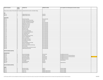
Abdominal: The anterior body trunk region inferior to the ribsAntecubital: The anterior surface of the elbowAxillary: The armpitBrachial: The armBuccal: The cheekCarpal: The wristCervical: The neck regionCoxal: The hipDeltoid: The roundness of the shoulder caused by the underlying deltoid muscleDigital: The fingers or toesFemoral: The thighFibular: The side of the legInguinal: The groinMammary: The breastManus: The handNasal: The noseOral: The mouthOrbital: The bony eye socket (orbit)Patellar: The anterior knee (kneecap) regionPelvic: The pelvis regionPubic: The genital regionSternal: The region of the breastboneTarsal: The ankleThoracic: The chestUmbilical: The navelPosterior Body LandmarksIdentify and appropriately label the following body surface regions in Figure 1 b:Cephalic: The headGluteal: The buttocks or rumpLumbar: The area of the back between the ribs and hips; the loinOccipital: The posterior aspect of the head or base of the skullPopliteal: The back of the kneeSacral: The area between the hipsScapular: The scapula or shoulder blade areaSural: The calf or posterior surface of the legVertebral: The area of the spinal columnBIO 113 Fall 2011 LAB 1Page 2
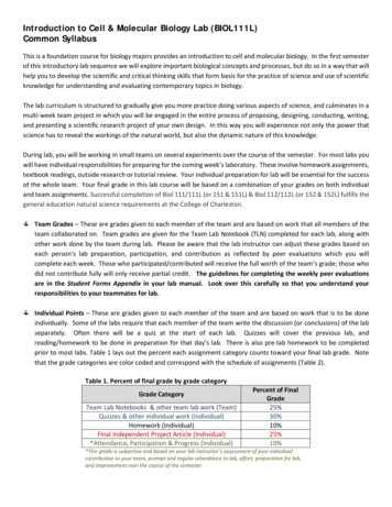
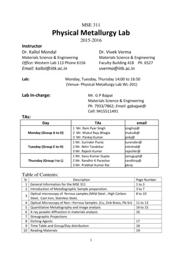
Figure 1. Surface Anatomy. Left image is anterior, Right image is posteriorBody Orientation and DirectionStudy the terms below, referring to Figure 2. Notice as you read that certain terms havea different meaning for a four-legged animal than they do for a human.Superior/inferior (above/below): These terms refer to the location of a structure alongthe long axis of the body. Superior structures appear above other structures, andinferior structures are always below other body parts.Anterior/posterior (frontlback): In humans the most anterior structures are those thatare most forward-the face, chest, and abdomen. Posterior structures are those towardthe backside of the body.BIO 113 Fall 2011 LAB 1Page 3
Medial/lateral (toward themidline/away from the midline ormedian plane): Medial structures arecloser to the body midline than arelateral structures.The terms described above assumethe person is in the anatomicalposition. The next four pairs of termsare more absolute. They do notrelate to a particular body position,and they have the same meaning inall vertebrate animals.Cephalad/caudad (caudal) (towardthe head/toward the tail): In humansthese terms are usedinterchangeably with superior andinferior. But in four-legged animals,they are synonyms of anterior andposterior, respectively.Figure 2. Anatomical terminology for orientation and directionDorsal/ventral (backsidelbelly side): Meaning "back," the term dorsal refers to theanimal's back or the backside of any other structures. The term ventral, meaning "belly,"always refers to the belly side of animals. In humans the terms ventral and dorsal areused interchangeably with the terms anterior and posterior, but in four-legged animalsventral and dorsal mean inferior and superior, respectively.Proximal/distal (nearer the trunk or attached end/farther from the trunk or point ofattachment): These terms locate various areas along the body limbs or an elongatedorgan such as the intestine. For example, the fingers are distal to the elbow; the knee isproximal to the toes.Superficial/deep (toward or at the body surface/away from the body surface or moreinternal): These terms locate body organs according to their relative closeness to thebody surface. For example, the skin is superficial to the skeletal muscles.Practice Using Correct Anatomical TerminologyBefore continuing, use a human torso model, a skeleton, or your own body to specifythe relationship between the following structures.1. The wrist is to the hand.2. The trachea (windpipe) is to the spine.BIO 113 Fall 2011 LAB 1Page 4
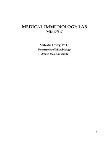
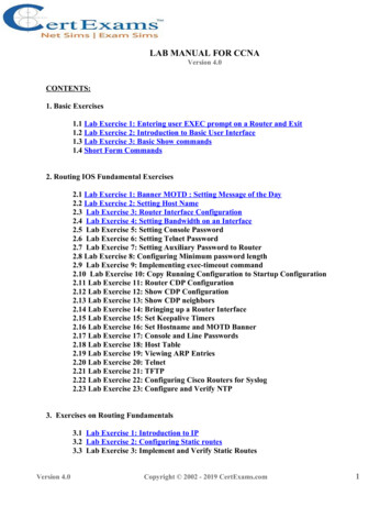
3. The brain is to the spinal cord.4. The kidneys are to the liver.5. The nose is to the cheekbones.6. The chest is to the abdomen.7. The skin is to the skeleton.Figure 3. Planes of the bodyBody Planes and SectionsThe body is three-dimensional. So, to observe its internal parts, it often helps to makeuse of a section, or cut made along an imaginary surface or line called a plane. Thereare three planes (Figure 3), or sections, that lie at right angles to one another.BIO 113 Fall 2011 LAB 1Page 5
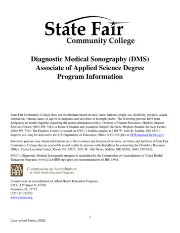
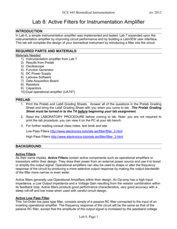
Sagittal plane: A plane that runs lengthwise or longitudinally down the length of thebody, dividing it into right and left parts, is a sagittal plane. If it divides the body intoequal parts, down the midline of the body, it is called a median, or midsagittal, plane.Frontal (coronal) plane: A longitudinal plane that divides the body (or an organ) intoanterior and posterior parts.Transverse plane: A plane that runs horizontally, dividing the body into superior andinferior parts. These sections are also commonly called cross sections.As shown in Figure 4, a sagittal orfrontal section of any nonsphericalobject, be it a banana or a bodyorgan, provides quite a differentview from a transverse section.Observing SectionedSpecimens1. Observe an entire (uncut)kidney and transversely andlongitudinally cut kidneys. Payclose attention to the differentstructural details you can see inthe samples.Figure 4. Longitudial and transverse sections2. Observe a sectioned gelatin-spaghetti mold (cooked spaghetti added to warm gelatinthat is then allowed to gel.) You should see spaghetti strands that have been cuttransversely (XS), longitudinally, and obliquely. Draw each of these spaghetti sectionsbelow.Body CavitiesThe axial portion of the body has two main cavities (Figure 5).Dorsal Body CavityThe dorsal body cavity consists of the cranial and spinal cavities. The cranial cavity,within the rigid skull, contains the brain. The spinal cavity, which runs within the bonyvertebral column, protects the spinal cord. The spinal cord is a continuation of the brain,and the cavities containing them are continuous with each other.BIO 113 Fall 2011 LAB 1Page 6
Ventral Body CavityLike the dorsal cavity, the ventralbody cavity is subdivided. Thesuperior thoracic cavity isseparated from the rest of theventral cavity by the musculardiaphragm. The heart and lungs,located in the thoracic cavity, areprotected by the bony rib cage.The cavity inferior to thediaphragm is the abdominopelviccavity. Although there is no furtherphysical separation of this part ofthe ventral cavity, some describethe abdominopelvic cavity in termsof a superior abdominal cavity,the area that houses the stomach,intestines, liver, and other organs,and an inferior pelvic cavity,which is partially enclosed by thebony pelvis and contains thereproductive organs, bladder, andrectum. Notice that the pelviccavity tips away from theabdominal cavity in a posteriordirection (Figure 5).Figure 5. Body cavitiesAbdominopelvic Quadrants and RegionsThe abdominopelvic cavity is quite large and contains many organs, so it is helpful todivide it up into smaller areas for study. The medical scheme divides the abdominalsurface (and the abdominopelvic cavity deep to it) into four approximately equal regionscalled quadrants, named according to their relative position-they are, right upperquadrant, right lower quadrant, left upper quadrant, and left lower quadrant (Figure 6).Another scheme, commonly used by anatomists, divides the abdominal surface andabdominopelvic cavity into nine separate regions by four planes, also shown in Figure 6.Although the names of these nine regions are unfamiliar to you now, with a littlepatience and study, they will become easier to remember. These regions are:BIO 113 Fall 2011 LAB 1Page 7
Umbilical region: The centermost region,which includes the umbilicus.Epigastric region: Immediately superior tothe umbilical region; overlies most of thestomach.Hypogastric (pubic) region: Immediatelyinferior to the umbilical region;encompasses the pubic area.Iliac regions: Lateral to the hypogastricregion and overlying the superior parts ofthe hip bones.Lumbar regions: Between the ribs and theflaring portions of the hip bones; lateral tothe umbilical region.Hypochondriac regions: Flanking theepigastric region laterally and overlying thelower ribs.ACTIVITYRead through the descriptions of these nine regions locate them in Figure 6. Be sure tonotice the organs they contain.Figure 6. Abdominopelvic surface and cavity.BIO 113 Fall 2011 LAB 1Page 8
Locate the regions of the abdominal surface on a torso model and on yourself beforecontinuing.Looking at Two Dimensional and Three Dimensional MorphologyCreating Serial Sections1. Obtain a hardboiled egg and removethe shell from the egg.Figure 7. Long and short axis andmeridian marks on hardboiled egg2. Using a paper towel, dry the shelledegg. Mark the short and long axismeridians using a Flair (water soluble)type marker (Fig. 7)3. Using the egg slicer, slice egg alongits long axis (Fig. 8).4. Arrange egg slices in order on apiece of paper (Fig. 9). You shouldcreate about 6 to 7 slices.5. Place grid on the first slice and alignaxis marks with the x and y axis (0,0)6. Draw the pattern seen in section oneon the serial section grid labeled“Section One” on Figure 9.Pleasethink about this: Seven slices shouldresult in 6 serial sections.7. Continue for each of the other serialsections. Keep the sections in order;label each in sequence.Figure 8. Hard-boiled egg in slicer along long axisFigure 9. Serial section arranged in order showing axis meridianmarksBIO 113 Fall 2011 LAB 1Page 9
Figure 10. Serial sections through hardboiled eggBIO 113 Fall 2011 LAB 1Page 10
Figure 11. Serial sections through hardboiled egg (continued)Visualizing a section in the 90 degree plane: ReconstructionThe sections you have just drawn are in a plane known as longitudinal. You will nowuse these sections to create a cross section. For this assignment you should choose aplane crossing each of your sections in which there is some of the egg yolk present. Forinstance, suppose you are going to draw the cross section at grid line 5 as shown inFigure 12. From each section at grid line 5 you would transfer the data to that sectionline in Figure 12. When you are done, you smoothly connect the dots and reconstructthe unknown cross section. Try this with your serial sections and reconstruct a 90degree cross section in Figure 10.Figure 12. Reconstruction of 90 degree cross section from serialsection data. Dots are data from single serial sections.BIO 113 Fall 2011 LAB 1Page 11
Figure 13. Reconstruction of 90 degree cross section of hardboiledeggBIO 113 Fall 2011 LAB 1Page 12
Surface Anatomy . Body surfaces provide a number of visible landmarks that can be used to study the body. Several of these are described on the following pages. Locating Body Landmarks . Anterior Body Landmarks . Identify and use anatomical terms to correctly label the following regions on Figure 1: