Transcription
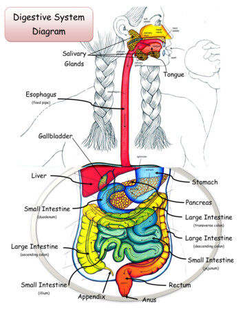
Digestive SystemDiagramSalivaryGlandsTongueEsophagus(food pipe)GallbladderLiverStomachPancreasSmall IntestineLarge Intestine(duodenum)(transverse colon)Large IntestineLarge Intestine(descending colon)(ascending colon)Small Intestine(jejunum)Small Intestine(illium)RectumAppendixAnus
On your Digestive SystemThe Digestive SystemCartoon Label these parts:MouthEsophagusSmall IntestineLarge IntestineSalivaMechanicalDigestionCirculatory SystemHydrochloric seEnzymes from Liverand eLarge IntestineCirculatoryDescending(Transverse Colon)ColonSystemWater and VitaminsKidneys#2#1
On your Digestive SystemThe Digestive SystemCartoon Label these enzymes(chemicals):SalivaHydrochloric leHydrochloric es from Liverand eLarge IntestineCirculatoryDescending(Transverse Colon)ColonSystemWater and VitaminsKidneys#2#1
On your Digestive System Cartoon The Digestive SystemLabel these:NutrientsWater and VitaminsMechanical DigestionChemical DigestionSalivaMechanicalDigestionHydrochloric es from Liverand eLarge IntestineCirculatoryDescending(Transverse Colon)ColonSystemWater and VitaminsKidneys#2#1
On your Digestive System Cartoon The Digestive SystemLabel these:#1 (Urine/pee pee)#2 (Solid Waste/poop)SalivaMechanicalDigestionHydrochloric es from Liverand eLarge IntestineCirculatoryDescending(Transverse Colon)ColonSystemWater and VitaminsKidneys#2#1
The Digestive SystemOn your Digestive System Cartoon Color all partsComplete the SUMMARY of DIGESTION(both of these can be done at home if needed)SalivaMechanicalDigestionHydrochloric es from Liverand eLarge IntestineCirculatoryDescending(Transverse Colon)ColonSystemWater and VitaminsKidneys#2#1
View the Video clip fromA.D.A.M.found on the Human Biology/Links page of ourwebsite (www.myscience8.com)DigestionAnswer all questions on the answer sheetClick here
View the Video clip fromA.D.A.M.found on the Human Biology/Links page of ourwebsite (www.myscience8.com)HeartburnAnswer all questions on the answer sheetClick here
View the Video clip fromA.D.A.M.found on the Human Biology/Links page of ourwebsite (www.myscience8.com)PeristalsisAnswer all questions on the answer sheetClick here
View the Video clip fromA.D.A.M.found on the Human Biology/Links page of ourwebsite (www.myscience8.com)SwallowingAnswer all questions on the answer sheetClick here
View the Video clip fromA.D.A.M.found on the Human Biology/Links page of ourwebsite (www.myscience8.com)UlcersAnswer all questions on the answer sheetClick here
The Digestive Systemis aGiant Food ProcessorMechanical DigestionFood is chopped and ground into smallpieces in the mouth.Chemical DigestionFood is broken down into simplenutrients by the chemical action ofenzymes.NutrientsCarbohydrates are broken downinto simple sugars (glucose) which isused by the cells for energy.Proteins are broken down into aminoacids (the building blocks of cells)which are used to repair old cells andbuild new cells (skin, blood, muscle,bone and nerve).Fats are stored for future use. Theycontain vitamins.
The Mouth Food is cooled or warmed to bodytemperature. Teeth chop and grind food and thetongue mashes the food. Saliva moistens the food and beginsbreaking down carbohydrates. The tongue moves the food to the backof the mouth to be swallowed.The Throat The Epiglottis closes off the wind pipe (trachea). Muscles push food into the esophagus.The Salivary Glands Produce saliva. Saliva is an enzyme(chemical) thatbegins thebreakdown ofstarches. Food becomes moistand “mushy” so thatit can be easilyswallowed. The foodis now called a Bolis.
3rd Molar(wisdom tooth)2nd Molar1st MolarIncisorsPremolarsCanine(Wisdom teeth)Your Teeth are specialized An adult has 32 teeth including 4 wisdom teeth. The Incisors are shaped like knives for cutting and slicing. The Canines have points for piercing and tearing. The Premolars and Molars have broad, bumpy surfaces for grinding.Tooth Anatomy Enamel is the hardest part of tooth. Made mostly of mineral. Dentin is softer than enamel. Contains some living cells. Pulp is also called the “nerve” of the cell. It is a soft tissuethat contains living nerve cells.
The Esophagus Connects the pharynx (throat) to the stomach. About 10 inches long. Flat when empty but changes shape to allowfood to travel to the stomach. Made of several layers of muscle that pushfood through to the stomach (peristalsis).Peristalsisis the name given for thewavelike muscle contractions found in theesophagus, small intestines and largeintestines. It is sort of like squeezingtoothpaste through a tube.Bottom’s up Imean down Imean .PeristalsisEsophagusYes, it is even possible todrink while upside down!!
The Stomach Food enters the stomach from the esophagus. Hydrochloric Acid is produced in the stomach to digest proteins andkill off bacteria. Pepsin (a digestive enzyme) is produced to help digest proteins. Mucus is produced by glands of the stomach to protect the stomachfrom its own acid. Sphincter muscles control both ends of the stomach to allow food toenter and exit. The stomach is made of 3 strong layers of muscle which mixes andmashes the food with digestive enzymes.An ulcer forms when the stomach’s protection breaks down its ownacid begin to eat through the stomach.StomachStomach
Small Intestine The longest part of the alimentarySmallIntestinecanal (digestive tract). Divided into 3 parts:Duodenum – first segmentJejunum – middle segmentIlleum – last segment Digestive enzymes from the liverand pancreas help to break downfood further. Nutrients are absorbed into thebody through the villi.DuodenumJejunumIlleum
The Liver, Gallbladder,andPancreas The Liver produces the enzyme (chemical) bileBile breaks down fats.Liver Bile is stored in the gallbladder and entersthe duodenum (1st part of small intestine)when needed. The Pancreas produces ½ to 1 liter of enzymes(chemicals) daily. These enzymes are used tobreak down carbohydrates as well as fats andPancreasproteins.The Liver: Stores vitamins Stores glycogen for energy Breaks down old red blood cells Removes poisons from the bodyEnzymes fromthe liver andpancreasenter thesmallintestine atthe duodenumLiverPancreasGallbladderg
Nutrients are absorbed throughthe small intestine where the bloodcarries them to all the cells of thebody.The Basic Nutrients are: Amino Acids Simple Sugars Fatty AcidsSmall IntestineThe inside lining of the small intestine containsVilli.These Villi tiny are fingerlike projections throughwhich the nutrients are absorbed into thebloodstream. The Villi capture nutrients as theymove through the small intestine.VilliBlood vesselsPhotograph of Villi magnified(very high power)Note; your microscopewill not show nearly thedetail as in this picture.Glands secretingdigestive enzymes
In theLarge Intestine: Indigestible parts of food move from thesmall intestine to the large intestine. Water and vitamins are absorbed back inthe blood to be reused.Large intestine The remaining waste passes to theRECTUM where peristalsis forces itthrough the ANUS and out of the body.TheLarge IntestineRectumis made of 3 parts: Ascending colonNote: The Appendix serves no Transverse colonuseful purpose. Perhaps it had a Descending colonrole in digesting rough foodsmany, many years ago.Large IntestineLarge Intestine(transverse colon)Large Intestine(descending colon)Large Intestine(ascending colon)AppendixAnusRectum
Try to swallow this some interesting facts about your digestive system. The average digestive tract(alimentary canal) is 27 feetlong! During a lifetime, a personwill process between 60,000to 100,000 pounds of food! Just the sight and smell offood begins the digestiveprocess (saliva in your mouth,esophagus begins to ripple,stomach produces digestiveenzymes) Your stomach can expand tohold 2 ½ pints of food. The liver is the body’s secondlargest organ weighing 3-4pounds. (the skin is thelargest organ) A meal takes between 15 to48 hours to completely digestand move through thealimentary canal.
WhichDigestive Systemorgan is shown in the x-ray?Hint:It stores Bile that wasproduced in the liver.(If this doesn’t help, do some other stations first)
Check out this x-ray:The digestive organ coloredyellow is probably the Small intestine Large intestine Heart PancreasThe digestive organ coloredpink is probable the Small intestine Large intestine Heart Pancreas(answer on your lab answer sheet)Hint: if you are notsure, do some otherstations first.Have a seat, Kermit. What I’m about to tellyou might come as a big shock.
This is Tommythe Torso(but he prefersElvis). Tommy is an expensive, hand painted modelHellothere!of the human torso. His organs are removable butmust be handled with care.Do This:1. Carefully remove the Liver, Stomach, and Intestines.2. Locate and identify the following parts and match themwith the numbers on the model:TongueSalivary numSmall IntestineLarge IntestineAppendixRectumChoose from these 37/138/139Place all answers on you lab answer sheetReturn all partsbefore leavingthis station.Ask if you needhelp.
YourSaliva contains the enzyme amylasewhich breaks down huge starch molecules intosmaller simple sugars.A cracker is mostly carbohydrate (starch) but ifyou leave it in your mouth long enough, it willbecome sugar and you will notice a sweet taste!!Try it!!!Do this:1. Take one unsalted cracker and chew but don’t swallow.2. Keep the bolus (chewed mush cracker) in your mouth for a minute.3. After you notice the sweet taste you may swallow. Yum!!Only onequacker percustomer!!Crackers arelocated on thefront lab table.
How many DigestiveSystem pig parts canyou find in this FetalPig Model?Locate and identify the following parts andmatch them with the numbers on the model:PancreasSmall IntestineGallbladderDuodenum (1st part of small intestine)Large Intestine (caecum)Large Intestine (spiral colon)Large Intestine (Descending colon)LiverChoose from these numbers:Stomach3611Esophagus4712591314
Fetal Pig Internal OrgansFront mach(first part of small intestine)Large IntestinePancreas(Spiral Colon)Large Intestine(Caecum)Small Intestine
Fetal Pig Internal OrgansBack viewLungsEsophagusStomachDiaphragmLiverSmall IntestineLarge Intestine(Descending colon)
Paper Model of Digestive SystemPancreasLiverGallbladderLarge IntestineStomachSmall mall IntestineEsophagusLarge Intestine
Build a paper model of the digestive systemthat looks like the picture below!!1. Color each part so that it looks very similar to the picture.2. Cut out each part carefully and tape it to the outline. Parts must be tapeddown in the proper order beginning with the pancreas.3. Cut out the outline with all of the parts. Find the place on your lab answersheet labeled “Tape Paper Digestive System Here” and tape your completedpaper digestive system in that place.LiverStomachGallbladderLarge IntestineSmall Intestine(Transverse Colon)(duodenum)PancreasLarge Intestine(descending Colon)Small Intestine(Illium)Small Intestine(Jejunum)Large Intestine(Ascending Colon)RectumAppendixAnus
On the computer Go to theHuman Biology/Links page of ourscience website (www.myscience8.com)Click on Digestive System Tour LabFind this page in the lab and click on the links.Answer all questions on your lab answer sheet:1.A Balanced logy/pc/learningsteps/ABDLC/launch.htmlAlso found atwww.myscience8.comHuman Biology/Links tmlAlso found atwww.myscience8.comHuman Biology/Links page
Write these steps ofdigestion in their properorder. They are allSummary of Digestionmessed up here. Hydrochloric acid and pepsin digest proteinsin the stomach. The stomach squeezes to mix food. Nutrients are absorbed into the blood by villi in the smallintestine. Water is absorbed from the food waste back into the body. The tongue pushes food to the back of the mouth where it isswallowed. Food is chopped and ground in the mouth. Bile (produced by the liver and stored in the gallbladder) entersthe small intestine to break down fats. Solid waste material is forced out of the body by action of bothvoluntary and involuntary muscles (if ya know what I mean). “Food” moves to the small intestine (through the duodenum). Waste (food) leaves the small intestine and enters the largeintestine. The food moves along the esophagus to the stomach.
The Basic Nutrients are: Amino Acids Simple Sugars Fatty Acids The inside lining of the small intestine contains Villi. These Villi tiny are fingerlike projections through which the nutrients are absorbed into the bloodstream. The Villi capture nutrients as they move through the small intestine. Villi Photograph of Villi magnified