Transcription
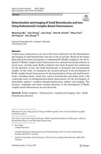
Topics in Current Chemistry(2022) VIEWDetermination and Imaging of Small Biomolecules and IonsUsing Ruthenium(II) Complex‑Based ChemosensorsMiaomiao Wu1 · Zexi Zhang1 · Jiaxi Yong1 · Peer M. Schenk2 · Dihua Tian1 ·Zhi Ping Xu1 · Run Zhang1Received: 30 September 2021 / Accepted: 27 May 2022 The Author(s) 2022AbstractLuminescence chemosensors are one of the most useful tools for the determinationand imaging of small biomolecules and ions in situ in real time. Based on the uniquephoto-physical/-chemical properties of ruthenium(II) (Ru(II)) complexes, the development of Ru(II) complex-based chemosensors has attracted increasing attention inrecent years, and thus many Ru(II) complexes have been designed and synthesizedfor the detection of ions and small biomolecules in biological and environmentalsamples. In this work, we summarize the research advances in the development ofRu(II) complex-based chemosensors for the determination of ions and small biomolecules, including anions, metal ions, reactive biomolecules and amino acids, witha particular focus on binding/reaction-based chemosensors for the investigation ofintracellular analytes’ evolution through luminescence analysis and imaging. Theadvances, challenges and future research directions in the development of Ru(II)complex-based chemosensors are also discussed.Keywords Ru(II) complexes · Chemosensors · Luminescent imaging · Ions · Smallbiomolecules detectionMiaomiao Wu, Zexi Zhang and Jiaxi Yong contributed equally to this work and should be regardedas joint first authors.This article is part of the Topical Collection “Metal Legand Chromophores for Bioassays,” edited byKenneth Kam-Wing Lo and Peter Kam-Keung Leung.* Run Zhangr.zhang@uq.edu.au1Australian Institute for Bioengineering and Nanotechnology, The University of Queensland,Brisbane, QLD 4072, Australia2School of Agriculture and Food Sciences, The University of Queensland, Brisbane, QLD 4072,Australia13Vol.:(0123456789)
29Page 2 of 45Topics in Current Chemistry(2022) 380:29AbbreviationsRu(II) Ruthenium(II)HPLC High-performance liquid chromatographyICP-OES/MS Inductively coupled plasma-optical emission spectroscopy/massspectrometrybpy 2,2′-BipyridineCT Computerized tomographyMRI Magnetic resonance imagingPET Positron emission tomographyCA Contrast agentLoD Limit of detectionϕ Quantum yieldsTGL Time-gated luminescenceIr(III) Iridium(III)Pt(II) Platinum(II)Au(I) Gold(I)Re(I) Rhenium(I)Os(II) Osmium(II)MLCT Metal-to-ligand charge transferILCT Intraligand charge transferLLCT Ligand-to-ligand charge transferMMLCT Metal–metal-to-ligand charge transferLMCT Ligand-to-metal charge transferMLLCT Metal-to-ligand-ligand charge transferLMMCT Ligand-to-metal–metal charge transferMC Metal centeredLC Ligand centeredFRET Förster resonance energy transferPeT Photo-induced electron transferdppz DipyridophenazineROS Reactive oxygen speciesRNS Reactive nitrogen speciesRCS Reactive carbonyl speciesRSS Reactive sulfur speciesF FluorideCH3COO AcetateCN CyanideH2PO4 PhosphateCl ChlorideBr BromideCH3CN AcetonitrileDMSO Dimethyl sulfoxideSCN ThiocyanateCys CysteineHcy HomocysteineGSH Glutathione13
Topics in Current Chemistry(2022) 380:29Page 3 of 4529HSO3 Hydrogen sulfiteS2 SulfidePPi PyrophosphateDPA Di(2-picolyl)amineATP Adenosine triphosphateUCNP Upconversion nanoparticleUCL Upconversion luminescenceNO Nitric oxideONOO PeroxynitriteHNO NitroxylO2 SuperoxideCy5 Cyanine 5 OH Hydroxyl radicalsH2O2 Hydrogen peroxide1O2 Singlet oxygenHOCl Hypochlorous acidDNP 2,4-DinitrophenylBFO Benzofurazan-1-oxideGO GlyoxalCO Carbon monoxideMGO MethylglyoxalFA FormaldehydeCBS Cystathionine β-synthaseCSE Cystathionine γ-lyaseH2S Hydrogen sulfideNBD 7-Nitro-2,1,3-benzoxadiazolesHis HistidineS/N Signal-to-noise1 IntroductionSmall biomolecules and ions are building blocks of living organisms and play indispensable roles in all biological processes, such as enzymatic reactions, metabolism, growth, adaptation, and various disease developments and progressions [1, 2].Determination and monitoring of the levels of these biomolecules and ions in situare essential for better understanding their biological roles in biomedical systems,thus contributing to early diagnosis and treatment assessment of various diseases[3–5]. The most common and typical methods, such as high-performance liquidchromatography (HPLC) and inductively coupled plasma-optical emission spectroscopy/mass spectrometry (ICP-OES/MS), have been developed for the determinationof small biomolecules and ions in vitro [6–8]. Onsite determination of these analytes in situ and in vivo, using these techniques, is not possible because the samplepreparation in solution is an essential step to ensure performing successful analysis[9]. Imaging technologies, such as computerized tomography (CT) [10], magneticresonance imaging (MRI) [11] and positron emission tomography (PET), have been13
29Page 4 of 45Topics in Current Chemistry(2022) 380:29widely used in clinical diagnosis [12], while these technologies cannot be directlyused for the determination of the concentration and/or activity of these analytes inbiological samples [13, 14]. This is mainly because the contrast agents (CAs) usedin these technologies are generally nonspecific; more importantly, these CAs hardlyrespond to small biomolecules and ions at molecular level because of their resolution and sensitivity limitations [15]. Other approaches to optical detection, suchas fluorescence and phosphorescence measurements, have also been successfullydeveloped and adopted in biomedical research and clinical diagnosis [16]. In contrast to conventional bioassay and imaging technologies, luminescence bioassayand imaging using advanced optical spectroscopic and imaging instruments are featured with high sensitivity and selectivity, fast response time and low cost, enablingtheir use in biological and biomedical investigations involving in vitro bioassay andin vivo luminescence bioimaging [17–20].Chemosensors are one of the most important tools for luminescence bioassayand imaging of small biomolecules and ions in situ in real time [21–23]. Luminescent chemosensors are normally designed as chemical compounds that can respondto targeted analytes through a unique binding/reaction (Fig. 1) [24, 25]. Generally,the chemosensors with reaction-based sensing mechanisms have higher selectivity, and the chemosensors with binding-based sensing mechanisms feature excellent reversibility for monitoring the targeted analyte in situ. As a result of theseresponse processes, the luminescence signals can be switched “ON” (Fig. 1A) or“OFF” (Fig. 1B). Of the luminescence switch “OFF” and “ON” responses, theemission switch “ON” chemosensors are preferable for imaging analysis becauseFig. 1 Design of chemosensors for the determination of analytes through the response mechanisms ofbinding and reaction, resulting in “OFF–ON” (A), “ON–OFF” (B), and ratiometric (C) luminescenceresponse to analytes13
Topics in Current Chemistry(2022) 380:29Page 5 of 4529the enhancement of the luminescence intensity can be easily observed by microscopy. The emission wavelengths of the chemosensors can also be shifted after theresponse processes (Fig. 1C) [25–27], allowing ratiometric luminescence detection and imaging of targeted analytes with the potential for precise and quantitativeanalyses. The changes of emission signals generally correspond to the concentrations of the targeted analyte and thus can be recorded for the analyte’s determinationof abundance by luminescence spectroscopes and/or microscopes. Because of theunique advantages of luminescence bioassays and imaging, enormous efforts havebeen devoted to the development of luminescent chemosensors for the detection of avariety of analytes in complicated biological and environmental systems in the pastfew decades.As shown in Fig. 1, luminescence chemosensors generally consist of three parts,including the luminophore, response unit and spacer, which links the luminophoreand response unit. Of all luminophores that are being used for the development ofchemosensors, fluorescent organic dyes are most widely investigated because oftheir high quantum yields (ϕ) and easy modification of chemical structures [28].Lanthanide chelates are another family of luminophores that have been successfullyemployed in the development of chemosensors [29, 30]. Compared with fluorescentorganic dyes, lanthanide chelate luminescence has high photostability, large Stokesshift and unique line-like emissions [29]. The prolonged lifetime of lanthanide chelates (microseconds to milliseconds) enables a background-free bioassay and imaging of targeted analytes through time-gated luminescence (TGL) measurement [28,31]. Transition metal complexes, particularly the luminescent ruthenium(II) (Ru(II))[32], iridium(III) (Ir(III)) [33], platinum(II) (Pt(II)), gold(I) (Au(I)) [34], rhenium(I)(Re(I)) [35] and osmium(II) (Os(II)) complexes with d 6, d8 and d10 electron structures, have also been studied when developing chemosensors for biomolecule andion detection and imaging [36–40]. Different from the fluorescent organic dyes thatemit from excited singlet state, phosphorescence of transition metal complexes isderived from excited triplet states [41]. The excited states of these transition metalcomplexes are more complicated than those of fluorescent dyes and mainly includemetal-to-ligand charge transfer (MLCT), intraligand charge transfer (ILCT), ligandto-ligand charge transfer (LLCT), metal–metal-to-ligand charge transfer (MMLCT),ligand-to-metal charge transfer (LMCT), metal-to-ligand-ligand charge transfer (MLLCT) and ligand-to-metal–metal charge transfer (LMMCT) [41–43]. Theexcited state-mediated emission properties of transition metal complexes are variedupon the changes of the metal center, local environment and particularly chemicalstructure of ligands, enabling transition metal complexes to be designed as the chemosensors through modulating these parameters [43].Of all transition metal complex-based luminophores, Ru(II) polypyridine complex, particularly the prototype of the Ru complex ([Ru(bpy)3]2 (bpy: 2,2′-bipyridine)) (Fig. 2A), has been one of the most popular molecules and widely investigated in the past few decades [44]. Ru(II) polypyridine complexes have octahedralsymmetry with three kinds of electronic transitions, including metal centered(MC), ligand centered (LC) and MLCT [41]. As shown in Fig. 2B, in this octahedral symmetry of Ru(II) complex, MC excited states are obtained for an electron transition from πM to σ*M orbitals, LC excited states are formed through an13
29Page 6 of 45Topics in Current Chemistry(2022) 380:29Fig. 2 Molecular structure of [Ru(bpy)3]2 prototype complex (A). Ru(II) polypyridine complexes’molecular orbital diagram and the corresponding LC, MC and MLCT electronic transitions (B)electron transition from πL to π*L, and MLCT excited states are produced by promotion of an electron from πM metal orbital to π*L ligand orbitals [45]. The lowest excited state MC can decay to the ground state through a fast radiationlessprocess. In contrast, the lowest excited states LC and MLCT undergo radiativedeactivation to the ground state, thus exhibiting intense luminescence at roomtemperature in a rigid matrix and fluid solution, respectively [41]. Consequently,the lowest excited state is 3MLCT for most luminescent Ru(II) polypyridine complexes in solution. Upon the excitation at about 450 nm (spin-allowed 1MLCT),the lowest spin-forbidden 3MLCT excited state is obtained after a fast intersystem crossing process and then emits orange to near infrared emission [46]. The3MLCT-based emission of Ru(II) polypyridine complexes displays unique photochemical and photophysical properties, including large Stokes shift (about150 nm), prolonged luminescence lifetime (hundreds of nanoseconds to microseconds level), high photostability and brightness by visible light excitation [41,47].The emission of Ru(II) complexes, including luminescence intensity and lifetime,can be fine-modulated by modifying the chemical structure of ligands, allowing theRu(II) complexes to be used for the development of chemosensors for biomoleculeand ion detection. As described in early review articles [42, 43, 48, 49], the responsemechanisms of luminescent Ru(II) complex chemosensors for target analytes mainlyinclude (1) photo-induced electron transfer (PeT), in which the Ru(II) polypyridinecomplexes are ideal electron donors and acceptors, (2) Förster resonance energytransfer (FRET), in which the Ru(II) complexes can serve as the energy donor andacceptor [50–53] for the energy transfer (ET), (3) distortion of ligand and the octahedral symmetry after binding/reaction with the target analyte and (4) changes ofthe local environment upon the analyte’s binding. In addition to the luminescenceresponse of Ru(II) complex-based chemosensors to the analyte [40, 54, 55], otherprerequisites for this family of chemosensors to be applied in bioassay and imaginginclude (1) capability of analyte determination in aqueous solution, (2) high sensitivity and selectivity, which allow the chemosensors to be used for targeted analyte detection even at extremely low concentration without non-specific binding of13
Topics in Current Chemistry(2022) 380:29Page 7 of 4529interference species, (3) low cytotoxicity, enabling targeted analyte determinationand imaging with minimum perturbation to the native micro-environment and (4)high cell membrane permeability to ensure the chemosensors are easily internalizedinto biological tissues.The last few years have witnessed a huge leap forward in the development ofRu(II) complexes as the chemosensors for the detection of various analytes. Basedon the above-described response mechanisms, hundreds of Ru(II) complex chemosensors for colorimetric and luminescent determination of anions, metal ions, smallbiomolecules and biomacromolecule have been available [42, 43]. For example, thetriplet nature of the emission state with long lifetime of Ru(II) complexes allowsthem to be used for oxygen sensing by monitoring the changes of their luminescence intensity and lifetime [56–58]. Some of the Ru(II) complexes have also beenrevealed to be nucleic acid sensitive [59, 60], thus serving as a “light switch” forDNA detection [61]. The Ru(II) complexes with dipyridophenazine (dppz) ligands(e.g., complex 1 ([Ru(phen)2(dppz)]2 ) are particularly interesting (Fig. 3A) [61].These complexes are non-luminescent (undetectably small quantum yield) in water,while the emission intensity is significantly increased when bound to DNA [62, 63],which allows the Ru(II) complexes (e.g., complex 2) to be used for imaging of DNAstructure and related mitosis progression (Fig. 3A). Subsequent research has alsorevealed that Ru(II) complexes (e.g., complex 3) can be used for the determinationof DNA mismatches and RNA (Fig. 3B) [64–67]. Since the investigation of cellularuptake and imaging of the Ru(II) complex by Barton’s group [68, 69], the application of Ru(II) complex chemosensors for sensing and imaging of intracellular biomolecules and ions has increasingly attracted interest in recent years.In this chapter, we wish to summarize recent examples of Ru(II) complex chemosensors for the detection of small biomolecules and ions in aqueous solution, witha particular focus on binding/reaction-based chemosensors for the investigation ofintracellular analytes’ evolution through luminescence imaging. Specifically, thechemosensors for the determination of reactive oxygen/nitrogen/carbonyl species(ROS/RNS/RCS), biothiols, amino acids, pH, metal ions and anions are summarized, followed by the discussion of these chemosensors for luminescence bioimaging. The advances, challenges and future research directions in the development ofRu(II) complex-based chemosensors will also be discussed.2 Ru(II) Complex Chemosensors for AnionsIn various chemical and biological processes, anions play important roles in thebody, such as blood pressure stabilization, blood purification, sugar level reduction, respiration and fatigue recovery. For anion sensing, the development of Ru(II)complex-based chemosensors has attracted enormous attention in the past few decades [70–72]. By virtue of their abundant photo-physical and chemical properties,hundreds of Ru(II) complexes have been designed and synthesized for the detection of various anions, such as fluoride (F ) [73–76], acetate (CH3COO ) [77], cyanide (CN )[78], phosphate ( H2PO4 ) [79–81], chloride ( Cl ) and bromide ( Br ).Similar to the design of other anion receptors [82], most Ru(II) complex-based13
29Page 8 of 45Topics in Current Chemistry(2022) 380:29Fig. 3 Molecular structures of examples of Ru(II) complexes 1 ([Ru(phen)2(dppz)]2 ) and 2 for DNAdetermination and imaging (A) and complex 3 ([Ru(Me4phen)2(dppz)]2 ) for detection of DNA mismatches. The luminescence spectra represent the emission of complex 3 with well-matched and mismatched DNA. Adapted with permission from Ref. [67]. Copyright 2016 American Chemical Societychemosensors are designed using the following three response mechanisms, including (1) the binding of the Ru(II) complex’s recognition unit with anions via hydrogen bonding and deprotonation [83], electrostatic and Lewis acid–base interactions[84], (2) specific reactions of the Ru(II) complex’s recognition unit with anions [85]and (3) displacement of metal ions from heterobimetallic Ru(II) complex [86]. Inthe following section, the Ru(II) complex-based chemosensors for anions will bediscussed according to their different response mechanisms.2.1 Response Based on Hydrogen Bonding, Electrostatic and Lewis Acid‑BaseInteractionsAlthough most Ru(II) complex-based anion chemosensors are developed throughthe mechanism of hydrogen bonding and electrostatic and Lewis acid-base interactions, the colorimetric and luminescent response of these Ru(II) complexes to anions can only be obtained in organic solvents, including acetonitrile ( CH3CN) anddimethyl sulfoxide (DMSO) [87, 88]. This is mainly because the hydrogen bonding,electrostatic and Lewis acid-base interactions are significantly inhibited by the watermolecules and other anions in the buffer solution [89]. This sub-section will discuss some examples of Ru(II) complex chemosensors for determination of anions inaqueous solution [90–92].13
Topics in Current Chemistry(2022) 380:29Page 9 of 4529Through modification of the [Ru(bpy)3]2 with amide containing a calixarene moiety (binding site), Maity et al. reported a Ru(II) complex (4) for C N and CH3COO determination (Fig. 4) [93]. Titration of complex 4 with C N and CH3COO in H2O–CH3CN (95:5) resulted in a remarkable luminescence quenchingand enhancement, respectively. The different response mechanisms of complex 4 to CN and CH3COO were investigated by 1H NMR spectra. The CN can bind toeach amide N–H and leads to the deprotonation to form HCN. The electron densityon the bpy ligand increased, and thus the intramolecular quenching was enhanced.In contrast, the weak interaction of bidentate C H3COO with two N–H protons ledto the formation of electron delocalization, in which the CH3COO pulls the electron density to itself and thus decreases the intramolecular quenching. The luminescence response of complex 4 to CN showed a LoD of 70 ppb.Mardanya and co-workers described a Ru(II) complex (5) with pyrene-biimidazole ligand as a chemosensor for highly selective CN detection in both CH3CNand aqueous media (Fig. 5) [94]. The imidazole N–H protons of the coordinatedligand were found to be highly acidic with pKa1 5.09 and pKa2 8.95. Deprotonation of these two N–Hs was found through hydrogen bonding interaction with CN , leading to the increase of electron density of the metal center. As a result, redshift of absorption and quenching of emission were obtained for complex 5 after CN binding. Detection limit of complex 5 to CN was determined to be 5.24 10–9and 4.67 10–9 M for colorimetric and luminescent analyses, respectively. A similar hydrogen bonding-based interaction has been employed for the developmentof Ru(II) complex 6 for selective detection of thiocyanate ( SCN ) (Fig. 5) [95]. Incomplex 6, the S CN interacts with N–H through a 1:1 binding mode, which hinders the photo-induced electron transfer (PeT) between the long pair electron of theFig. 4 Molecular structure of Ru(II) complex 4 and its response mechanism to CN and CH3COO 13
29Page 10 of 45Topics in Current Chemistry(2022) 380:29Fig. 5 Molecular structures of Ru(II) complexes 5 and 6 as the chemosensors for CN and SCN , respectivelyN atom and the Ru(II) complex. The increase of luminescence intensity was thusrecorded for SCN detection in CH3CN-HEPES buffer solution (1:1, v/v, pH 7.2).In an early study, Lin et al. reported a Ru(II) complex (7) for highly selectivedetection of F in water by naked eye and luminescence (Fig. 6) [96]. Complex 7was developed by incorporating a Schiff-base ligand with two bpy ligands. In thepresence of F , the conversion of quinonehydrazone moiety to azophenol couldoccur, resulting in a remarkable red shift of absorption spectra from 475 to 580 nmand a solution color change from orange to blue-violet. The binding of F also ledFig. 6 Molecular structure of Ru(II) complexes 7 and 8 as chemosensors for F . The test paper preparedby complex 7 was then used for F detection in aqueous solution. Adapted with permission from Ref.[96]. Copyright 2006 Royal Society of Chemistry13
Topics in Current Chemistry(2022) 380:29Page 11 of 4529to the increase of luminescence intensity at 630 nm. Although the spectrometricresponses were measured in CH3CN, the test paper prepared by staining of complex 7 also showed color changes in aqueous solution. In a later study, the samegroup modified the 1,10-phenanthroline-5,6-dione ligand to produce complex 8 for F detection (Fig. 6) [97]. In the presence of F , a similar red shift of absorptionspectra (from 467 to 580 nm), color change (from yellow to magenta) and luminescence enhancement were obtained, which were attributed to the F -mediated hydrogen bonding and deprotonation of N–H. The test papers were also prepared for F detection in aqueous solution with ten times higher sensitivity (LoD 1 ppm) thancomplex 7.2.2 Response Based on Specific ReactionsCompared with the above-discussed response mechanism of hydrogen bonding andLewis acid-base interactions, the chemosensors developed by the mechanism ofchemical reaction have high sensitivity and selectivity [78, 98, 99]. The interferencefrom water is also minimized because specific chemical reactions are involved inthe sensing mechanism of these chemosenosors. Aldehyde is a strong electron-withdrawing group that can quench the MLCT emission of Ru(II) complex. In a previous study, a Ru(II) complex (9, [Ru(CHO-bpy)3]2 ) with three aldehyde functionalized bpy ligands was designed and synthesized by Zhang et al. as the chemosensorfor biothiol (cysteine-Cys and homocysteine-Hcy) detection in DMSO-HEPESbuffer [100]. In a later study, Zhang et al. found that the nucleophilic addition ofaldehyde can also be triggered by hydrogen sulfite (HSO3 ) in acidic buffer [24],Fig. 7 Molecular structures of Ru(II) complexes 9 and 10 and their reactions with HSO3 and CN ,respectively13
29Page 12 of 45Topics in Current Chemistry(2022) 380:29thus allowing complex 9 to be used as a chemosensor for HSO3 detection in phosphate-buffered saline (PBS) buffer (50 mM, pH 5) (Fig. 7) [101]. The reaction of HSO3 and complex 9 led to the formation of 9-SO, accompanied by an increase ofluminescence at 620 nm. The result of HSO3 detection in wine and sugar samplesshowed that complex 9 has good precision and accuracy for H SO3 in food samples.Using 5-formyl-2,2′-bipyridine as the ligand, Zhu et al. prepared a Ru(II) complex(10) as the luminescence chemosensor for CN detection (Fig. 7) [102]. Similar to[Ru(CHO-bpy)3]2 , complex 10 in CH3CN-H2O (6:4) showed weak emission at700 nm. The nucleophilic attack by C N led to the formation of 10-CN, accompanied by a blue shift and increase of emission at 618 nm. Complex 10 with a detection limit of 0.75 μM was then used for the preparation of a test paper for naked eye CN detection.In addition to aldehyde, nucleophilic addition between the azo (N N) group and HSO3 has recently been exploited for the development of chemosensors for HSO3 detection (Fig. 8A) [24, 85]. Owing to the PeT from the Ru(II) center to the attachedazo-2,4-dinitrobenzene (DNB), Ru(II) complex 11 (Ru-azo) exhibited weak emission in 25 mM PBS buffer of pH 7.4. The HSO3 triggered reaction with the azogroup led to the formation of 11-SO3 (Ru-SO3), accompanied by an enhancementin luminescence at 635 nm. More interestingly, complex 11 has a long emission lifetime of 258 ns, which enabled its use for background-free luminescence detectionFig. 8 Molecular structure of Ru(II) complex 11 and its reaction with HSO3 (A). Detection of HSO3 inrhodamine (RhB)-contaminated wine samples by steady-state luminescence (B) and TGL (C) analyses.Adapted with permission from Ref. [85]. Copyright 2020 Royal Society of Chemistry13
Topics in Current Chemistry(2022) 380:29Page 13 of 4529of HSO3 through a TGL mode. In a wine sample containing rhodamine (spiked asthe artificial background signal), steady-state luminescence analysis of HSO3 failed(Fig. 8B). TGL analysis eliminated the background signals and allowed for HSO3 detection with high accuracy and precision (Fig. 8C).2.3 Response Based on Displacement of Metal IonsBy modifying the ligands with additional coordination sites, the produced Ru(II)complexes are capable of binding to other metal ions, such as C u2 , Co2 , Zn2 2 and Hg , to form heterobimetallic complexes. The heterobimetallic Ru(II) complexes thus can be used as chemosensors for anion sensing through a displacementapproach. Different from the hydrogen bonding-based anion sensing approach, thedisplacement-based response mechanism also allowed the chemosensors to be usedfor anion detection in water because the water molecules are not involved in the displacement processes. Among various heterobimetallic Ru(II) complexes, the onewith Cu2 (Ru(II)–Cu(II)) has been widely studied because Ru(II) complex luminescence can be quenched by Cu2 binding through electron and energy transfers. Then, Cu2 can be displaced in the presence of several anions, including sulfide ( S2 ), CN and pyrophosphate (PPi) (Fig. 9A).By coupling di(2-picolyl)amine (DPA) to one bpy ligand, Zhang et al. synthesized a Ru(II) complex (12) and then demonstrated the corresponding heterobimetallic Ru(II)–Cu(II) complex as the chemosensor for S 2 detection (Fig. 9B) [86].Complex 12 showed high luminescence (ϕ 3.07%) in HEPES buffer (pH 7.2),while the luminescence was completely quenched upon binding to Cu2 througha 1:1 coordination stoichiometry. In the presence of S 2 , Cu2 was then displacedto form the original complex 12, which was accompanied by the recovery of luminescence. This heterobimetallic Ru(II)–Cu(II) complex showed high sensitivity to S2 (LoD 20.7 nM), allowing its use for S 2 detection in three wastewater samples. In a similar work, Li et al. modified the bpy ligand with a Cu2 receptor,1,4,7,10-tetraazacyclododecane (cyclen), and then demonstrated the Ru(II) complex(13) for sequential C u2 and S 2 detection (Fig. 9B) [103]. Compared with complex12, complex 13 has better selectivity to C u2 binding, while the formed 13-Cu is not2 as sensitive as 12-Cu for S sensing.The displacement approach has also been employed for the development ofRu(II) complex chemosensors for C N . In 2017, two Ru(II) complexes (14 and 15)were synthesized by Zheng and co-workers, and the corresponding heterobimetallicRu(II)–Cu(II) complexes were used for CN detection in 20 mM HEPES buffer (pH7.2) (Fig. 9B) [104]. A recovery of luminescence was observed after the displacementof Cu2 from 14-Cu to 15-Cu to form [Cu(CN )X]n and complexes 14 and 15. TheLoD for C N was then determined to be 0.36 and 0.87 μM using 14-Cu and 15-Cuas the chemosensors, respectively. Similarly, Zhang et al. reported a Ru(II) complex(16) for PPi detection in 2018 (Fig. 9B) [105], in which one Cu2 from the 16-Cu wasdisplaced by the addition of two PPi. The chemosensor 16-Cu showed high sensitivity(LoD 0.58 nM) for PPi detection in 10 mM HEPES buffer (pH 7.4).13
29Page 14 of 45Topics in Current Chemistry(2022) 380:29Fig. 9 The principle of displacement-based Ru(II) complex chemosensors for anion detection (A) andthe molecular structures of Ru(II) complexes 12–163 Ru(II) Complex Chemosensors for pHSimilar to most pH sensors, Ru(II) complex chemosensors for pH have mainly beendeveloped through the mechanism of protonation and deprotonation of several functional groups, such as imidazole [106, 107], hydroxyl (–OH) [108–110], carboxyl(–COOH) [111], pyridine [112, 113] and others [114]. As a result of the protonation-deprotonation process, the molecular structures and the electron density distribution of Ru(II) complex are changed, leading to the variations of absorption andemission intensity/wavelength. In this section, the progress in the development ofRu(II) complex chemosensors for pH will be briefly discussed.As described above, deprotonation of N–H of Ru(II) complex 5’s 2,2′-biimidazole ligand occurs under basic conditions or binding with CN [94]. In 2020,Tormo et al. also reported the use of imidazole-based ligand for the developmentof Ru(II) complex chemosensors for pH [115]. In later research, deprotonation of13
Topics in Current Chemistry(2022) 380:29Page 15 of 4529imidazole N–H under neutral and b
37.Lo KK-W, Louie M-W, Zhang KY (2010) Design of luminescent iridium(III) and rhenium(I) polypyridine complexes as in vitro and in vivo ion, molecular and biological probes. Coord Chem