Transcription
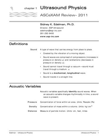
chapter 1Ultrasound PhysicsASCeXAM Review- 2011Sidney K. Edelman, Ph.D.Director, ESP nc.comDefinitionsSoundA type of wave that carries energy from place to place. Created by the vibration of a moving object. Sound waves are comprised of compressions (increases inpressure or density ) and rarefactions (decreases inpressure or density ). Sound cannot travel through a vacuum—sound musttravel through a medium. Sound is a mechanical, longitudinal wave. Sound travels in a straight line.Acoustic VariablesAcoustic variables specifically identify sound waves. Whenan acoustic variable changes rhythmically in time, a soundwave is present.PressureDensityDistanceEdelman · Ultrasound PhysicsConcentration of force within an area. Units: Pascals (Pa)Concentration of mass within a volume. Units: kg/cm 3Measure of particle motion. Units: cm, feet, milesUltrasound Physics · 1
WavesTransverse Wave Particles move in a direction perpendicular (at right angles) tothe direction of the wave:vibrationof particlesdirection of propagationLongitudinal Wave Particles move in the same direction as the wave:direction of propagationvibration of particlesCompressions are regions of higher density & pressureRarefactions are regions of lower density & pressureSeven parameters that describe sound S peWavelengthAcoustic variables identify sound wavesAcoustic parameters describe the particular features ofsound waves2 · Ultrasound PhysicsESP, Inc.
PeriodDefinitionThe time required to complete a single cycle.Period can also be described as the time from the start of acycle to the start of the next cycle.ExampleThe period of the moon circling the earth is 28 days.The period of class in high school may be 50 minutes.UnitsTypical Values sec, seconds, hours—all units of time0.1 to 0.5 sec0.0000001 to 0.0000005sec1 x 10 -7 to 5 x 10 -7 secDetermined ByChanged by SonographerSound sourceNotimeone periodFrequencyDefinitionUnitsDetermined ByChanged by SonographerEdelman · Ultrasound PhysicsThe number of certain events that occur in a particular timeduration.In diagnostic ultrasound, the frequency of a wave is describedas the number of cycles of an acoustic variable that occurin one second.1per second, --------------------- , Hertz, HzsecondSound sourceNoUltrasound Physics · 3
UltrasoundAudible SoundInfrasoundTypical ValuesNoteA wave with a frequency exceeding 20,000Hz.This frequency is so high that it is not audible.Heard by man, a frequency between 20Hz and 20,000Hz.A frequency less than 20Hz.This frequency is so low that it is not audible.Range from 2MHz to 10MHzFrequency is inversely related to penetration.Frequency is related to axial resolution, higher frequencyimproves image quality.Examplecycles 01seconds 023145263784There are 8 cycles that occur in 4 seconds; the frequency of8this wave is --- or 2Hz.4Frequency and PeriodRelated, period and frequency are reciprocals.This is also called an inverse relationship (when one goesup, the other down).In reality, frequency is derived from period.Equationfrequency (Hz) period (sec) 11period (sec) cy (Hz)As frequency increases,period decreases.As frequency decreases,period increases.If period is unchanged,frequency is alsounchanged.1frequency (Hz) -----------------------------------period (sec)Remember to use complementary units:» sec & Hz» msec & kHz4 · Ultrasound PhysicsESP, Inc.
Amplitudeconcerned with the strength of a sound beam.DefinitionUnitsThe difference between the average value and the maximumvalue of an acoustic variable. The variation of an acousticvariable.Those of the acoustic variables: Pressure–PascalsDensity–grams/cubic cmParticle motion–cm, inches, units of distanceAmplitude can also be expressed in decibels, dB. Determined ByChanged by SonographerSound source (initially)YesAmplitude decreases as sound propagates through the body.The difference between maximum and minimum values of anacoustic variable is called the peak-to-peak amplitude.Amplitude is half of the peak-to-peak litudeEdelman · Ultrasound PhysicsUltrasound Physics · 5
PowerDefinitionUnitsDetermined ByChanged by SonographerThe rate that work is performed, or the rate of energytransfer.Watts! Sound source (initially)Units for power:think of a light bulb or stereo!YesPower decreases as sound propagates through the body.IntensityDefinitionThe concentration of energy in a sound beam.The beam’s power divided by the beam's cross sectional area.Intensity depends upon both the power and the crosssectional area of the beam:power watts watts intensity ---------------- ---2 cm 2 beam area cm Equation UnitsDetermined ByChanged by Sonographerwatts/square cm or watts/cm 2 (watts from power, and cm 2from beam area.)Sound source (initially)YesIntensity decreases as sound propagates through the body.Intensity is the key parameter for bioeffects & safety.WavelengthDefinitionThe length or distance of a single cycle.Similar to the length of a single boxcar in a train of infinitelength.UnitsDetermined ByChanged by Sonographermm or any unit of lengthBoth the source and the mediumNoWavelength influences axial resolution (image quality).Typical ValuesEquation6 · Ultrasound Physics0.1–0.8mmpropagation speed mm/ s wavelength (mm) ----------------------------------frequency (MHz)ESP, Inc.
Higher frequency also meansshorter wavelengthLower frequency also meanslonger wavelength.Wavelengths in SoftTissueRule Higher FrequencyLower FrequencyShorter WavelengthLonger WavelengthIn soft tissue, sound with a frequency of 1MHz has awavelength of 1.54mm.In soft tissue, divide 1.54mm by the frequency in MHz.Wavelength (mm) 1.54 / frequency (MHz)Propagation SpeedDefinitionUnitsDetermined ByThe rate that sound travels through a medium.meters per second, mm/ sMedium onlyDensity and stiffnessChanged by SonographerTypical ValuesNo Average speed of all sound (regardless of frequency) inbiologic or “soft tissue:” 1.54km/s 1,540m/s 1.54mm / s Edelman · Ultrasound PhysicsMediumSpeed (m/s)Air330Lung300 - 1,200Fat1,450Soft Tissue1,540Bone2,000–4,000Ultrasound Physics · 7
Rule of ThumbsDensity and Speed — opposite directions Density is related to weightStiffness and Speed — same direction Stiffness is related to‘squishability’» General Rule: gas (slower) liquid solid (faster)Note:EquationAll sound, regardless of the frequency, travels at the samespeed through any specific medium. This means thatsound with a frequency of 5MHz and sound with afrequency of 3MHz travel at the same propagation speedif they are traveling through the same medium.mspeed ----- frequency (Hz) x wavelength (meters) s The Skinnydirectlyrelated toeach otheradjustableamplitudepowerintensity8 · Ultrasound PhysicspeperiodfrequencySinverselyrelated toeach othereddetermined by sound sourcedetermined by mediumdetermined by bothWa velengthESP, Inc.
chapter 2Pulsed UltrasoundBasic Rulethe pulse doesn’t changethe listening time changes when depth of view is alteredPulse DurationDefinitionUnitsDetermined ByThe time from the start of a pulse to the end of that pulse,only the actual time that the pulse is “on”. sec—all units of timeSound sourcePulse duration is determined by the number of cycles ineach pulse and the period of each cycle.Changed by SonographerNo, does not change when sonographer alters imaging depth.Pulse duration is a characteristic of each transducer.Typical ValuesIn clinical imaging, pulse duration ranges from 0.5 to 3 secs.In clinical imaging, a pulse is comprised of 2–4 cycles.timeEquationPulse Duration (msec) # cycles in pulse period (msec)# cycles in pulsePulse Duration (msec) requency (kHz)Pulse Repetition PeriodDefinitionUnitsDetermined ByEdelman · Ultrasound PhysicsPulse repetition period is the time from the start of one pulseto the start of the next pulse. It includes both the timethat the pulse is “on” and the “dead time.”msec—all units of timeSound sourcePulsed Ultrasound · 9
Changed by SonographerYesThe operator changes only the “listening time” (whenadjusting the depth of view), never the pulse duration.Typical ValuesEquationIn clinical imaging, the PR period has values from 100 secto 1 msec.PRP 13 s/cm X depth of view (cm)Deeper ImagingPulse Repetition FrequencyDefinitionPRF is the number of pulses that occur in one second.HINT: PRF is only related to depth of view, it is not related tofrequency.UnitsDetermined ByChanged by SonographerTypical valuesNotesEquationShallow image, higher PRF.Deeper image, lower PRF.Hertz, hz, per secondSound sourceYesIn clinical imaging, from 1,000–10,000Hz (1-10kHz) The PRF depends upon imaging depth. PRF (hz) 77,000 (cm/s) / depth of view (cm) As imaging depth increases, PRF decreases (inverserelationship). In most cases, only one pulse of US travels in the body atone time. Thus, as imaging depths changes, PRF changes.Since the operator determines the maximum imagingdepth, the operator alters the PRF.Thus, the operator also adjusts the pulse repetitionperiod.10 · Pulsed UltrasoundESP, Inc.
Relationship Pulse repetition period and pulse repetitionfrequency are reciprocals (inverse relationship-whenone parameter goes up, the other goes down). Therefore,pulse repetition period also depends upon imaging depth.Equationpulse repetition period X PRF frequency 1Duty FactorDefinitionUnitsThe percentage or fraction of time that the system transmitsa pulse. Important when discussing intensities.» Maximum 1.0 or 100%Unitless!» Minimum 0.0 or 0%If the duty factor is 100% or 1.0, then the system is alwaysproducing sound. It is a continuous wave machine.If the duty factor is 0%, then the machine is never producinga pulse. It is off.Determined ByChanged by SonographerTypical valuesSound sourceYesFrom 0.001 to 0.01 (little talking, lots of listening)As we know, the operator adjusts the maximum imagingdepth and thereby determines the pulse repetition period.Therefore, the operator indirectly changes the duty factorwhile adjusting imaging depth.NoteCW sound cannot be used to make anatomical images.If an ultrasound system is used for imaging, it must usepulsed ultrasound and, therefore, the duty factor must bebetween 0% and 100% (or 0 and 1), typically close to 0.Equationpulse duration (msec)duty factor (%) ----------------------------------------- 100pulse repetition period (msec)Spatial Pulse LengthDefinitionUnitsDetermined ByEdelman · Ultrasound PhysicsThe length or distance that a pulse occupies in space. Thedistance from the start of a pulse to the end of that pulse.mm, meters—any and all units of distanceBoth the source and the mediumPulsed Ultrasound · 11
Changed by SonographerExampleTypical ValuesNoteNoFor SPL, think of a train (the pulse), made up of cars(individual cycles). The overall length of our imaginarytrain from the front of the locomotive to the end of thecaboose.0.1 to 1mm.Important - it determines axial resolution (image quality).Shorter pulses create higher quality images.EquationHintSpatial Pulse Length (mm) # of cycles wavelength (mm)If we know the length of each boxcar in the train and weknow the number of cars in the train, then we know thetotal length of the train!distanceParameters of Pulsed WavesParametersBasic UnitsUnitsDetermined ByTypical Valuespulse durationtimesec, secsound source0.5–3.0 secpulse repetition periodtimesec, msecsound source0.1–1.0 msecpulse repetition frequency1/time1/sec, Hzsound source1–10 kHzspatial pulse lengthdistancemm, cmsource & medium0.1–1.0 mmduty factornonenoneSound Source0.001–0.01By adjusting the imaging depth, the operator changes the pulse repetition period,pulse repetition frequency, and duty factor.The pulse duration and spatial pulse length are characteristics of the pulse itself and areinherent in the design of the transducer system. They are not changed by sonographer.12 · Pulsed UltrasoundESP, Inc.
chapter 3Interaction ofSound and MediaAttenuationDefinitionThe decrease in intensity, power and amplitude of a soundwave as it travels.The farther US travels, the more attenuation occurs.UnitsIn soft tissueThree ComponentsdB, decibels (must be negative, since the attenuationcauses intensity to decrease)Attenuation of sound in soft tissue is1) directly related to distance traveled, and2) directly related to frequency. This is why we are ableimage deeper with lower frequency ultrasound.1. Absorption (sound energy converted into heat energy)2. Scattering3. ReflectionMediaNoteHint Air—much, much more attenuation than in soft tissue Bone—more than soft tissue, absorption & reflection Lung—more than soft tissue, due to scattering Water—much, much less than soft tissueAir Bone & Lung Soft Tissue WaterAttenuation of sound in blood is approximately equal to thatin soft tissue.Attenuation and speed are totally unrelated.Overall attenuation is increased when:1) frequency increases or2) path length increasesEdelman · Ultrasound PhysicsInteraction of Sound and Media · 13
Reflection and ScatteringReflectionOccurs when propagating sound energy strikes a boundarybetween two media and some returns to the transducer.Specular ReflectionReflections from a very smooth reflector (mirror) are specularSpecular reflections also occur when the wavelength is muchsmaller than the irregularities in the boundary. NoteDiffuse (Backscatter)ReflectionSpecular reflectors are well seen when sound strikes thereflector at 90 . Specular reflectors aren’t well seen whenthe wave strikes the reflector at angles other than 90 .Sound returning towards the transducer that is disorganizedand random.Occurs when the boundary has irregularities that areapproximately the same size as the sound's wavelength.ScatteringIf the boundary between two media has irregularities (with asize similar to or a bit smaller than the pulse'swavelength), then the wave may be chaotically redirectedin all directions.Rayleigh ScatteringIf a reflector is much smaller thanthe wavelength of sound, the sound isuniformly diverted in all directions.Higher frequency sound undergoesmore Rayleigh scattering. A red bloodcell is a Rayleigh scatterer.Rayleigh scattering is proportional to frequencyAttenuation & ImagingDepth4Attenuation ultimately limits the maximum depth from whichimages are obtained. The goal in diagnostic imaging is touse the highest frequency that still allows us to image tothe depth of the structures of clinical interest. That is whywe use 2–10 Mhz sound tion(back to transducer)SpecularDiffuse or BackscatterScattering(in all directions)RayleighScatter14 · Interaction of Sound and MediaESP, Inc.
ImpedanceCharacteristic of the medium only.Impedance is not measured, it is calculated.UnitsTypical ValuesRayls, often represented by the letter “Z”.Between 1,250,000 and 1,750,000rayls (1.25 - 1.75Mrayls)Reflection of an ultrasound wave depends upon a difference in theacoustic impedances at the boundary between the two media.Reflection and TransmissionIncident IntensityThe intensity of the sound wave at the instant prior to strikinga boundary.Reflected IntensityThe portion of the incident intensity that, after striking aboundary, changes direction and returns back from whereit came.Transmitted IntensityThe portion of the incident intensity that, after striking aboundary, continues on in the same general direction thatit was originally traveling.UnitsW/cm 2 (they are all ReflectedIntensityEquationincident intensity reflected intensity transmitted intensity“Conservation of energy” occurs at a boundary.Reflection With Normal IncidenceWith NORMAL incidence: Reflection occurs only if the two media at the boundaryhave different acoustic impedances.With greater impedance differences between the two media,the greater the IRC and the greater amount of reflection.Z2 – Z1 2% Reflection --------------------Z2 Z1Edelman · Ultrasound PhysicsInteraction of Sound and Media · 15
Transmission With Normal IncidenceWith NORMAL incidence:These are simply reflection questions, whatever remains aftertransmission, must be reflected!Reflection & Transmission With Oblique IncidenceExtremely complex physics regarding transmission &reflection with obliquity.I Don’t Know!Transmission and reflection may or may not occur withoblique incidence, but there are no “simple” rules.What we DO know with oblique incidence:RememberWith oblique incidence, weare uncertain as to whetherreflection will occur. Simplysay “I don’t know!”Incident Intensity Transmitted ReflectedReflection Angle Incident AngleIncidentir? Reflection? TransmissionSpecular reflections arise when the interface is smooth.RefractionDefinitionRefraction is transmission with a bend.Occurs when two conditions are met :1. oblique incidence & 2. different propagation speedsEquationThe physics of refraction are described by Snell's Law.propagation speed 2sine (transmission -------------------------------- ----------propagation speed 1sine (incident angle)16 · Interaction of Sound and MediaESP, Inc.
ExamplesIf propagation speed 2 is less than propagation speed 1, then thetransmission angle is less than the incident angle. If propagation speed 2 is greater than propagation speed 1, thetransmission angle is greater than the incident angle.Medium 2 Slower1 Same Speeds21I Medium 2 Faster21I 2IT1T2T3Range EquationWhen know the time-offlight, we can determinethe distance.Time-of-flightUltrasound systems measure “time-of-flight” and relate thatmeasurement to distance traveled.Since the average speed of US in soft tissue (1.54 km/sec) isknown, the time-of-flight and distance are directly related. The time needed for a pulse to travel from the transducer to thereflector and back to the transducer is called:» the go-return time or the time-of-flight Equationsgo-return time s speed mm s distance to boundary (mm) -----------2In soft tissue:mmdistance to boundary (mm) time s 0.77 ---------- sThe 13 Microsecond RuleEdelman · Ultrasound PhysicsTime-of-FlightReflector DepthTotal DistanceTraveled13 s1cm2cm26 s2cm4cm39 s3cm6cm52 s4cm8cmInteraction of Sound and Media · 17
Axial ResolutionResolutionThe ability to image accurately (accuracy, not merely quality)Axial ResolutionThe ability to distinguish two structures that are close to eachother front to back, parallel to, or along the beam’smain axis.SynonymsUnitsWith shorter pulses, axialresolution is improved.longitudinal or axialmm, cm — all units of distanceThe shorter the pulse, the smaller the number, the better thepicture quality.Changed by sonographerNo, a new transducer is needed to change axial resolution.Note“Short pulse” means a short spatial pulse length or a shortpulse duration.Ultrasound transducers are designed by the manufacturers tohave a minimum number of cycles per pulse, so that thenumerical value is low and the image quality is superior.Typical ValuesHintEquation0.05–0.5mmpulse duration & pulse duration are determined by theexcitation voltage of the transducer crystalspatial pulse length (mm) Axial resolution (mm) ---------------------------2For soft tissue:0.77 # cycles in pulseAxial resolution (mm) -------------------frequency (MHz)Less ringing (fewer cycles in pulse)Higher frequency (shorter wavelength)18 · Interaction of Sound and MediaESP, Inc.
Lateral ResolutionDefinitionthe minimum distance that two structures are separated byside-to-side or perpendicular to the sound beam thatproduces two distinct echoes.SynonymsLateral Angular Transverse AzimuthalLATA resolution.Unitsmm, all units of lengthsmaller number, more accurate imageNoteSince the beam diameter varies with depth, the lateralresolution also varies with depth.The lateral resolution is best at the focus or one near zonelength (focal depth) from the transducer because thesound beam is narrowest at that point.See question #65 in thecomprehensive exam.NoteWhen two side-by-side structures are closer together thanthe beam width, only one wide reflection is seen on theimage.NoteLateral resolution is usually not as good as axial resolutionbecause US pulses are wider than they are short.Be aware of the term “POINT SPREAD ARTIFACT”HintLateral resolution isapproximately equal tobeam diameter.Lateral resolution at a variety ofdepths can be assessed with atest phantom by measuring thewidth of a reflection created bya pin in the phantom.imageWider reflections at depthsfurther away from the focusexhibit poor lateral resolution.Edelman · Ultrasound PhysicsInteraction of Sound and Media · 19
FocusingResults in:1. a narrower “waist” in the US beam.2. a decrease in focal depth (the focus is shallower).3. a reduction in the size of the focal zone.Effective mainly in the near field and the focal zone.HINT (maybe)FOCAL DEPTH (Diameter2) / (4 x wavelength)Electronic FocusingPhased array technology provides dynamic, variable(adjustable) focusing or multi-focusing.Transducer lCaseActive ElementThe piezoelectric crystal.CaseProtects the internal components from damage and insulatesthe patient from electrical shock.WireEach active element in a transducer requires electrical contactso that the voltage from the US system can excite thecrystal to vibrate thereby producing an ultrasonic wave.Similarly, during reception the sound wave deforms thecrystal, producing a voltage. The voltage is sent back tothe ultrasound system for processing into an image.Matching LayerRecall that impedance differences result in reflection atboundaries. The matching layer has an impedancebetween those of the skin and the active element toincrease the percentage of transmitted US between theactive element and the skin. Gel’s impedance is inbetween those of the matching layer and the skin.The matching layer is one-quarter wavelength thick.Impedances: PZT matching layer gel skin20 · Interaction of Sound and MediaESP, Inc.
Damping Element orBacking MaterialA material that is bonded to the active element that limits the“ringing” of the PZT. Commonly made of epoxy resinimpregnated with tungsten.Damping material - advantages:» shortens spatial pulse length, pulse duration» decreases numerical value of axial resolution» improves axial resolution & picture accuracyDamping material – also causes:» decreased transducer's sensitivity» increased bandwidth (range of frequencies) in thepulse - also called wide bandwidth » decreased “Q” factor. Imaging probes are low-QBandwidth and Quality FactorBandwidthcenterfrequencypowerIt is uncommon for a transducer to emit a sound beam withonly a single pure frequency. Rather, the pulse is morelike a sound ‘click’ and contains a range of frequenciesbelow and above the main frequency.The bandwidth is the range of frequencies between thehighest and the lowest frequency emitted from thetransducer.frequencyQuality FactorA unitless number representing the degree of damping.Imaging transducers are low-Q transducers whencompared to therapeutic transducers because imagingtransducers use backing material.The Q-factor of typical imaging transducers can beapproximated by the number of cycles in the pulseproduced by the transducer (approximately 2 - 4).Equationresonant frequency (MHz)quality factor ---------------------------bandwidth (MHz)When Q-factor is low (imaging probe):1. damping is effective2. pulse length & duration are short3. bandwidth is wide4. axial resolution is improvedEdelman · Ultrasound PhysicsInteraction of Sound and Media · 21
Transducer FrequenciesWhat determines the resonant frequency of a transducer?ContinuousWaveTransducersSound wave's frequency equals the frequency of the voltageapplied to the PZT by the machine's electronics.Electrical FrequencyPulsedTransducersSound’s FrequencyThe pulse repetition frequency (PRF) is determined by thenumber of electrical pulses the US machine delivers to theactive element.The frequency of the US for a pulsed txr is determined by 2 factors:1. the thickness and2. the propagation speed of the piezoelectric material.» propagation speed for PZT is approx. 4-6 mm/ s.EquationNote22 · Interaction of Sound and MediaHigher FrequencyLower Frequencythin crystalthick crystalfast PZTslow PZTmaterials propagation speedfrequency (MHz) -----------------------------------2 thickness (mm) The thickness of the PZT crystal equals ½ of thewavelength of sound in the crystal. The thickness of the matching layer is ¼ of thewavelength of sound in the matching layer. ESP, Inc.
Sound BeamsBeam width. RULE: Narrow beams create better imagesAs sound travels, the width of the beam changes:» starts out at exactly the same size as the transducerdiameter,» gets progressively narrower until it reaches its smallestdiameter, and then» it divergesfocusfocal depthfar zonenear zonefocal zoneFocus or Focal PointThe location where the beam reaches its minimum diameter.Focal Depth - the distance from the transducer face to thefocus. Also called focal length or near zone length.Near Zone(Fresnel Zone)Far Zone(Fraunhofer Zone)Focal ZoneNoteThe region or zone in between the transducer and the focus.Sound beams converge in the near zoneThe region or zone deeper than the focus, beyond the nearfield. Sound beams diverge in the far zone.NEAR ZONEFAR ZONEshort namelong nameFresnelFraunhoferThe region surrounding the focus where the beam is “sort ofnarrow” and the picture is relatively good.For an unfocused continuous wave disc transducer:At the end of the near zone, the beam diameter is ½ thetransducer diameter.At two near zone lengths, the beam diameter is equal tothe transducer diameter.Edelman · Ultrasound PhysicsInteraction of Sound and Media · 23
Focal DepthDefinitionDistance from transducer to the focal point.Determined by two factors:1. transducer diameterand2. frequency of the ultrasound.HINTCompared to beams with a shallow focus, beams with a deepfocus have a lower intensity at the focus.SHALLOW FOCUSDEEP FOCUSsmall diameterlarge diameterlow frequencyhigh frequency2Equation transducer diameter frequencyFocal Length (cm) ----6Sound Beam DivergenceDefinitionDescribes the spread of the sound beam in the deep far zone.Determined by two factors:1. the transducer diameterand2. the frequency of the ultrasoundHintNumerical question: In the far field, beam is narrow (lateralresolution is best) with large diameter, high frequencysound. In the far field, beam is wide (lateral resolution isworst) with small diameter, low frequency sound.LESS DIVERGENCEMORE DIVERGENCEnarrower beam in far fieldwider beam in far fieldlarger diameter crystalsmaller diameter crystalhigh frequencylow frequencyimproved lateral resolutionin far fielddegraded lateral resolutionin far fieldLarger diameter crystals producinghigher frequency sound producebeams that diverge less in the farfield.Smaller diameter crystals producinglower frequency sound producebeams that diverge substantially inthe far field.24 · Interaction of Sound and MediaESP, Inc.
Mechanical ScanningCrystalsScanhead contains one active elementSteeringThe active element is moved by a motor, oscillating crystalor mirror through a pathway, automatically creating ascan plane.FocusingConventional or Fixed: curvature of the PZT or an acousticlens focuses the beam at a specific depth Transducer ArraysArrayA collection of active elements in a single transducer.ElementA single slab of PZT cut into a collection of separate piecescalled elements.ChannelThe electronic circuitry is connected to each element.Phased ArraysMeaningCrystals, Steering &FocusingEdelman · Ultrasound PhysicsAdjustable focus or multi-focus; achieved electronically.A collection of electric pulses, separated by miniscule timedelays (10 ns is delivered to all of the transducer’selements in various patterns. The patterns focus & steerthe US beam during transmission. Thus, focusing andsteering are electronic.Interaction of Sound and Media · 25
Real Time Imaging & Temporal ResolutionReal-Time ImagingThe production of a motion picture. Frames displayed in arapid fashion to give the impression of constant motion.Machines displaying both real-time imagesand Doppler at one time are called duplex.Determined By Temporal resolution depends only upon frame rate. Moreimages per second improves temporal resolution. UnitsFrame rate has units of Hertz, or “per second”Determined by 2 factors1. Imaging depth - shallower image depth» higher frame rate; improved temporal resolution2. Number of pulses per frame - fewer pulses» higher frame rate; improved temporal resolutionHINTFrame rate is limited by 2 factors:1) the speed of sound in medium, and/or2) the depth of view.Imaging DepthWith regard to maximum imaging depth, what will create aframe in less time?Shallow depth of view makes a frame faster, andimproves temporal resolution.NoteIf imaging depth of view is doubled (for example from 6cm to12cm), the frame rate will be halved.Shallow Imaging (less depth)26 · Interaction of Sound and MediaDeep Imaging (more depth)Higher frame rateLower frame rateBetter temporal resolutionPo
Edelman · Ultrasound Physics Ultrasound Physics · 7 Wavelengths in Soft Tissue In soft tissue, sound with a frequency of 1MHz has a wavelength of 1.54mm. Rule In soft tissue, divide 1.54mm by the frequency in MHz. Wavelength (mm) 1.54 / frequency (MHz) Propagation Speed