Transcription
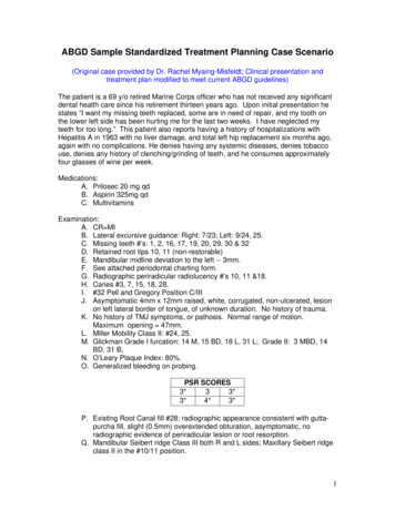
ABGD Sample Standardized Treatment Planning Case Scenario(Original case provided by Dr. Rachel Myaing-Misfeldt; Clinical presentation andtreatment plan modified to meet current ABGD guidelines)The patient is a 69 y/o retired Marine Corps officer who has not received any significantdental health care since his retirement thirteen years ago. Upon initial presentation hestates “I want my missing teeth replaced, some are in need of repair, and my tooth onthe lower left side has been hurting me for the last two weeks. I have neglected myteeth for too long.” This patient also reports having a history of hospitalizations withHepatitis A in 1963 with no liver damage, and total left hip replacement six months ago,again with no complications. He denies having any systemic diseases, denies tobaccouse, denies any history of clenching/grinding of teeth, and he consumes approximatelyfour glasses of wine per week.Medications:A. Prilosec 20 mg qdB. Aspirin 325mg qdC. MultivitaminsExamination:A. CR MIB. Lateral excursive guidance: Right: 7/23; Left: 9/24, 25.C. Missing teeth #’s: 1, 2, 16, 17, 19, 20, 29, 30 & 32D. Retained root tips 10, 11 (non-restorable)E. Mandibular midline deviation to the left -- 3mm.F. See attached periodontal charting form.G. Radiographic periradicular radiolucency #’s 10, 11 &18.H. Caries #3, 7, 15, 18, 28.I. #32 Pell and Gregory Position C/IIIJ. Asymptomatic 4mm x 12mm raised, white, corrugated, non-ulcerated, lesionon left lateral border of tongue, of unknown duration. No history of trauma.K. No history of TMJ symptoms, or pathosis. Normal range of motion.Maximum opening 47mm.L. Miller Mobility Class II: #24, 25.M. Glickman Grade I furcation: 14 M, 15 BD, 18 L, 31 L; Grade II: 3 MBD, 14BD, 31 B,N. O’Leary Plaque Index: 80%.O. Generalized bleeding on probing.PSR SCORES3*33*3*4*3*P. Existing Root Canal fill #28; radiographic appearance consistent with guttapurcha fill, slight (0.5mm) overextended obturation, asymptomatic, noradiographic evidence of periradicular lesion or root resorption.Q. Mandibular Seibert ridge Class III both R and L sides; Maxillary Seibert ridgeclass II in the #10/11 position.1
CLINICAL PHOTOS2
3
4
5
ABGD Sample Standardized Treatment Planning Write-UpI. DIAGNOSIS/PROBLEM LISTA. CHIEF COMPLAINT:“I want my missing teeth replaced, some are in need of repair, and my tooth onthe lower left side has been hurting me for the last two weeks. I have neglectedmy teeth for too long.”B. MEDICAL/SYSTEMIC1. ASA I2. Meds: Prilosec, Aspirin3. Total joint replacement of left hip six months agoC. ORAL PATHOLOGY1. Asymptomatic 4mm x 12mm raised white lesion of unknown durationand no history of trauma on left lateral border of tongue.D. PERIODONTAL1. Generalized moderate chronic periodontitis w/localized severe periodontitis2. Ineffective oral hygiene.3. Localized slight calculus & generalized plaque.4. Miller Mobility Class II: 24, 25 (Hopeless)5. Glickman Grade I: 14M, 15 BD, 18 L, 31L; Grade II: 3MBD, 14BD, 31B.6. Marginal ridge discrepancies: 13/14, 14/15.7. Biologic width violation and poor crown/root ratio #28.E. ENDODONTICS1. Radiographic periradicular lesion associated with mesial root #18.Diagnosis consistent with necrotic pulp and symptomatic apicalperiodontitis.2. #28 - 0.5mm overextended obturation.F. ORAL SURGERY1. Pneumatization of maxillary sinus.2. #32 complete bony impaction; Pell and Gregory Position C/III -- No clinicalor radiograohic pathology evident.3. Nonrestorable roots: #10 & 11.G. RESTORATIVE1. Generalized tooth decay2. Carious lesions: #3 DOF, #7 MI.2. Defective restorations/recurrent decay: #15, 18 and 28.6
3. Cervical abfraction/abrasion lesions: Moderate: 4, 5, 13, 14, 23; Severe:#12, 214. Caries Risk: HighH. OCCLUSION1. Angle’s borderline class 3 right cuspid relationship2. Compromised anterior guidance due to mutilated dentition.3. Uneven occlusal plane due to supererupted and mesially tilted teeth.4. Inadequate dentition for optimum masticatory function.5. Cervical abrasion/abfraction – generalized.6. Lateral excursive guidance: Right: 7/23; Left: 9/24, 25. No protrusiveinterferences noted.I. PROSTHODONTICS1. Missing teeth of concern: #’s 2, 10, 11, 19, 20, 29 & 302. Mandibular left and right posterior edentulous ridges Seibert Class IIISeibert Class II on Maxillary arch #10/11 area.3. #18 FVC with mesial open margin/decay,4. #28 FVC with insufficient post length with distal open margin/decay.5. Biologic width violation and poor crown/root ration of #28.6. Hopeless 24,257. Unesthetic appearance8. Mesial tilt 18, 319. Occlusal plane discrepanciesII. TREATMENT OBJECTIVE/OVERVIEWThe main objective for this particular case is to restore the patient to a state ofoptimum dental health, function and esthetics that can be maintained by the patient.Not to be forgotten will be the need to address the patient’s chief complaint at themost reasonable and opportune time during the various phases of the treatment plan.In order to achieve this goal, the following areas must be addressed .1. Oral Surgery consultation -- biopsy tongue lesion.2. Orthopedic consult – antibiotic prophylaxis for total joint replacmement.3. Diagnostic wax-up for occlusal analysis using 20 template.4. Extract #10 &11, 24, 25 and deliver Maxillary/Mandibular TTPs.5. Actively treat and control the carious disease process through education,diet modification, elimination of active lesions and defective restorations,use of fluoride, Xylitol products, and chlorhexidine.6. Achieve and maintain periodontal health through the reinforcement of oralhygiene, effective home care, and professional in-office treatment.Scaling and root planning will be performed as initial therapy, followed bya 6 week reevaluation. Periodontal maintenance therapy will beperformed every three months according to current accepted therapeuticprotocols.7. #12 elective NSRCT; #18 NSRCT; #28 NSRCT for use as overdentureabutment.7
8. Restore generalized abfraction/abrasion lesions.9. Reevaluate the patient’s disease risk assessment prior to progressing tothe “corrective phase” of treatment.10. Develop a right side mutually protected occlusal scheme and a left sidegroup function.11. #3, 18 & 31 crown lengthening surgery and mandibular anterior pocketreduction procedure. Option for crown lengthening procedure on #12 ifrequired to maintain biological width – to be determined after electiveNSRCT.12. Surveyed gold crowns # 3,18 & 31. Surveyed PFM #1213. RBB 23-2614. Maxillary Class 3 Lateral Rotational Path RPD and Mandibular Class 3Mod 1 RPD to replace missing teeth to increase patient’s chewingefficiency and provide stability of occlusion. Due to the patient’s currentfinancial status and personal preference, removable partial dentures willbe selected as the treatment of choice vice implants.15. Three month recall for periodontal maintenance, caries assessment,endodontic, prosthodontic, and biopsy follow ups.III. TREATMENT PLAN WITH RATIONALEA. EMERGENCY PHASETreatment#18 PulpectomyRationale-Relieve patient of pain, prevent progression to acuteapical abscess, and to gain patient trust andconfidence. The tooth displays a painful response tobiting and percussion, and demonstrates an apicalradiolucency. Calasept a calcium hydroxide intracanaldressing, placed between appointments for itsantimicrobial affect; Glass Iononmer temp to minimizemicroleakage.B. SYSTEMIC PHASETreatmentOrthopedic ConsultRationale-Total joint replacement within a two year periodplaces patient at a potential increased risk ofhematogenous total joint infection. Response fromconsult Orthopedic surgeon recommends antibioticprophylaxis of Amoxicillin 2g orally 1 hour prior toeach dental procedure.8
C. PREPARATORY PHASETreatmentOral Surgery consult for lingual lesion – biopsyRationale- To determine etiology and treatment options.Alginate impressions for TTP’s 10, 11, 24, 25-To maintain esthetic appearance during initial phaseof treatment; affords time for reevaluation of tissueresponses and patient compliance during thepreparatory phase to treatment. .Odontectomy #10, 11, 24, 25 w/ridge preservation-To eliminate non-restorable and grossly carious rootsas well as hopeless periodontally involved teeth. Hardand soft tissue ridge preservation utilizing Bio-Ossosteoconductive xenograft material to minimizeosseous defects and compromised esthetic outcomes.Resorbable collogen membrane to contain particlesand preserve graft.2% Lidocaine with 1:100,000 epinephrine (will beused for all dental procedures requiring localanesthesia)-Amide; 60 min pulpal, 3-4 hrs soft tissue anesthesia,low occurrence of allergic reactions.Deliver maxillary/mandibular temporary treatmentpartial dentures-Acrylic TTPs to provide an acceptable estheticappearance and to maintain space.Occlusal analysis and diagnostic wax-upAlginate impressions (3-sets)Hanau wide-view semi-adjustable articulator-To evaluate initial presentation of tooth-to-tooth andarch-to-arch relationships -- CO/MI discrepancy. Todevelop the most practical, protective, and functionalocclusal scheme; evaluate final restorationcontours/esthetics; fabricate stents for tooth reductionguides and provisional restorations. Develop customguide table to establish/preserve anterior guidance forduplication by the laboratory. Fabricate custom trays.Presentation of case to patient.-Inexpensive, quick and easy, good accuracy ofsurface detail. One set each for diagnostic mounting,diagnostic wax-up, and RPD designs-Class 3 Arcon semi-adjustable articulator;recommended when multiple restorations, FPD’s orRPD’s are going to be fabricated and there is a greaterneed for accuracy. Accepts a facebow transfer; canadjust horizontal and lateral condylar guidance;adjustable anterior guide table.9
Face Bow-Transfers the relationship of the maxillary teeth andthe transverse horizontal axis to the articulator, therebyduplicating the arc of closure exhibited by the patient.Protrusive and lateral bite records-Used to set the horizontal condylar inclination andBennett angle to simulate excursive movements.Diagnostic mountings-Aids in evaluating maxillary/mandibular occlusalrelationships and occlusal discrepancies. Equilibrationof duplicate casts aids in determining the extent ofocclusal adjustment needed to establish a coincidentMI/CO relationship and elimination/reduction ofexcursive interferences; survey hard/soft tissueundercuts, evaluate retentive areas and for claspdesign, evaluate guide planes, plan for surveyedcrowns.Occlusion will be restored utilizing a 20 occlusaltemplate-Reconstruction to a 20 template could beaccomplished in this case without requiring significantextensive restorative effort. Reasonable compromisebetween esthetics, chewing efficiency and cusp height;minimizes damaging lateral stresses.-The jaw-to-jaw relationship in which the condylesarticulate with the thinnest avascular portion of theirrespective discs with the complex in the anteriorsuperior position against the slopes of the articulareminences, regardless of any tooth-to-toothrelationship; A repeatable and verifiable position fromwhich to restore the occlusion; Long Centric Freedom to close the mandible either into centricrelation or slightly anterior to it without varying thevertical dimension at the anterior teeth.Restore in Long Centric OcclusionDevelop a mutually protected occlusal scheme onthe right side-Natural cuspid teeth are present and functional. Morepredictable treatment outcome, minimal adaptationrequired by the neuromuscular system, decreasedmuscle activity, reduced wear and stress on posteriorteeth, increased stability. Stable occlusal position,stable excursive contacts10
Develop a group function occlusal scheme on theleft side-Missing left canine, impractical to restore in canineguidance; widens distribution of forces; minimizesstresses on RPD framework, remaining teeth andresidual ridge.Medical Model of Caries Control-Risk assessment/combat S. Mutans infection,Modified O’Leary plaque index, Establish daily oralhygiene regimen, diet analysis and counseling, 2%NaF in-office tx of 4 applications over 4 weeks –antimicrobial; Prevident 5000 toothpaste –remineralization; Peridex regimen – antimicrobial;Xylitol gum – antimicrobial; Restore carious teethand defective restorations to eliminate seeding lesions;Reevaluation to determine patient compliance andefficacy of treatment.Scaling and Root Planning-Root plane areas with probing depths 4.3mm,remove accretions and endotoxins, alter subgingivalmicroflora, detoxify root surfaces, resolve gingivalinflammation, reduce probing depths throughestablishment of a long junctional epithelium, establishbiologically acceptable root surface for reattachmentand/or regeneration procedures; 4-6 wk follow-up toevaluate tissue responseNSRCT # 18;Elective endo retreat #28.Elective endo #12Step-Down-Step-Back Technique-Periradicular lesion #18, previously initiated therapyat emergency phase. Initial presentation consistentwith symptomatic apical periodontitis.-To use as overdenture abutment w/3i locatorattachment.-Severe abrasion has resulted in visualization of thebuccal root canal. For best long term prognosis andthe need to establish proper mesial undercut for arotational path RPD via a surveyed PFM, electiveNSRCT is recommended.-Pre-flare with Gates-Glidden, Anti-curvature filing tomaintain integrity of canal walls, instrumentation ofcoronal 1/3 improves access to the apical 1/3. Earlyflaring improves irrigation and reduces apical debrisextrusion. Instrument within 0.5-1mm of radiographicapex to maintain anatomical apical constriction and11
control zone.5.25% Sodium hypochlorite irrigation;RC Prep-Baceriocidal, dissolves residual tissue, lubricates, aidsin debridement.-Oxygenating/chelating agent, lubricant, facilitatesimproved debridement by keeping dentinal debris insuspension.Lateral condensation with Gutta-Percha and Roth’s -Comparable or superior to alternative techniques,proven apical seal. Gutta-percha is biocompatible,Sealercapable of a three dimensional fill, easy to retreat andradiopaque. Roth’s sealer fills voids in gutta-percha,improves apical seal, radiopaque, fills lateral canalsand aids in placement of gutta-percha points.Perma-flo Purple orifice sealer-Provides interim coronal seal minimizing chance ofmicroleakage and recontamination.Cavit access closure-Provides adequate temporary seal, easy to use,inexpensive.Operative restorations:-Rubber dam isolation for all operative procedures,provides isolation, tissue retraction, improvedvisualization, protects the patient from foreign bodyinhalation, provides increased infection control byreducing aerosol contaminants.#18 Core amalgam-Tytin spherical alloy for enhanced early compressivestrength, high copper content and superior mechanicalproperties for core substructures. To be placed inpreparation for full gold crown prep and surveyedcrown.3 & 15 (Amalgam)- Dispersalloy (admix) amalgam, excellentcompressive strength and mechanical properties,adequate working time, condenses well to developcontacts.7 (Composite Resin)-Acid etch (37% phosphoric acid); PQ1 bonding agent 61MPa bond strength, radiopaque, bonds todentin/enamel, cast metal, porcelain, amalgam andcomposite. Amelogen Plus composite restorativematerial microhybrid composite with 0.7micron maxparticle size, radiopaque, superior polish, 76% fill by12
weight, increased wear resistance and strength, usedfor Class 1 through 6 restorations.Cervical lesions (resin modified glass ionomer).-Fuji II LC excellent retention to dentin, fluoriderelease, biocompatibility, chemical bond to enameland dentin, enhanced esthetics compared to chemicalcure glass ionomers, improved resistance to acidsolubility, favorable resistance to micro-flexure forcescreated by bruxism.#12 Custom gold post/core-To achieve maximum retention and resistance formfor a surveyed PFM restoration; Type 4 gold preventscorrosion products which might compromise estheticsover the long haul; strong, durable and time tested asbeing the standard to beat. Fabricate using directmethod with Duralay acrylic resin material; threedimensional fit ensures good retention. Cement withKetac Cem which provides good compressive strength,adheres to tooth structure, fluoride release and ease ofuse.Reevaluation of caries/periodontal disease controland biopsy site.-If the disease processes are not under control, thepatient will be placed in a “holding pattern” andtreatment will not progress to the “corrective phase.”This process is vital for any acceptable long-termprognosis. Evaluate diet analysis, O’Leary PlaqueIndex, periodontal charting, new incipient or cariouslesions, patient compliance.D. CORRECTIVE PHASETreatment#21-27 Undisplaced flapRationale-Increased visibility and accessibility to root deposits,eliminate or reduce pocket depth by resection of thepocket wall, remove bacterial plaque & calculus,detoxify roots, alter subgingival microflora, resolvegingival inflammation, minimize further attachmentloss, prepare biologically acceptable root surface forreattachment and/or regeneration, reduction in probingdepths.#18 & #31 Crown lengthening(possibly #12 if indicated)-Allow for proper tooth preparation, impressionprocedures, placement of attachments and to preservebiologic width (3mm between gingival margin andbone crest). Allow 4-6 weeks for healing prior tomanipulation of tissues for final restoration.13
#18 & #31 Surveyed full gold crowns; # 12Surveyed PFM crown-Full coverage restorations to protect coronal integrity,provide for enhanced fit, function, and retention ofRPD frameworks. PFM #12 w/metal occlusalcoverage to provide optimum wear characteristicsagainst opposing dentition, accuracy of fit for RPDframework, and overall esthetics. Provisionalrestorations made with “Jet” acrylic for acceptablestrength and resistance to wear; cemented with Tempbond NE which provides adequate retention.Permanent restorations cemented with Ketac Cemwhich provides good compressive strength, adheres totooth structure, fluoride release, ease of use, lowsolubility, coefficient of expansion similar to toothstructure. PVS impression material for all finalimpressions; ease of use, exceptional dimensionalstability, accuracy, and hydrophilic properties.RBB 23-26-Eliminate RPD modification space and to enhanceesthetic outcome; conservative preparation preservingtooth structure; acceptable long term prognosis.Cement with Panavia 21#28 3i Locator abutment-3i cemented “Locator” abutment provides excellentretention of RPD’s and CD’s, ease of placement,simple maintenance, greatly enhanced function,excellent durability, superb patient satisfaction. KetacCem glass ionomer cement for ease of use andmechanical properties commensurate with the clinicalapplication.Maxillary Kennedy Class III Lateral RotationalPath RPD, Mandibular Kennedy class III mod IRPD-Taking into consideration Ante’s Law, the Law ofBeams, angulation of abutment teeth, the patient’sdesires, and the overall expense of replacing missingteeth, removable partial dentures will be the treatmentof choice for this case. This treatment will alsoenhance masticatory function, afford limitedimprovement of esthetics and stabilize the occlusion.Framework metal: Ticonium (ease of casting);Lucitone 199 Resin (Butadiene styrene reinforcedPMMA); Denture teeth: 20-degree IPN;Polyvinylsiloxane impression material for exceptionaldimensional stability, accuracy and hydrophilicproperties. Follow-up appointments scheduled for 2days, 1 week, 1 month and then 6 month intervals.See attached lab script for RPD design.14
E. MAINTENANCE PHASETreatment3-6 month recall for periodontal/caries disease control.Eval endodontic treatment at 6 mos, 1 year and 2 yearintervals. Perform scaling/root planning as needed,fluoride treatment and OHI.Rationale-Control disease processes to afford the patient the bestlong term prognosis for treatment rendered. Evaluatepatient compliance and efficacy of home care. Adjustrecall schedule to fit requirements and needs of thepatient. Generally it takes approximately three monthsfor periodontal pathogens to repopulate pockets.Studies support that the patient can maintainattachment levels for three months with continuedperiodontal maintenance and follow-up.Assess fixed, removable and implant prostheticrestorations at all recall appointments-Check for changes associated with the wearing ofRPDs, such as fit, stability, retention, tissues, andhealth of implant.Eval biopsy site at each recall visit-Monitor for recurrence of any pathology.IV. PROGNOSISSince the beginning of treatment, and also noted during the reevaluation period, thepatient has displayed a marked improvement in compliance and overall efficacy of homecare. In light of the patient’s newfound interest to ensure treatment success over the longhaul, and the fact that the periodontal and caries disease process appear to be stable andunder control, it is expected that both the short and long term prognosis for this case areconsidered to be good.15
LAB SCRIPTMaxillary Class 3 Lateral Rotational Path RPD349MDCCC .01” DBCC .01” MB.02” DproximalLingualLingualTooth #RestsDirectRetainerIndirectRetainerGuide PlaneMajor Connector: Palatal StrapTooth #RestsDMetal: TiconiumMandibular Class 3 Mod 1 RPD182128MM3i LocatorAbutmentCC .01” DBCC .01” de PlaneMDMajor Connector: Mandibular lingual bar12M.02” MproximalM31MCC .01” DBLingualMMetal: Ticonium16
17
MEDICAL CONSULTDate:To:Orthopedic SurgeonFrom: David Smith, D.D.S.Mr. Jones, a 69 yr old male, in good general health, presents to our dental facilitywith a medical history of having a total joint replacement of the left hipapproximately six months ago while under your care. It is generallyrecommended that he receive Amoxicillin 2g orally one hour prior to each dentalprocedure for the first two years following such a procedure. Please advise as towhat you recommend prior to initiating comprehensive dental treatment toinclude dental prophylaxis, endodontics, routine fillings, tooth extractions, andperiodontal surgery.18
ABGD Sample Standardized Treatment Planning Case Scenario (Original case provided by Dr. Rachel Myaing-Misfeldt; Clinical presentation and treatment plan modified to meet current ABGD guidelines) The patient is a 69 y/o retired Marine Corps officer who has not received any significant de