Transcription
Downloaded from http://cshperspectives.cshlp.org/ on March 28, 2015 - Published by Cold Spring Harbor Laboratory PressAstrocyte Regulation of Blood Flowin the BrainBrian A. MacVicar1 and Eric A. Newman21Djavad Mowafaghian Centre for Brain Health, Department of Psychiatry, University of British Columbia,Vancouver, British Columbia V6T 2B5, Canada2Department of Neuroscience, University of Minnesota, Minneapolis, Minnesota 55455Correspondence: ean@umn.eduNeuronal activity results in increased blood flow in the brain, a response named functionalhyperemia. Astrocytes play an important role in mediating this response. Neurotransmittersreleased from active neurons evoke Ca2þ increases in astrocytes, leading to the releaseof vasoactive metabolites of arachidonic acid from astrocyte endfeet onto blood vessels.Synthesis of prostaglandin E2 (PGE2) and epoxyeicosatrienoic acids (EETs) dilate bloodvessels, whereas 20-hydroxyeicosatetraenoic acid (20-HETE) constricts vessels. Therelease of Kþ from astrocyte endfeet may also contribute to vasodilation. Oxygen modulatesastrocyte regulation of blood flow. Under normoxic conditions, astrocytic Ca2þ signalingresults in vasodilation, whereas under hyperoxic conditions, vasoconstriction is favored.Astrocytes also contribute to the generation of vascular tone. Tonic release of both 20HETE and ATP from astrocytes constricts vascular smooth muscle cells, generating vesseltone. Under pathological conditions, including Alzheimer’s disease and diabetic retinopathy, disruption of normal astrocyte physiology can compromise the regulation ofblood flow.strocytes have an intimate relationship withblood vessels in the brain as their endfootprocesses completely envelop all cerebral bloodvessels. The interplay between astrocyte endfeetand the cerebral vasculature is an area of intenseinvestigation as work over the past 10 years hasshown that astrocytes are involved in the regulation of cerebral blood flow (CBF). MatchingCBF to the metabolic demands of brain activity is essential for healthy brain function. Themaintenance of brain homeostasis and cognitive processing requires substantial energyexpenditures relative to the rest of the humanbody. It is estimated that 20% of total bodyAenergy consumption is caused by brain activity, although the brain represents only 2% ofthe total body weight. Calculations of the brainenergy budget have estimated that the greatestproportion of the energy expenditure in thebrain is attributed to synaptic transmission(Howarth et al. 2012). This suggests that synaptic transmission will be preferentially impactedby reductions in the energy supply to the brain.Oxidative metabolism in the brain is almostexclusively owing to aerobic glycolysis, indicating that glucose and oxygen are the primary energy fuels in the brain (Magistretti et al.2000). Therefore, the roles of astrocytes in reg-Editors: Ben A. Barres, Marc R. Freeman, and Beth StevensAdditional Perspectives on Glia available at www.cshperspectives.orgCopyright # 2015 Cold Spring Harbor Laboratory Press; all rights reservedAdvanced Online Article. Cite this article as Cold Spring Harb Perspect Biol doi: 10.1101/cshperspect.a0203881
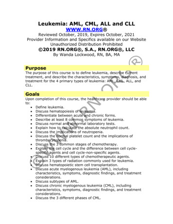
Downloaded from http://cshperspectives.cshlp.org/ on March 28, 2015 - Published by Cold Spring Harbor Laboratory PressB.A. MacVicar and E.A. Newmanulating CBF is likely to be of fundamental importance in providing appropriate and consistent energy supply to support brain function.There are two important levels at which CBFis regulated. Basal CBF, which is remarkablyhigher than the rest of the body (Magistretti etal. 2000), is regulated so that the brain receivesan adequate supply of blood at all times. Autoregulatory mechanisms ensure that CBF doesnot vary in the face of changes in systemic bloodpressure. CBF is also regulated in response tobrain activity. It has long been recognized, sincethe classic papers of Mosso (1880) and Roy andSherrington (1890), that the brain has an intrinsic ability to rapidly regulate its own blood supply in response to local energy requirements,a response named functional hyperemia. Thishomeostatic response is important for providing increased delivery of glucose and oxygen attimes of intense activity and metabolic demand.HISTORICAL OVERVIEW OF FUNCTIONALHYPEREMIAFunctional hyperemia was first described in the1880s by Angelo Mosso (1880), who observedchanges in brain volume in patients with skulldefects, allowing direct observation of the cortical surface. Mosso found that sensory stimuliproduced an increase in brain volume, representing increased cortical blood flow. A decadelater, Roy and Sherrington (1890) showed thatstimulation of sensory nerves in dogs producedincreases in cortical blood flow. They speculatedthat “the chemical products of cerebral metabolism can cause variations of the caliber of thecerebral vessels: that in this reaction the brainpossesses an intrinsic mechanism by which itsvascular supply can be varied locally in correspondence with local variations of functionalactivity.”In the late 1800s, around the same time thatfunctional hyperemia was first observed, astrocyte morphology was described by Virchow(1858), Golgi (1894), Ramón y Cajal (1995)(first published in 1897), and others. They observed that astrocytes contacted both blood vessels, which are enveloped by astrocyte endfeet,and neurons (Fig. 1). Thus, astrocytes are ide2ally positioned to mediate neurovascular coupling, relaying signals from neurons to bloodvessels. The association of astrocytes with bloodvessels led Ramón y Cajal to suggest that thesecells might regulate blood flow in the brain.Ramón y Cajal wrote, “The perivascular neuroglial cells live only in the proximity of the capillaries of the gray matter, to which they sendone or more thick appendages inserted in theouter side of the endothelium. Each capillarygives insertion to thousands of these pseudopods (endfeet), which diverge in all directions.The object of such elements is to evoke, bycontraction of the aforementioned appendices,local dilations of the vessels” (Ramón y Cajal1895). Ramón y Cajal’s suggestion that astrocytes regulate blood flow, albeit by an incorrectmechanism, was prescient.More recently, astrocyte regulation of bloodflow has been investigated directly. Paulson andNewman (1987) proposed that astrocytes mediate functional hyperemia by a Kþ siphoningmechanism, releasing Kþ onto blood vesselsfrom their endfeet in response to neuronal activity. Later, Zonta et al. (2003) suggested thatneurovascular coupling is mediated by the release of prostaglandins from astrocytes. Theyshowed that prostaglandin E2 (PGE2) release,resulting from increases in glial Ca2þ, evokesvessel dilation.Since the publication of Zonta et al. (2003),the role of astrocytes in mediating functionalhyperemia has been studied intensely. Severalmechanisms of glial control of CBF have beenproposed and tested. These mechanisms are discussed in the following sections.NEUROVASCULAR COUPLINGIt was originally hypothesized that the controlof CBF flow was mediated by a negative feedback mechanism, whereby the generation ofmetabolites from active neurons (such as CO2)were the signals that resulted in increased CBF(Roy and Sherrington 1890). However, it is nowrecognized that brain activity itself can increaseCBF. Indeed, CBF increases to such an extentthat more oxygen is provided to active brainregions than is consumed. Recordings of tissueAdvanced Online Article. Cite this article as Cold Spring Harb Perspect Biol doi: 10.1101/cshperspect.a020388
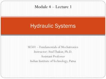
Downloaded from http://cshperspectives.cshlp.org/ on March 28, 2015 - Published by Cold Spring Harbor Laboratory PressAstrocyte Regulation of Blood Flow in the BrainFigure 1. Drawing of brain astrocytes by Santiago Ramón y Cajal. (A,B) Astrocytes, the darker cells in thedrawing, contact both neurons, (C,D) the lighter cells, and (F ) a blood vessel. As suggested by Ramón y Cajal,astrocytes are ideally situated to mediate signaling from neurons to blood vessels and to increase cerebral bloodflow (CBF) in response to neuronal activity.pO2 levels during activation of cortical synapticactivity show that pO2 increases rapidly abovebasal levels during and after repetitive synapticactivity (Offenhauser et al. 2005; Devor et al.2011). This oversupply of oxygen is the basisof the BOLD (blood oxygen level– dependent)effect in fMRI (functional magnetic resonanceimaging), whereby increased levels of blood oxy-gen are detected by the change in the magneticproperties of hemoglobin as the proportion ofoxygenated hemoglobin increases and the deoxygenated form is decreased. It is now recognized that synaptic and brain activity itself leadsto enhanced CBF and that astrocytes are keyplayers in mediating neurovascular coupling,the transduction of brain activity to alterationsAdvanced Online Article. Cite this article as Cold Spring Harb Perspect Biol doi: 10.1101/cshperspect.a0203883
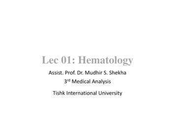
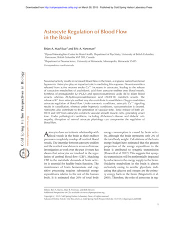
Downloaded from http://cshperspectives.cshlp.org/ on March 28, 2015 - Published by Cold Spring Harbor Laboratory PressB.A. MacVicar and E.A. NewmanArteriolesmoothmuscleAAAstrocytemGluRCa2 K GCa2 nNOSNOO2 and glucoseNONOO2 andglucoseFigure 2. Summary of signaling pathways that mediate neurovascular coupling in the brain. Synaptically releasedglutamate acts on N-methyl-D-aspartate receptors (NMDARs) in neurons to increase [Ca2þ]i, causing neuronalnitric oxide synthase (nNOS) to release nitric oxide (NO), which activates smooth muscle guanylate cyclase.Raised [Ca2þ]i may also (dashed line) generate arachidonic acid (AA) from phospholipase A2 (PLA2), which isconverted to prostaglandins (PG) that dilate vessels. Glutamate also raises [Ca2þ]i in astrocytes by activatingmetabotropic glutamate receptors (mGluR), generating arachidonic acid, and three types of AA metabolites:prostaglandins and EETs in astrocytes, which dilate vessels, and 20-HETE in smooth muscle, which constrictsvessels. An increase of [Ca2þ]i in astrocyte endfeet may also activate Ca2þ-gated Kþ channels (gk(Ca), alternativeabbreviation, BK), releasing Kþ, which dilates vessels. (From Attwell et al. 2010; reprinted, with permission,from the authors.)in CBF. Although direct signaling from neuronsto blood vessels contributes to the regulation ofCBF (Attwell et al. 2010), astrocytes play an important role in mediating neurovascular coupling (Fig. 2).MECHANISMS OF ASTROCYTE-MEDIATEDNEUROVASCULAR COUPLINGArachidonic Acid – Mediated NeurovascularCouplingWork over the past decade has shown that neurovascular coupling is mediated, to a significantdegree, by a Ca2þ-dependent astrocytic mechanism. The link between astrocyte Ca2þ and vascular changes was best shown by uncaging ofCa2þ in astrocytes in brain slices, retinal explants, and in vivo. These experiments showed4unequivocally that Ca2þ transients in astrocytescan induce dilations and constrictions in adjacent arterioles. Mulligan and MacVicar (2004)showed that arterioles display constrictions asopposed to the dilations, reported by Zonta etal. (2003), when Ca2þ transients were evokedwith two-photon Ca2þ uncaging in endfeet adjacent to arterioles. The arteriole responses wereblocked by inhibiting phospholipase A2, theenzyme that liberates arachidonic acid (AA)from membrane lipids, and by inhibiting theconversion of AA to the vasoconstrictive lipid,20-hydroxyeicosatetraenoic acid (20-HETE). InCa2þ uncaging experiments in vivo, Takano etal. (2006) showed that arteriole dilations in thecortex were triggered by astrocyte Ca2þ transients via a mechanism sensitive to the inhibition of cylclooxygenase-1 (COX-1), one of theenzymes that synthesizes PGE2 from AA. Ta-Advanced Online Article. Cite this article as Cold Spring Harb Perspect Biol doi: 10.1101/cshperspect.a020388
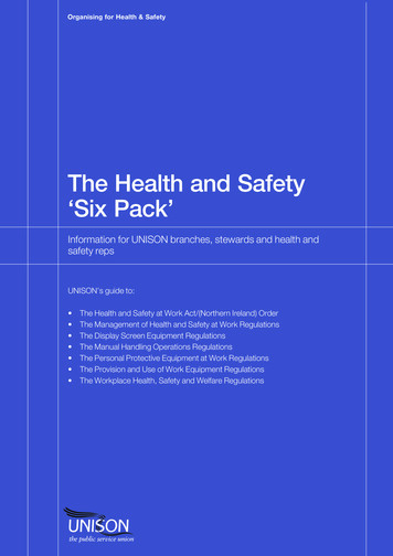
Downloaded from http://cshperspectives.cshlp.org/ on March 28, 2015 - Published by Cold Spring Harbor Laboratory PressAstrocyte Regulation of Blood Flow in the Braintrienoic acids (EETs), that are also synthesizedfrom AA. Subsequent work by Mishra et al.(2011) showed that astrocyte-evoked vasodilation in the retina is mediated by synthesis ofboth EETs and PGE2.Part of the explanation for the divergentobservations that both dilations and constrictions could be triggered by astrocyte Ca2þ signaling came from the work of Gordon et al.(2008). In this study of brain slices, the investigators reported that the polarity of the vascular response to astrocyte Ca2þ transients wasdependent on the level of pO2 in the superfusion solution. Arteriole constrictions thatwere evoked by Ca2þ uncaging in astrocytes inkano et al. also showed that COX-1 was expressed in the endfeet of cortical astrocytes,supporting the role for PGE2 synthesis by astrocytes. These results suggested that PGE2 is released from astrocytes to cause arteriole dilations similar to the mechanism proposed byZonta et al. (2003) (Fig. 3). The first hints ofthe complexity of astrocyte vascular couplingcame from work in the retina explant by Meteaand Newman (2006). They showed that, in thesame preparation, Ca2þ uncaging in astrocytesand Müller cells (the principal retinal glial cells)could trigger both arteriole constrictions via20-HETE synthesis and dilations via the formation of other vasoactive lipids and epoxyeicosa-Low O2High O2Ca2 AACYP4ACa2 AAPLA2PLA2COXGlycolysisGlycolysisPGTAstrocyte endfoot20-HETEAdenosineCa2 LactateAdenosineSmooth muscle cellConstrictionPGE2PGE2Ca2 DilatationVessel lumenFigure 3. Summary of the modulation of arachidonic acid metabolite-mediated neurovascular coupling bytissue pO2 and lactate. Intracellular Ca2þ transients activate PLA2 to liberate arachidonic acid (AA) from plasmamembrane lipids. In high pO2, AA is converted to 20-HETE, which increases intracellular Ca2þ in smoothmuscle cells, causing vasoconstriction (left side). Extracellular adenosine that can inhibit Ca2þ entry intosmooth muscle cells is also lower in high tissue pO2, further promoting vasoconstriction. In lower pO2 (rightside), the vasodilation pathway dominates and AA is converted to PGE2 by COX-1 and is released by diffusion.Extracellular PGE2 is cleared from the extracellular space by the prostaglandin transporter (PGT), which isexpressed in astrocytes and neurons. When pO2 decreases, glycolysis is enhanced and extracellular levels oflactate increase. Extracellular lactate attenuates PGE2 uptake by PGT leading to higher extracellular PGE2 andenhanced relaxation of smooth muscle tone and vasodilation. In addition, higher levels of extracellular adenosine reduces smooth muscle cell constriction via A2A activation. (From Gordon et al. 2011; reprinted, withpermission, from the authors.)Advanced Online Article. Cite this article as Cold Spring Harb Perspect Biol doi: 10.1101/cshperspect.a0203885
Downloaded from http://cshperspectives.cshlp.org/ on March 28, 2015 - Published by Cold Spring Harbor Laboratory PressB.A. MacVicar and E.A. Newmanhigh O2 (95%) solutions were reversed to arteriole dilations in more physiological (20%) solutions. Oxygen modulation of neurovascularcoupling occurred because low pO2 increasedextracellular lactate concentrations, which, inturn, reduced the uptake and clearance of PGE2by the prostaglandin transporter. In high pO2,the conversion of AA to 20-HETE predominates, leading to constriction, whereas, in lowpO2, the PGE2 dilation pathway is dominant(Fig. 3). In addition, the increased adenosinetone in low pO2 solutions reduced the vasoconstriction. A similar switch from dilation toconstriction was observed in the retina explantwhen tissue pO2 was altered from a low O2 levelto a high level (Mishra et al. 2011). However,this O2-modulatory effect on retinal blood flowwas not observed in vivo when animals weremade hyperoxic. The results of these experiments indicated that the polarity of astrocytemodulation of CBF may reflect the metabolicstate of the brain tissue and may be modifiedby the level of the extracellular lactate concentration. It is intriguing that a link between themagnitude of CBF changes and lactate levels hasbeen observed in awake human subjects in anfMRI study (Mintun et al. 2004), in which anincreased lactate/pyruvate ratio was observedto augment blood flow in the physiologicallyactivated human brain. In addition, a nuclearmagnetic resonance (NMR) spectroscopy studyshowed a remarkably strong correlation between functional hyperemia and brain lactatelevels (Lin et al. 2010).The dilation or constriction of arteriole diameter as a result of Ca2þ transients in astrocyte endfeet has been replicated in numerouslaboratories over the past 10 years. It is clearthat Ca2þ-dependent activation of PLA2 in astrocytes is an important initial step in the formation of vasoactive lipids that are synthesizedfrom AA and can trigger either arteriole dilationsor constrictions. The reliance on PLA2 as thecritical first step in this pathway has been supported by studies in PLA2 knockout transgenicmice, in which the ability of astrocyte Ca2þ signals to modify CBF is lost when the Ca2þ-dependent form, cPLA2, is not expressed (He et al.2012). However, controversies remain concern6ing when Ca2þ signals are generated in astrocytesby synaptic activity and whether these Ca2þ signals precede the functional hyperemia responsein vivo. Among the points of contention are:† Glutamate mGluR5 receptors can mediateastrocyte Ca2þ signaling in juvenile but notin adult mice (Sun et al. 2013) and rats (Duffy and MacVicar 1995). It is not clear whichreceptors generate astrocytic Ca2þ signals inadults.† Several neurotransmitters, besides glutamate,evoke Ca2þ signaling in astrocytes (Schipkeand Kettenmann 2004) and could also leadto the modulation of CBF. However, releaseof these neurotransmitters is not consistently associated with neuronal activity as is glutamatergic signaling.† It is well established that neuronal activ-ity results in Ca2þ signaling in astrocytes invivo. What is not clear, however, is how consistent this astrocytic Ca2þ signaling is andwhether it precedes changes in CBF. Somestudies have shown rapid Ca2þ signaling inastrocytes that precedes arteriole dilation(Winship et al. 2007; Lind et al. 2013). Otherstudies show that Ca2þ signaling occurs infew astrocytes and develops slowly after vasodilation has begun (Nizar et al. 2013;Bonder and McCarthy 2014; Paukert et al.2014). These divergent results leave openthe question of whether rapid functional hyperemia responses are mediated by a Ca2þdependent astrocyte mechanism.Potassium-Mediated Neurovascular CouplingNeurovascular coupling may also be mediatedby the glial release of Kþ onto blood vessels.When the extracellular Kþ concentration([Kþ]o) is increased from a resting level of 5 mM to 15 mM, blood vessels dilate. Potassium-induced vasodilation is mediated byan increase in the conductance of inwardly rectifying Kþ channels on vascular smooth muscle cells (Filosa et al. 2006; Haddy et al. 2006)and by activation of the Naþ-Kþ ATPase on thesmooth muscle cells (Bunger et al. 1976; HaddyAdvanced Online Article. Cite this article as Cold Spring Harb Perspect Biol doi: 10.1101/cshperspect.a020388
Downloaded from http://cshperspectives.cshlp.org/ on March 28, 2015 - Published by Cold Spring Harbor Laboratory PressAstrocyte Regulation of Blood Flow in the Brain1983), both of which result in hyperpolarizationand relaxation of the muscle cells. However,larger Kþ increases, above 15 mM, depolarizevascular smooth muscle cells, resulting in vasoconstriction (Girouard et al. 2010). However,[Kþ]o in the brain normally reaches 12 mMonly during intense seizure activity and hasnot been observed to exceed 15 mM except during pathological processes, such as spreadingdepression or stroke (Vyskocil et al. 1972; Somjen 2001).Active neurons release Kþ into the extracellular space, resulting in an increase in [Kþ]o.Light stimulation produces slow, transient[Kþ]o increases of 1 mM in the cat visual cortex (Singer and Lux 1975; Connors et al. 1979)and in the synaptic layers of the cat and frogretina (Dick et al. 1985; Karwoski et al. 1985).Higher [Kþ]o elevations are evoked by directelectrical stimulation of afferent pathways andby induction of seizure activity in the brain(Heinemann and Lux 1977).Paulson and Newman (1987) proposed thatneurovascular coupling is mediated by a feedforward glial cell Kþ siphoning mechanism(Newman et al. 1984). According to this model,the diffusion of Kþ through the extracellularspace from neurons to blood vessels is enhancedby a Kþ current flow through astrocytes. The[Kþ]o increase as a result of neuronal activitygenerates an influx of Kþ into astrocytes andresults in astrocyte depolarization. Depolarization, in turn, induces a Kþ efflux from cell endfeet, which have a high density of Kþ channels(Newman 1984, 1986). Because astrocyte endfeet terminate on blood vessels, the Kþ effluxoccurs directly onto the vessels. Computer simulations of Kþ dynamics (Paulson and Newman1987) show that glial Kþ siphoning results in aKþ increase at the vessel that is larger and morerapid than would occur solely by Kþ diffusionthrough the extracellular space. Potassium siphoned from astrocytes onto blood vesselscould mediate neurovascular coupling.More recently, the glial Kþ siphoning hypothesis of neurovascular coupling was testedin the retina (Metea et al. 2007). The results donot support the hypothesis. Glial cells were depolarized by current injection through patchpipettes. This depolarization should evoke Kþefflux from the cell endfeet and result in vasodilation. Although vessels dilated in response tobath-applied increases in [Kþ]o, current injection did not induce vasodilation. In a second testof the Kþ siphoning hypothesis, light-evokedvasodilation was assessed in mice in whichKir4.1 Kþ channels were genetically knockedout. Kir4.1 is the principal Kþ channel in retinalglial cells (Kofuji et al. 2000) and Kþ siphoningfluxes should be nearly abolished in Kir4.1knockout animals. However, light-evoked vasodilation was as large in the knockout animalsas in wild-type controls. The experiments showthat glial cell Kþ siphoning does not contributesignificantly to neurovascular coupling in theretina.Although neurovascular coupling may notbe mediated by Kþ siphoning, it could be mediated by a related Kþ mechanism. Astrocytesexpress big potassium (BK) Ca2þ-activated Kþchannels as well as Kir channels (Price et al.2002). When neuronal activity evokes Ca2þ increases in astrocytes, BK Kþ channels will open(Filosa et al. 2006). BK channels are also modulated by arachidonic acid metabolites andCa2þ-dependent increases in EETs will openthe channels (Gebremedhin et al. 2003; Dunnand Nelson 2010). BK channel opening will result in an efflux of Kþ from glial cell endfeetonto blood vessels, which could lead to vesseldilation. Genetic and pharmacological block ofBK channels results in a reduction of whiskerstimulation-evoked blood flow increases in thecortex, supporting this Kþ-glial BK channel hypothesis of neurovascular coupling (Filosa et al.2006; Girouard et al. 2010).REGULATION OF BLOOD FLOWBY CAPILLARIESWithin the brain vascular network, regulationof blood flow occurs at the level of arterioles,with neuronal activity leading to arteriole dilation and increased blood flow. Recent workhas shown, however, that capillaries can actively regulate blood flow as well. Sensory stimulation leads to active dilation of brain capillaries(Chaigneau et al. 2003; Lacar et al. 2012; HallAdvanced Online Article. Cite this article as Cold Spring Harb Perspect Biol doi: 10.1101/cshperspect.a0203887
Downloaded from http://cshperspectives.cshlp.org/ on March 28, 2015 - Published by Cold Spring Harbor Laboratory PressB.A. MacVicar and E.A. Newmanet al. 2014), which could play an important rolein regulating blood flow as a significant fraction of total vascular resistance likely residesin these small vessels (Blinder et al. 2013; Hallet al. 2014). Active regulation of blood flow bycapillaries has also been shown in the retina,where flickering light stimulation results in theselective dilation of specific capillaries (Kornfield and Newman 2014).Active control of capillary diameter is mediated by pericytes and their processes, whichsurround capillaries. Pericytes, the only cellsassociated with these vessels that express contractile proteins (Nehls and Drenckhahn 1991;Hughes and Chan-Ling 2004; Hamilton et al.2010), can actively dilate and constrict in response to vasoactive agents (Schonfelder et al.1998; Peppiatt et al. 2006; Puro 2007; Hamiltonet al. 2010). Hall et al. (2014) have shown thatpericytes in the somatosensory cortex activelyrelax in response to whisker-pad stimulation.The resulting capillary dilation precedes the dilation of upstream arterioles, demonstratingthat pericytes play an important role in controlling blood flow.There are still many unanswered questionsconcerning how neurovascular signaling mechanisms control pericyte tone. It is possible thatsome of the same glial and neuronal signalingmechanisms that control the tone of smoothmuscles cells surrounding arterioles also modify pericyte tone. For example, Hall et al. (2014)showed that glutamate-induced pericyte relaxation is mediated by activation of PGE2 receptors and by nitric oxide (NO) inhibition ofthe synthesis of the vasoconstrictor 20-HETE.These are two of the same astrocyte-mediatedsignaling mechanisms that control the contractile state of arteriole smooth muscle cells.ASTROCYTE REGULATION OF VASCULARBASAL TONEThe supply of oxygen and nutrients to the brainis controlled by the basal tone of blood vessels,as well as by changes in tone that occur duringfunctional hyperemia. Basal vascular tone iscontrolled by a number of mechanisms. Theseinclude extrinsic innervation from the auto8nomic nervous system, intrinsic innervationfrom subcortical neurons and cortical interneurons, and the local release of vasoactive agentsfrom vascular cells (Hamel 2006). A numberof vasoactive agents modulate cerebrovasculartone, including noradrenaline, released by sympathetic terminals and by locus coeruleus neurons (Bekar et al. 2012); serotonin, released byraphe nucleus neurons (Cohen et al. 1996); andneuropeptides, released from local interneurons(Cauli et al. 2004).Vasoconstricting agents released from astrocytes also contribute to the generation of vascular tone. As discussed above, Ca2þ signalingin astrocytes results in the production of thevasoconstrictor 20-HETE, as well as the vasodilators PGE2 and EETs. 20-HETE is a wellknown vasoconstrictor of arteries and arterioles(Imig et al. 1996) and is thought to act by inhibiting Ca2þ-activated Kþ channels and by activating voltage-gated Ca2þ channels on vascular smooth muscle cells (Zou et al. 1996). Catcerebral arterial smooth muscle cells express cytochrome P450 4A enzymes and produce thevasoconstrictor 20-HETE, which enhances Ltype Ca2þ currents (Gebremedhin et al. 1998).In addition, Cyp450 4A2, the enzyme that synthesizes 20-HETE in smooth muscle cells, isinhibited by NO, a potent vasodilator (Oyekanet al. 1999). These studies indicate that a balancebetween 20-HETE and NO levels is importantunder some circumstances for setting the basaltone of cerebral blood vessels (Gordon et al.2007; Metea and Newman 2007). This conclusion is supported by observations, in brain slicesand in vivo, that blocking NO release leads to avasoconstriction that is 20-HETE synthesis dependent (Zonta et al. 2003; Mulligan and MacVicar 2004). The importance of this pathway insetting basal tone was shown in a recent studythat described a very long-lasting (2 h) decreasein CBF following spreading depression that wascaused by increased 20-HETE in the cerebralcortex (Fordsmann et al. 2013).Astrocytes also generate vascular tone bythe tonic release of ATP (Kur and Newman2013). Hippocampal astrocytes tonically releaseATP, resulting in extracellular ATP levels of 10 mM (Pascual et al. 2005), whereas astro-Advanced Online Article. Cite this article as Cold Spring Harb Perspect Biol doi: 10.1101/cshperspect.a020388
Downloaded from http://cshperspectives.cshlp.org/ on March 28, 2015 - Published by Cold Spring Harbor Laboratory PressAstrocyte Regulation of Blood Flow in the Braincytes and Müller cells in the retina release ATP inresponse to cellular Ca2þ increases (Newman2001). Released ATP tonically constricts arterioles by activating P2X1 receptors on vascularsmooth muscle cells. Vascular tone is loweredwhen extracellular ATP levels are reduced byenzyme degradation, when P2X1 receptors areblocked with purinergic antagonists, and whenglial cells are selectively poisoned with the toxinfluorocitrate (Kur and Newman 2013).ASTROCYTES AND FUNCTIONALHYPEREMIA IN PATHOLOGYAlzheimer’s DiseaseAlzheimer’s disease is characterized, classically, by extracellular accumulation of amyloidplaques and intracellular protein inclusions,now known as neurofibrillary tangles (Alzheimer 1907; Kosik et al. 1986; Goedert et al.1988; Graeber and Mehraein 1999). In addition,there are recent observations of physiologicaland phenotypic changes in the cells surrounding plaques in mouse models of Alzheimer’sdisease. For example, spontaneous intercellularCa2þ waves and higher resting Ca2þ levels inastrocytes have been observed (Kuchibhotlaet al. 2009), in addition to abnormal synchronous neuronal hyperactivity and signs of profound microglia activation (Terry et al. 1991;Akiyama et al. 2000; Wyss-Coray 2006; Vennetiet al. 2008). These changes in astrocyte Ca2þsignaling could generate inappropriate neurovascular changes as there are several vasoactivelipids, described in this review, that are generated in response to Ca2þ signals in astrocytes.These alterations in both neuronal and astrocyte Ca2þ signaling could synergistically leadto impaired vascular control and possibly leadto constrictions of arterioles and tissue hypoxia(Mulliga
changes in brain volume in patients with skull defects, allowing direct observation of the cor- . recognized that brain activity itself can increase CBF. Indeed, CBF increases to such an extent .