Transcription
THE BLOOD
BLOOD One of the largest organs of the body. An average 70 kg man has almost 5Lblood (5.5 kg). Blood circulates throughout the bodyand supports the functions of all otherbody tissues.
Blood integratestissues and organsand providea special means ofcommunications.
THE FUNCTIONS OF BLOOD Respiration: transport of O2 and CO2. Transport: hormone, nutrients, metabolic waste. Excretion of metabolic wastes to the kidney, lungsand skin. Regulation of body temperature by distribution ofbody heat. Defense against infections (WBCs, antibodies). Maintenance of acid-base balance. Nutrition: transport of absorbed food material.
PHYSICAL PROPERTIES OF BLOOD Specific gravity:- Whole blood:- Plasma:Viscosity:Mass:Blood volume:1.055 - 1.0651.024 - 1.0285-6 times that of water.6-8% of the body weight. 8% of body weight. 86% ml/kg body weight.5-6L in adults[Infants have a larger blood volume in proportion tobody weight than adults].Osmotic pressure: 7-8 atmosphere at body temperature.
Composition of Blood Formed Elements (45%),i- Red blood cellsii- White blood cellsiii- ) Fluid medium i.e. the plasma (55%).
Gross composition of Plasmaand Blood CellsConstituentsPlasmaRed blood cellsWater91-95%65%Solid8-9 %35%Protein6-8 gm %31-33%Specific gravity1.026
Haematocrit or Packed Cell Volume in)heparin)PlasmaPlasmaPCVPCVPCVPCV 0.450.45L/LL/L 0.410.41L/LL/LPCVPCV isisanaemiaanaemia isispolycythaemiapolycythaemia
SERUMNo anticoagulantWhole bloodClot formationCentrifugeClot Clear yellowishfluid (serum)
ERYTHROCYTE SEDIMENTATION RATE(ESR) Rate of Sedimentation of Erythrocytes. ESR at 20 3 C (Westergress Method)Male 0-5 mmFemale 0-7 mm-ESR - Non-specific indicator of infection.ESR - For monitoring status of chronicinflammatory diseases.
HaemopoisisThe process of formation of blood.ErythropoisisFormation ofErythrocytesThrombopoisisFormation ofThrombocytesLeucopoisisFormation ofLeucocytes
ProcessProduct ErythropoesisRBC LeucopoiesisWBC GranulopoiesisGranulocytes LymphopoiesisLymphocytes MegakaryocytesPlatelets
Site of HaemopoisisFetal Life1-2 m2-6 m1-9 mfrom 4 mAt BirthAdult lifeYolk SacSpleenLiverBone marrowBone marrowBone marrow
Erythropoisis—A two stagedifferentiation systemStage 1: From Pluripotent stem cellsto committed cellsStage 2:From committed cells to therecognisable precursors
ErythropoisisStem cell (pluripotent)Committed stem cells( CFU)PronormoblastBasophil normoblastPolychromatic normoblast I and IIOrthochromatoblastReticulocytesErythrocytes
Control of Erythrocyte SynthesisCellular hypoxiaOxygen sensorE-Releasing factorSerumErythropoietin (E)KidneyErythropoietin (E)SerumStem cellBone Marrow
ErythropoisisStem cellPronormoblastBasophilic normoblastPolychromatophilic normoblastOrthochromatic normoblastReticulocytesMature red cell
Current model of the feedback circuit which regulate therate of RBC synthesis to the need for O2 in the peripheral tissues.(Surface receptors and intracellular secondary messenger):Bone marrowstem cellErythroid tissueDNA, mRNAErythropoiteinRenal vaccRenal O2ConsumptionRed cell massAtm.O2CardiopulmonaryfunctionBlood volumeHb concentrationO2 affinityErythropoietin O2SensorProduce EpoKidney(Liver, Macrophages)
Erythropoiesis The proliferation and differentiation of cellsfrom pleuripotent noncommitted stem cellsof the bone marrow. Two main compartments:
Erythropoiesis compartmentsErythropoiesis compartmentsThe erythroid progenitor cell compartmentThe erythroid precursor cell (erythron)
1.The erythroid progenitor cells The earliest recognizable committedprogenitor for erythroid cells is the CFUGEMM. The next are the BFU-E. The final progenitor cells are the CFU-E.
2. The erythroid precursor cell It is the morphologically recognizableerythroid cell within the normal bonemarrow.
Cells involved in erythropoiesis 1.Pluripotent stem cell:– Most primitivehaemopoietic cell.– Extensive capacityto proliferate.– Mature into other cell types: Multipotent myeloid stem cell. Lymphoid stem cell.
Cells involved in erythropoiesis 2. Pronormoblast:–––––––earliest recognized cell in erythron.A large cell.Basophilic cytoplasm.Has a large nucleus.High conc. Of m.RNA.1% of protein is Hb.1000 receptor/cell for Epo.
Cells involved in erythropoiesis 3. Basophilic normoblast:– Nucleolus is lost.– The golgi apparatus remainsprominent.
Cells involved in erythropoiesis 4. Polychromatophilicnormoblast:– Hb production.– smaller nuclear:cytoplasmicratio.– Chromatin is more clumped andcondensed.
Cells involved in erythropoiesis 5. Orthochromatic normoblast:– cytoplasm is more eosinophilic.– Final nucleated stage.– 300 receptors/cell for Epo.
Cells involved in erythropoiesis 6. Reticulocyte:– No nucleus.– Reticular networkes of polyribosomes.– It enters blood stream and circulate for 1-2days they become mature RBC.– Rounder, faintly polychromatic, largerdiameter than RBC.– 95% of cell protein is Hb.– No receptors for Epo.
Maturation Maturation of the proerythroblast within the bone marrowto the end of the basophilic erythroblast stages takes about60 hrs. Maturation of the polychromatophilic erythroblast takesabout 30 hrs, the late erythrocyte stage about 50 hrs andthe reticulocyte in the steady state remains in the bonemarrow for 40-48 hrs.
Transcription factorsfor differentiation and maturation Random process. Epo prevent programmed cell death. Commitment of haemopoietic cells to the erythroid lineageinvolves action of several transcription factors including:TAL1, LMO2, and GATA-2. Genes for α- and β- chain of Hb are activated andcontrolled by cis-acting DNA sequence.– Other specific proteins: glycophorin A and EpoR.– Transferrin and genes of haem synthesis are also activated by cisacting mechanism.
Transcription factorsfor differentiation and maturation Cis-acting control DNA is activated by trans-acting factor. Trans-acting NF-E2 binds to specific sequence in locuscontrol region for β-globulin, probably also to α-globulin. Other cis-acting promoter regions are involved in theregulation of genes coding for enzymes of haem synthesisincluding: porphobilinogen deaminase, ferrochetalase andδ-amino laevulinic acid synthetase. The m.RNA for these factors disappear afterproerythroblast stage.
Transcription factorsfor differentiation and maturation Upon binding Epo, cell surface EpoR dimerizes andactivates specific intracellular kinases including: Janusfamily tyrosine protein kinase-2, phosphoinositol-3 kinase,and mitogen-activated protein kinase, and the RASpathway. Other trans-acting DNA binding proteins are the erythroidKruppel factor and the human stem cell leukemia genes.
Intrautreine erythropoiesisand postnatal changes Erythropoiesis occurs in two distinct wavesduring embryogenesis:– The primitive wave in the extra-embryonic sacof the 14-19 day human embryo.– The definitive wave in the fetal liver and spleenare the main sites of erythropoiesis in the 2ndtrimester of pregnancy and the fetal bonemarrow in the 3rd trimester.
Intrautreine erythropoiesisand postnatal changes The placenta activates fetal erythropoiesis by producingfactors that stimulate erythropoiesis. The major site of Epo gene expression in the fetus is in thekidney. After birth, during the 1st 4yrs of life, nearly all themarrow cavities contain red haemopoietic marrow withvery few fat cells. By the age of 25yrs the no. of fat cellsincrease.
Erythropoiesis in adults Erythropoiesis occurs within thehaemopoietic marrow. Pregnancy is characterized by increasederythropoiesis within the maternal and fetalcompartments.
BloodCellsPlasma
Blood Cells
BLOOD CELLS Erythrocytes Leukocytes(Red blood cells)(White blood cells)– Granulocytes Neutrophils Basophils Eosinophils– Monocytes– Lymphocytes T B Megacaryocyte(Platelets)
NORMAL RANGESRBC Men4.6 – 6.2 x 10¹²/l. Women4.2 – 5.4 x 10¹²/l Total number of red cells in circulation 2.5x10¹³WBC Men and women 5-7 x 10⁹/l.Platelets Men and women 250 x 109/lHb Men14 – 16 g/l Women12 – 16 g/lPCV (Haematocrit) Men0.42 – 0.52 l/l Women0.37 – 0.47 l/l
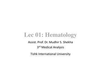
Red blood cells(Erythrocytes) BiconcaveBiconcavedisks:disks:-- DiameterDiameter-- Thickness:Thickness:-- --99µmµm11--22µmµm 8888fl.fl. oodblood vessels.vessels. ContainContainhaemoglobinhaemoglobin( ( 33%).33%). NoNonucleusnucleusorormitochondria.mitochondria. Function:Function: lRange:Range:12-- 5.55.5 1.01.0xx101012/L/L12-- 4.8/L4.8 1.01.0xx101012/L
Red blood cells(Erythrocytes) Deliver oxygen to tissues and CO2 fromtissues to lungs. Synthesis is increased by erythropoietin Red cell life span is 120 days. Senescent red cells are destroyed by spleenand replaced by juvenile cells released bybone marrow. An average 70 Kg adult male produces2.3x106 red cell/sec.
ERYTHROPOIETIN A polypeptide hormone.Glycoprotein of 166 a.a. (mol. li 34 Kdl).Major regular of human erythropoiesis.Synthesized mainly in kidney, released in response tohypoxia and arts on bone marrow. Interacts with progenitor of red cells (BFU – E) viaspecific receptors causing proliferation anddifferentiation. Also interacts with late progenitor cell (CFU – E) tocause proliferation and differentiation. Requires cooperation of other factors e.g. interleukin-3and insulin like growth factor.
ERYTHROCYTE STRUCTURE Biconcave shape. Spherical. Simple structure:– Membrane surrounding cytoplasm.– Almost 95% of solutes in cytosalhaemoglobin.is No intracellular organnels Non-nucleated Has a cytoskeleton, which plays animportant role in determining shape. Has deformability due to special structureof cytoskeleton
ErythrocytesErythrocytes Composition:Composition:-- MajorMajorcation:cation:KK -- OtherOthercation:cation: NaNa ,,CaCa ,,MgMg -- ,3diphosphoglyceratediphosphoglycerate
HAEMOGLOBIN
TYPES OF HAEMOGLOBINSIn AdultsHbHbFHb A2::: 97% 1%2.5 – 3.5%α2 β2α2 γ2α2 δ2At BirthHbFHb A::During Embryonic lifeHb Gower 1Hb Gower 2Hb Portlandβ2 γ2α2 β2
HAEMOGLOBIN IN THE RED CELLSHaemoglobin Major solute in red cells. Globular protein Conjugated protein: globin haem. Made of 4 subunits (Quarternary structure)4 globins 4 haems haemoglobin. Binds O2 to haem group to formoxyhaemoglobinHb 4 O2 Hb (O2)4.Contd .
GLOBIN CHAINS OF HAEMOGLOBINAmino acids in globin chainsα Globinβ-like globin chains:141 a.a.146 a.a.Structure of globin chains– Globular, compact structure– 75% α-helices– Have a hydrophobic cavity for bindingheme.
HaemoglobinGlobin ChainsHeam Group
HEME GROUP Protoporphyrin IX Has tetra pyrolle rings linked together bymethylene bridges. Fe coordinates with 4 N of the 4 pyrolle rings:– Bind with coordinate covalent bond to HistidineF8.– Binds to O2 between Fe and His E7. If Fe is oxidized to ferric (Fe ) the Hb isknown as met Hb, which cannot binds O2.
STRUCTUREOF HEMEGROUP
HAEMOGLOBIN IN THE RED CELLSHaemoglobin .Contd Allosteric protein: has 4 O2 binding sites O2 binding curve of Hb is sigmoidal. Shows cooperative effect: i.e. binding ofsome O2 molecules makes it easy forother O2 molecules to bind. O2 affinity of Hb is affected by pO2,pCO2,, H , 2,3 DPG.
HAEMOGLOBIN IN THE RED CELLSHaemoglobin .Contd Affinity for O2 depends on partial pressure ofO2, CO2, and H , 2,3 DPG level. Binds CO2 to N-terminal of β-globin chain toform carbamino Hb. Carboxy Hb.Hb 4CO Hb (CO)4.Has high affinity for CO
THE BOHR EFFECTIn Lungs: High PO2, H , CO2 high affinity of Hb forO2 (O2 dissociation curve shifts to left).In Tissues Low P O2, H , CO2, 2,3 DPG Lowaffinity of Hb for O2 (O2 dissociation curve shiftsto right)
THE BOHR EFFECTLungsCO2 Exhaled2CO2 2H2O2H2CO3PeripheralTissuesCarbonicAnhydraseHCO3 - 2H Hb 4O24O24O2Hb 2H 2H 2HCO32H2CO3-2CO2 2H2OGenerated by TCA CycleCarbonicAnhydrase
O2 Dissociation curve of HaemoglobinSaturation%1005026pO2(Torrs)
BINDING OF 2,3 DIPHOSPHOGLYCERATE 1 molecule of 2,3 DPG /Hb molecules 2,3 DPG binds between 2 β-chains of HbA. It is formed from 1,3 DPG (a glycolyticintermediate). In peripheral tissues level of 2,3 DPG is high. Itbinds Hb and decreases affinity for O2. HbF cannot bind 2,3 DPG and has higheraffinity for O2.– O2 can be transported from mother to fetalblood
Red Cell Metabolism
SUMMARY OF RED CELLMETABOLISM Highly dependant on glucose as energy source. Glucose is metabolized by:– Glycolysis ( 95%)– Pentose phosphate pathway ( 5%) Glycolysis produces lactate ATP– 2,3 DPG regulates O2 affinity of Hb. PPP produces NADPH, necessary for keeping redcells in reduced state. No synthesis of glycogen, fatty acids, proteins ornucleic acids in red cellsContd .
SUMMARY OF RED CELLMETABOLISM Contd Reduced glutathione is important as it keeps the:– red cells and other proteins in reduced state.– reduces oxidizing radicals (peroxides) generatedin red cells2GSHG-S-S-GH2O22H2O Iron of Hb is kept in reduced state (Ferrous, Fe )by NADH-dependant methaemoglobin reductase.
GLUCOSE TRANSPORTERS IN RED CELLMEMBRANE Glucose uptake by red cells is byfacilitated diffusion. Proteins involved in facilitated diffusion of glucose are glucose transporters ( 2%to membrane protein of RBC). Almost 7 different glucose transportershave been identified in different tissue. Glucose transporters in red cellsmembrane are insulin-independent.
Glucose Metabolism in Erythrocytes95% Oxidised by glycolysisGlucose5% Oxidized by pentosephosphate pathways
The role of glycolysis in the functional requirements ofmature red cells:FunctionEMP- Maintenance of shapeATP- Membrane structure andFunctionPPPGSH- Regulation of O2 transport2,3-DPGATP- Reducing potentialNADPGSHNADPH
PRODUCTION OF POWERFUL OXIDANT INRED CELLS DURING METABOLISM During metabolism, there is production of:– Superoxides (O2): O2 e O2– Hydrogen peroxide (H2O2)O2 O2 2H H2O2 O2– Peroxyl radicals (ROO)– Hydroxyl radicals (OH*) These oxidizing radicals are highly reactivemolecules and can react with proteins, nucleicacids, lipids and other mol. to alter their structureand produce tissue damage. Red cell need several reducing reactions to keep itin reduced state and protect it from damage byoxidizing radicals.
PROTECTION OF RED CELLSFROM HAEMOLYSISBy: Super oxide dismutaseO2- O2- 2H H2O2 O2 Catalase:H2O2 2H 2H2O Glutathione2GSH RO – OH GSSG H2O ROHGlutathioneOxidised Glutathione
Glucose-6-Phosphate Dehydrogenase(G-6-PD)- G-6-PD is the first enzyme of the Pentose Phosphate Pathway.- Catalyses the following reaction:G-6-P NADP 6-Phosphogluconolactone NADPH H - NADPH is necessary for the red cell integrity and stability.- Co-enzyme for glutathione reductase which converts oxidisedglutathione to reduced glutathione. This reduces oxidising radiclesand protects red cells from damage.-Deficiency of G-6-PD leads to hemolytic anaemia under oxidativestress(e.g. antimalarial drugs, fava beans, infections, diabeticacidosis)
Other Blood Cells
PLATELETS (Thrombocytes) Discoid, anucleated cells with agranularcytoplasm.- Diameter 3 µm- Thickness 1 µm- Volume 7 fl. 250x109 platelets/litre. Synthesis increased by thrombopoietin. Synthesised from megakaryocytes.Contd .
PLATELETS (Thrombocytes)Contd. Survival in circulation 10-12 days. Primary role:- in haemostasis: stick to the edgesof wounds and form a plug to arrestblood loss. Platelets also involved in development ofatherosclerosis and hence can lead tothrombosis.
White Blood Cells (Leucocytes)Two Main Groups:i. The Phagocytes ----------Play a role in protecting thea- Granulocytes:the body against infection by- Neutrophilsphagocytosis.- Eosinophils- Basophilsb- Monocytesii. The Lymphocytes (immunocytes)--Function in protecting.a- B-Lymphocytes-----------------Provide humoral immunity.b- T- Lymphocytes----------------Provide cellular immunity.Total leucocytes: 4.00-11.0x 10⁶/l
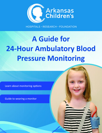
GRANULOCYTES Have numerous lysosomes and granules(secretory vesicles). Also known as polymorphonuclear leukocytes(PMN) as they have multilobular nuclei Types of granulocytes:– Neutrophils,– basophils and– eosinophilsare distinguished by their morphology and stainingproperties of their granules.
FUNCTIONS OF GRANULOCYTESNeutrophils:Phagocytose bacteria and playa major role in accurateinformation.Basophils:Resemble mast cells andcontain histamine and heparin –play a major role in immunologichypersensitivity reaction.Eosinophils:Involved in certain allergicreactions and parasitic infection.
GranulocytesNeutrophilsEosinophilsBasophils
NEUTROPHILSResponsible for acute inflammatory responseIncreasevascularpermeabilityCause entry ofactivatedneutrophilsinto tissuesCauseActivationofPlateletsBy: Platelet, activating factor (PAF) Eicosanoids (various prostaglandinsand leukotriens)Spontaneoussubsidence(resolution) ofinvading organismthat have beendealt withsuccessfully
FUNCTIONS OF MONOCYTES Monocytes are precursors ofmacrophages, which are activelyinvolved in phagocytosis.
FUNCTIONS OF LYMPHOCYTESB-Lymphocytes: Synthesize and secreteantibodies (humoral immunity)T-Lymphocytes: Involved in cellular immunemechanism e.g- killing virally infected cellsand some cancer cells.- activate B cells to makeantibodies.Lymphocytes
PLATELETS Involved in coagulationof bloodPLATELETS
Haemolysis of Erythrocyte
HaemolysisHaemolysed red cells 120 daysFree HaemoglobinIn Reticuloendothelial cells
Haemolysis of Erythocytes After a life span of 120 days, erythrocytes are haemolysed In:- Spleen- Bone marrow- Other REC Signal for haemolysis:- Loss or alteration of:- Cytoskeleton structure- Active ion pump- Membrane lipids- Membrane glycoproteins Most intracellular components are reutilized.
Fate of HaemoglobinHaemHaemoglobinGlobins
Haemolyzed RBCRE SystemHaemoglobinMet haemoglobinGlobin Heme Fe COBLOODAminoacidsTransferrin - Fe BiliverdinBilirubinBloodLiverBilirubin - AlbumincomplexAlbuminApotransferrinBonemarrowFe reutrilizationBilirubin
In R.E.S.Heme OxygenaseBiliverdin CO Fe Heme2H NADPH - CytochromeReductase3O2 3NADPH H 3 NADP NADPHBilirubinReductaseBILIRUBIN
In LiverBilirubin UDP Glucuronic acidGlucuronylTransferaseGlucuronyl TransferaseBilirubinMonoglucuronide UDPUDPglucuronic acidBilirubin DiglucuronideTo the Bile
Daily excretion of Bile Pigments250-350 ng Bile Pigment excreted in Feces/day1-2 mg Bile Pigment excreted in urine/dayPlasma Level of BilirubinTotal Bilirubin 17 µmol/LDirect Bilirubin 2 µmol/L
Disorder of Bile Pigment MetabolismCauses:1.2.3.4.An increase load of bilirubin arriving at the liver:- due to increased red cell destruction.- Absorption of large haematoma.Defective uptake and transport by the liver cells:- Gilbert’s disease.Disturbance of conjugation:- Liver cell destruction.- Reduced glucuronyl transferase activity.- Neonatal jaundice.- Crigler-Najjar Syndrome- Gilbert’s disease.Disturbance of excretion of conjugated bilirubin:- Liver cell destruction.- Intra and extrahepatic cholestasis.- Dubin-Johnson Syndrome
Jaundice Elevation of bile pigments in blood. Bile pigments escape into tissues - yellowcolouration. Due to:- Production of bile pigments.- Failure of liver to conjugate and excretebile pigments.- Decreased excretion of bile pigmentdue to obstructive of bile duct.
Types of Jaundice Hemolytic or prehepatic. Hepatic. Obstructive as posthepatic. Congenital non-hemolytic.
Hemolytic Jaundice Increase destruction of erythrocytes.Formation of bilirubin Elevation of serum bilirubin.e.g.- In hemolytic anemia- Infection- G-6-PD deficiency
Hepatic Jaundice Caused by liver dysfunction. Results from damage to parenchymal cells. Decreased conjugation of bilirubin.e.g.- Liver poisons (chloroformphosphorus, CCl 4)- Toxins.- Hepatitis virus.- Engorgement by hepatic vessels incardiac failure- Cirrhoses.
Obstructive Jaundice Results from blockage of the hepatic orcommon bile duct. Passage of blood into liver cell is normal. Conjugation of bilirubin in liver is normal. Failure of conjugated bilirubin to be excretedby bile capillaries. Bilirubin reabsorbed by hepatic veins andlymphatics.
Congenital Neonatal Hyperbilirubinaemia Activity of glucuronyl transferase in liver. Conjugation and excretion of bilirubin. Unconjugated level in blood. Often occurs in neonatal period. Treated by phototherapy.
Congenital Hyperbilirubinaemia Gilbert’s Disease:-Defective bilirubin transport into liver cells.Occassionally reduced glucuronyl transferase activity.Elevated plasma unconjugated bilirubin (20-35 µmol/L)Harmless. Crigler-Najjar Syndrome:- Deficiency in glucuronyl transferase.- Significantly elevated plasma unconjugated bilirubin(350 µmol/L)- Hyperbilirubinaemia in first few days of life.- Kernicterus in newborn.Contd .
Congenital HyperbilirubinaemiaContd . Dubin-Johnsons Syndrome-Defective excretion of conjugated bilirubin.Mildly raised conjugated bilirubin.Bilirubin in urine.Harmless.
Types of Bilirubin present in different JaundiceDefectTypes of Bilirubin- Increased production- Haemolytic diseaseUnconjugated bilirubin- Reduced liver uptake of bilirubin- Drug competitionUnconjugated bilirubin- Reduced conjugation of bilirubin- Developmental defect- Drug competition- Inherited enzyme defects(Gilbert’s disease,Criglar Najjar disease)Unconjugated bilirubinContd .
Types of Bilirubin present in different JaundiceContd .DefectTypes of Bilirubin- Decrease secretion of conjugatedbilirubin- Drug competition- Inherited defects.Mainly conjugatedbilirubin- Obstruction of biliary tree(cholestasis)- Within the liver(liver cirrhosis, drugsside-effects)- Outside the liver(gallstone, neoplasm)Mainly conjugatedbilirubin
Anaemia
Anaemia Decrease in the level of:- Haemoglobin or/and- RBC count or/and- PCV (Hematocrit)
Classification of AnaemiaClassified mainly in two ways:1. According to the morphology of theaverage red cells.2. According to the pathophysiologicmechanism of the red cell production.
Classification of AnaemiaBy Red Cell Morphology:An anaemic state with altered or normal red cell morphologyi.e. MCV and MCH:MCV: denotes the mean corpuscular volume ofred cells (Normal 85-100 f/l)MCH: denotes mean corpuscular hemoglobin(Normal 27-32 pg).
Mean Corpuscular Haemoglobin (MCH) MCH expresses the amount of haemoglobinin red blood cell in picogram (pg).Hb (g/dl blood)RBC Count X 1012/l Normal range 27-32 pg MCH
Mean Corpuscular HaemoglobinConcentration (MCHC) MCH expresses the amount of haemoglobin inRBC. It is expressed in gm/deciliter.Hb (g/dl)PCV (l/l) MCHC g/dl Normal Range 30-35 g/dl
Classification of Anaemia Contd.(a) Normocytic Normochromic Anaemia(MCV 85-100 fl) (MCH 31-35 pg)1.Acute bleeding.2.Haemolytic anaemia:(a) Extracorpuscular defects immune and non-immune.(b) Intracorpuscular defects, membrane,and metabolicdefects and hemoglobinopathies.(c) Combined defects.3.Marrow failure associated with hypoproliferation ofhematopoietic cells:(a) Aplastic anaemia.(b) Pure red cell aplasia.(c) Anaemia of chronic renal failure.(d) Anaemia of endocrine disease.(e) Toxic depression of bone marrow.
Classification of Anaemia Contd.(b) Microcytic - Hypochromic Anaemias(MCV 87 fl) (MCH 30 pg)1.2.3.(c)Iron deficiency.Thalassaemias.Sideroblastic anaemia:(a) Reflactory.(b) Reversible.(c) Pyridoxine - responsive.Macrocytic - Normochromic Anaemias:(MCV 103 fl)(MCH 31-35 pg)1.2.Megaloblastic anaemia:(a) Vit. B12 deficiency.(b) folic acid deficiency.(c) Others.Non-Megaloblastic Macrocytic Anaemia.
Classification of Anaemias(Contd )2. According to the pathophysiologic mechanism:(A) Increased loss of RBCs- Acute bleeding(B) Increased destruction of RBCs:- Haemolytic anaemia(C) Decreased production of RBCs:1. Marrow failure associated with hypo-proliferationof hematopoietic cells.2. Marrow failure associated with ineffectiveerythropoiesis.
Causes of Anaemias1.Dyshaemopoietic anaemias:Due to insufficient bloodproduction.2.Haemolytic anaemias:Due to excessiveintra-vascular destruction.3.Haemorrhagic anaemias:Due to extravascularblood loss.4.Anaemias of unknown causes.
Dyshaemotopoietic AnaemiasDeficiency of Factors Essential for Erythropoiesis.- Iron deficiency.- Trace metal (copper) deficiency.- Haemopoietic principle deficiency- Extrinsic factor (Vit. B12)- Intrinsic factor (in gastric juice)- Other vitamin deficiencies:- Folic acid deficiency.- Pyridoxine deficiency.- Riboflavin deficiency.- Nicotinic acid deficiency.- Internal secretion deficiency:- Thyroid hormone deficiency- Pituitary hormone deficiency- First class proteins deficiency:- Milk and milk product.- Eggs.- Meat proteins.
Causes of Deficiency in Factors Essential for Erythropoiesis-Food Intake Defect - (Nutritional Anaemias):- Deficiency of:- Proteins- Iron and other metals.- Vitamin C- Vitamin B12- Folic acid-Defect of Digestion - due to impaired gastric function:- Achlorhydria- Deficiency of intrinsic factor- Presence of autoantibodies-Defects of absorption and transport:- Fatty diarrhea, sprue, coeliac disease, diarrhea- Transferrin deficiency, ceruloplasmin deficiency.
Causes of Deficiency in the Factors (Contd.)--Defects of storage:- Liver damage.Failure to utilize the factors essential for haemopoiesis:- Failure of iron utilization.- Sepsis.- Chronic infection (TB, Syphilis)- Nephritis.- Cachexia of malignant disease.- Leukaemia.- Liver cirrhosis.Toxic and aplastic conditions:- Idiopathic aplastic anaemia.- Damage by Benzol, X-rays, Radium.
Haemolytic AnaemiasCauses:Infections:- Sepsis and septicaemia:- Streptococcus, Clostridium, Welchi(qas gangrene)- Typhoid fever- Viral infection- Poisons:- Chronic lead poisoning- Acute lead poisoning- Chemicals (phenylhydrazine, saponins)- Snake venoms- Allergic haemolytic anaemia:- Pollens or vegetables
Haemolytic Anaemias (Contd.)Causes:Paroxysmal haemoglobinuria.- Intravascular haemorrhage due to cold exertion.Hereditary Intracorpuscular Defects:- Abnormal haemoglobins (Hb S, Hb C).- Thalassaemias ( α and β)- Enzyme deficiency (G-6-PD and PK deficiency)Hereditary abnormalities in corpuscular shape:- Congenital haemolytic icterus.(Hereditary spherocytosis)- Hereditary Elliptocytosis.Hereditary Defects of Unknown cause:- Familial non-spherocytic haemolytic anaemia
Haemorrhagic Anaemias---Acute haemorrhage:- Accidents- SurgeryChronic haemorrhage:- Epistaxis, Menorrhagia- Haemorrhoids- Bleeding duodenal ulcerHaemorrhagic disease:- Congenital coagulation defects:- Haemophilia (Def. of factor VIII)- Christmas disease (Def. of factor IX)- Acquired coagulation defects:- Vitamin K deficiency- Liver disease- Congenital platelet defects:- Familial thrombocytopenia- Acquired platelet defects:- Irradiation- Drugs (cytotoxic drugs)
Anaemias ofUnknown Causes Refractory anaemias. Anaemia secondary to otherdiseases. Anaemia due to exertion.
Pernicious Anaemia(Addisonial Megaloblastic Anaemia) Most common megaloblastic anaemia. Due to the absence of intrinsic factor in the gastric juice(atrophy of gastric mucosa). Intrinsic factor is needed for absorption of Vit. B12. Vit. B12 necessary for Haematopoises. Intrinsic factorIneffective erythropoiesisVit. B12 absorptionMegaloblastic red cells
TheBloodPlasma
The Blood Plasma-Contains 91-95% water.- Solutes in plasma range from 5-9%- Proteins are the major solute in theplasma and their level ranges from6-8 gm %.
Principal Inorganic Constituents of Human Blood PlasmaAnionsConcentration ulfateIodine,totalProtein odiumIronCopper*These concentrations are in micrograms per 100 0*
anic ConstituentsConstituentsofof --88Contd.Contd.
anic ConstituentsConstituentsofof --1717
Plasma Proteins
TOTAL PLASMA PROTEINS The normal serum protein level is 63-83 g/L. The type of proteins in serum include:a. Albuminb. Globulinsα globulin:α1 & α2 globulinsβ globulin:β1 & β2 globulinsγ globulinsc. Fibrinogen Under different pathological conditions the proteinlevels depart from the normal range.
Functions of Plasma proteins Transport: e.g.- Transferrin transports iron.- Ceruloplasmin transports coppe
BLOOD One of the largest organs of the body. An average 70 kg man has almost 5L blood (5.5 kg). Blood circulates throughout the body and supports the functions of all other body tissues. Blood integrates tissues