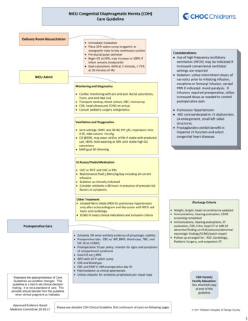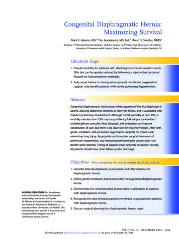
Transcription
3/14/2016CONGENITAL AND ACQUIRED HEARTDISEASES.DR Manori GamageMBBS MD DCH MRCPCHcongenital heart diseasesCONGENITAL HEART DISEASES.Factors responsible for congenital heart Diseases are:1. Prenatal factors:Mothers suffering from infectious diseases: rubella.Mother's alcoholism.Maternal age over 40 years.Mothers suffering from diabetes mellitus who require insulin.2 Types.1. Acyanotic heart diseases.Mothers taking sedative drugs or herbs.2. Genetic factorsChildren born before suffering from CHD.2. Cyanotic heart diseases.Father / mother suffering from CHD.Chromosomal abnormalities eg Down syndrome.Born with other congenital abnormalities.Acyanotic heart diseases.VSD.ASD.PDA.Coarctation of aorta.Cyanotic heart diseases.TOF.TGA.Complex heart diseases.1
3/14/2016Ways of presentation.Detection of murmur.- Neonatal examination.- SMI.- When visit for other illnesses.Heart failure.Cyanosis.- Neonatal.- LaterNursing Diagnosis for Congenital Heart DiseaseRisk for decreased cardiac outputAltered Growth and Development due to inadequateoxygen and nutrients to the tissues.Risk for infectionPoor physical status.Altered family processes due to children with heartdisease.Risk for injury (complications) due to the heart conditionand therapy.Failure to thrive.Recurrent respiratory tract infections.SABE.Nursing interventionsDeliver oxygen and prevent hypoxiaGive afterload lowering medications as instructed.Give diuretic as instructed.Provide frequent rest periods and periods ofuninterrupted sleep.Encourage quiet activities.Give a balanced diet high in nutrients, to achieveadequate growth.Monitor height and weight.Encourage the family to participate in the care process.Teach families to recognize the signs of complicationsRheumatic heart diseasePathophysiology : Autoimmune conditionTriggered by Group A beta haemolyticstreptococcus infectionAuto antibodies act against heart(Pancarditis)Acquired heart diseasesDiagnosis : Modified Jones criteria.Symptoms :FeverArthralgiarditis:CarditisPolyarthritis2
3/14/2016Management :Short term – Eliminate streptococcal infection(oral Penicillin for 10 days)High dose Aspirin to reduce inflammationInvestigation :Bed restProlonged PR interval on ECGIncreased erythrocyte sedimentation rate [ESR]Presence of C-reactive proteinLong term – Prophylaxis against SABE(monthly IM Benzathine penicillin)LeukocytosisSurgical correction of damaged valvesComplications : SABEHeart failurePrevention– Reduce overcrowding.Prompt treatment with antibiotics in PharyngitisKawasaki diseaseVasculitis affecting the coronary arteriesDiagnosed by using clinical criteriaa. Fever 5 dyasComplicationsCoronary artery aneurysmb.Skin rashManagementc.Red lips and red tongueIV immunoglobulind.Pedal oedemaAsprin ( initially – high dose - later low dose )e.ConjuctivitisClinical featuresInfective endocarditisInfection of the endocardiumRisk factors-Neonates-Congenital heart lesions-Acquired heart lesions (valve)-Cardiac prosthesesPersistent feverChanging murmursPallorSpleen enlargementClubbingArthritisDiagnosis2 D echoBlood culture3
3/14/2016Management1.IV antibiotics – 6 weeks2.Manage any complications develop3.Prevention – when there are risk factors start onprophylaxisMedical and Surgicalproblems of theGastrointestinal tractDr Manori GamageMedical problems.Vomiting.Loose stool.GORD.Constipation.Gastritis.HepatitisIBDMBBS MD DCH MRCPCHVomiting.Forceful ejection of stomach content through mouth is known asvomiting.CAUSES Infants- GORDFeeding problems.Infections.Intestinal obstruction.Inborn errors of metabolism.Pre school childrenInfection.Appendicitis.Intestinal obstruction.Testicular torsion.School age and adolescentsGastroenteritis.Systemic infections.PUD.Hx : Find out the causeEx : Assess the level of dehydrationMx : Treat the underlying causeSymptomatic treatment with antiemeticsproper rehydrationand( depending on the level of dehydration,water, ORS, 0.9% NaCl)testicular torsion.4
3/14/2016Loose stoolsWatery diarrhoeaAcute gastroenteritis.Food poisoning.Systemic infections.IBSToddlers diarrhoea.Acute gastroenteritisCommonest causeViral – Rota virusBacterialE. ColiMainly presents with severe watery diarrhoeaBlood and mucus diarrhoeaDysentery. (Shigella, e-coli )Hx : Assess frequency, severity, associatedfeatures, complications (hydration level,electrolyte imbalance), look for a possible cause.Ex : level of dehydration.IBD.Ix : Serum electrolytes.Stool full report.Intussusception.Level of dehydrationa.No dehydrationb.some dehydrationc.Severe dehydrationMx : Rehydration(ORS, IV fluid)Antibiotics (If needed)Avoid anti motility drugs.Depending on history and examination we categorizethem in to each.ASKLOOKFEEL5
3/14/2016GORDRehydrationDepending on the degree of dehydrationIs the involuntary passage of gastric content in to theoesophagus.No dehydration- maintenance on going lossessSome dehydration- maintenance on going deficitSevere dehydration – maintenance on going deficitCAUSES :Functional immaturity of LOS.Hiatus hernias.Ix : 24 hour oesophageal pH monitoringHx : Recurrent regurgitation, Vomiting.Heart burn.Recurrent chest infections.Neuro developmental disorders.Ex : Adequate growth except in very severe cases.(gold standard)Mx : Uncomplicated – Reassurance andpositioning after feeds.Significant - Acid suppression with PPI or H2 receptorblockers.Surgical management.Constipation.Less frequent bowel opening with hard consistency.CAUSES : FunctionalHirsch sprung disease.Hypothyroidism.Anorectal abnormalities.Precipitated by : low fluid and fiber intake.Hx : Frequency, consistency, features of underlying cause,involuntary soiling, toilet habits.Ex : Anal fissures.Mx : life style modifications. (toilet training)dietary advices ( high fluid and fiberintake)Laxatives.6
3/14/2016Gastritis.CAUSES : Autoimmune.H-pylori infection.Drugs (NSAID, Steroids)Hx : Regurgitation.Heart burn.Epigastric pain.Ex : Nothing significantlook for associated factors.Ix : UGIE and biopsy.Mx : PPIH-pylori eradication therapy.Nausea , Vomitting.Drug history.Diaphragmatic hernia.Hx : Difficulty in breathing.Ex :Respiratory distress.Scaphoid abdomen.Mediastinal shift.Audible bowel sounds in chest.Ix : Chest and abdominal x ray.Mx : Large NG tube insertion and suction to prevent distensionof intrathoracic bowel.Surgical repair.Hirschsprung disease.Hx : Constipation.Abdominal distention.Delayed passage of meconium.Bile stained vomiting (later).Ex : Distended abdomen.DRE (narrowed segment)Ix : Suction rectal biopsy.Mx : Surgical management.7
3/14/2016Acquired.Intussusception.Hx : Insolvable crying.Severe colicky pain.Red current jelly stool.Refused feeding.Vomiting.Ex : sausage shaped mass palpable in the abdomen.Abdominal distention and shock.Ix : USS abdomen.Mx : Rectal air/ hydro insufflation.Surgical management.Hypertrophic pyloric stenosisHypertrophy of the pylorus cause gastric outletobstruction.Clinical featuresPresent during 2-7 weeks of lifeProjectile vomitingPoor weight gainVisible peristalisisManagementStabilize the babyCorrect hydrationCorrect electrolyte imbalancesSurgical correctionDiagnosisUS Scan of the abdomenAppendicitis.Hx : Abdominal pain (umbilical area RIF)Vomiting.Fever.Anorexia.Ex : Febrile.Ix : USS abdomen.FBC, CRP.Mx : Appendicectomy.Conservative management.RIF rebound tenderness and guarding.8
3/14/2016Intestinal obstruction.Hx : Vomiting.Abdominal distention.Constipation.Ex : Distended abdomen.Exaggerated bowel sounds.Ix : Supine abdominal x ray.Serum Electrolytes.Mx : Nil by mouth.NG tube decompression.Hydration and electrolyte correction.Surgical or conservative management.Viral hepatitisHepatitis AFeco oral routeOccur due toHepatitis virus A ,B, C,or EClinical featuresSelf limitingManagementNauseaIsolateVomitingSymptomatic managementAbdominal painNortificationJaundiceHealth educationDark colour urineMusculoskeletal systemAbnormalitiesDR Manori GamageMBBS MD DCH MRCPCHMusculoskeletal abnormalities inchildrenCongenitalClub footDevelopmental dysplasia of the hipMyopathiesAcquiredFracturesTransient synovitisSeptic arthritisosteomyelitis9
3/14/2016Club foot2 typesDiagnosis is by clinical examinationExclude any associated hip anomaliesexamine spine to see any associated anomalyPositional talipescan be corrected to neutral position with passive manipulationTalipes equinovarusA complex deformity. Entire foot is inverted, supinatedForefoot is adductedManagement:If positionalReassurancephysiotherapyHeel rotated inwards and plantar flexionIf severePOP castsurgical correctionDevelopmental dysplasia of the hipSpectrum of disorders of the hip jointDuring managementRanging from dysplasia to subluxation to frank dislocation of the hip fromacetabulumRelieve maternal anxiety is very importantIf Baby has a POP castlook after the cast and look after the limb with the cast isvery importantDiagnosisEarly detection is important.FracturesNeonatal screening is performed byBarlow’s manaeuver- to check whether hip is dislocableOtolani manaeuver -to relocate the dislocated hip into acetabulumConfirmed by USSManagement :Respond to conservative managementComplete or incomplete break on a bone resulting fromthe application of excessive forcecommonest sites:ClavicleHumerusHarness, Splinting to keep hip flexed and adductedFemurif not responding – corrective surgerySupracondylar fracture of the humerusForearm fractures of radius, ulnar10
3/14/2016Causes for fracturesClinical features of fracturesBirth traumaShoulder dystociaPain and swelling at the fracture siteFallen on outstretched handTenderness close to the siteDirect blow on boneDeformity of the affected limbInherited diseases of bones –osteogenesis imperfectaBleeding and bruising at the siteTumours of bonesLoss of pulse distal to the fractureNumbness, tingling or paralysis distal to the fracturesiteTransient synovitis(irritable hip)DiagnosisClinical examinationX rayMost common cause of acute hip painCommonest between 2-12 yearsManagementPain reliefImmobilizationSplintsPOP castsReductionTraction reductionManipulation under anaesthesiarehabilitationSeptic arthritisSerious infection of the joint spaceCan lead to bone destruction if not treatedEarly diagnosis is importantCan give rise to infection of the underlying bone OsteomyelitisJoint is, erythematous, warm, tender joint, reducedmovementsFollows/accompanies by a viral infectionSudden onset pain in hip or limpLimited internal rotation and reduced range of movementsDiagnosisUSSManagementRestPain reliefDiagnosisWBCCRPBLOOD CultureUSSXraysManagementPain reliefAntibiotics- should give a prolonged course11
3/14/2016Genito-urinary disorders inchildrenWays of presentation OedemaReduced urine output.DR Manori GamgeMBBS MD DCH MRCPCHHaematuriaPolyuriaHypertensionOther urinary symptomsWays of assessment of urinary systemBlood gasUrine culture and ABSTUS ScanImaging studiesUrine analysisX- RaySerum ElectrolytesBlood investigationsDMSABlood ureaDTPAMCUGSerum creatinineCommon problems seen among childrenUTI1.UTIIt is an infection of the urinary tract.2.Nephrotic syndromeWhy important?3.Nephritic syndromeKidneys are growing4.HaematuriaIf damagedCRFScarringHTN12
3/14/2016Ways of presentationDepending on the age, presentation will varyNeonate/Infant- FeverOlder children-Fever-FTT-Urinary symptoms-Irritability-Vomiting-Jaundice-Abdominal painHow to diagnose ?UFR – SupportiveUrine culture- Gold standardHow to collect U.Culture is very important-VomittingSuprapubic aspirationMethod of collection-MSU – Clean catch-Catheter-Suprapubic aspiration-Bag – Not availableTreatmentNursing interventionsAcute-Do investigations-Short course of antibiotics -7 to 10 days-Post treatment culture ABST-Long term plan will be discussed, dependingAssessmentSupport investigationsAdminister medication as prescribedHealth education (Very important)onAgeway of presentationUnderlying other problems13
3/14/2016Nephritic SyndromeFeaturesIxUFR- OliguriaUrine culture- HypertensionRenal functions-HaematuriaUS scan KUBFollowing Streptococcal infection.ComplicationsMx-Acute renal failureInput/ Output chart-Hypertensive encephalopathyDaily weight-Heart failureDaily urine protein testBP chartOral Penicillin – 10 daysAntihypertensive (If BP high)Nursing interventionNephrotic syndrome1.Assessment - Oedema- BP- UOP2.Supportive treatment3.Administer medications prescribed4.Input/Output5.Urine ward test daily6.Health education/Psychological supportFeatures- Gross oedema- Proteinuria- Reduced serum Albumin- Increased serum Cholesterol14
3/14/2016PresentationIxOedemaUFRFrothy urineSerum proteinSerum CholesterolRenal functionsUrine culture & ABSTMxComplicationsHypovolemic shockGeneralSpecificRenal vein thrombosis- Isolate- O.PrednisolonePeritonitis-Input / Output- O. PenicillinIncreaced risk of other infections-Daily weightIf ascitis present-U. ward test-BP chartNursing interventionsAssessmentsOdemaVolume statusComplicationsSupportive treatmentMedications on prescribeMonitor the vitals/ other parameters Health education15
3/14/2016Pre renal causesAcute renal failureHypovolaemia- GastroenteritisPre renalRenalPost renal-Burns-Sepsis-Haemorrhage-Nephrotic syndromeCirculatory failureRenal causesPost renal causesVascular-HUS-Vasculitis-Embolism-Congenital (PUV)Tubular-Acquired ( Blocked urinary catheter)-Acute tubular necrosis-Toxic-ObstructionObstructionGlomeruler – GlomerulornephritisInterstitial nephritisCKDAssociated problemsIncreased Blood ureaIncreased PotassiumAcidosis Dialysis NaHCO3When GFR is 15ml/min per 1.73m2Causes- Structural malformations-Glomerular nephritis-Unknown16
3/14/2016MxNutritionTreat acidosisTreat anaemiaANY QUESTIONS ?DialysisTransplantationThank you !17
disease. Monitor height and weight.Risk for injury (complications) due to the heart condition and therapy. Nursing interventions Give diuretic as instructed. Provide frequent rest periods and periods of uninterrupted sleep. Encourage quiet activities. Give a balanced diet high in nutrients, to achieve adequate growth.
