Transcription
Quality DepartmentGuidelines for Clinical CareInpatientGallstone-RelatedDiseases GuidelineTeamTeam LeadersBen E Biesterveld, MDSurgeryHasan B Alam, MBBSSurgeryTeam MembersWilliam T Repaskey, MDInternal MedicineSteven L Kronick. MD, MSEmergency MedicineBenjamin Pomerantz, MDInterventional RadiologyMichael David Rice, MDGastroenterologyInitial ReleaseMay, 2014Most Recent Major UpdateAugust, 2020Inpatient ClinicalGuidelines OversightMegan R Mack, MDDavid H Wesorick, MDApril Proudlock, RNEvaluation and Management of GallstoneRelated Diseases in Non-Pregnant AdultsPatient Population: Adult patients with suspected or confirmed biliary colic, acute cholecystitis,choledocholithiasis, cholangitis, or mild gallstone pancreatitis. Excluded are patients who: are pregnantpatients, have a history of bypass surgery or biliary surgery, or have acute pancreatitis. For these conditions,consult appropriate subspecialists.Objectives: Create an evidence-based guideline for the management of gallstone-related diseases thatprovides prompt and appropriate service to patients, reduces unnecessary diagnostic testing, and improvespatient outcomes.Key PointsClinical Presentation. Patients presenting with upper abdominal pain or jaundice should be evaluated forgallstone-related disease.Diagnosis.The evaluation for gallstone-related disease is summarized in Table 1. The evaluation routinely includes:1. Complete physical exam2. Laboratory evaluation – CBC, comprehensive metabolic panel, lipase3. Imaging – Right upper quadrant (RUQ) ultrasoundFor most patients with acute cholecystitis, diagnosis can be based on history, physical findings, laboratorytests, and ultrasound (see Table 3 for the sonographic diagnostic criteria). In rare cases where the diagnosisremains uncertain after this evaluation, additional imaging modalities may be necessary.Treatment.The treatment of gallstone-related diseases is summarized in the Figure.Biliary ColicLiterature Search ServiceTaubman Health SciencesLibrary For more information:734- 615-8201Minimally symptomatic or with symptoms that resolve: provide reassurance, education on avoidanceof triggers (eg, dietary fat). Provide direct referral to elective surgery (Priority Gallbladder Clinic forsurgery within 2 weeks at University of Michigan, see Appendix). [II-C*] Moderate to severe symptoms: consult surgery. Perform non-urgent laparoscopic cholecystectomyduring same visit [II-C*]. Timing of surgery determined by patient preference and operating roomavailability. Regents of the Universityof MichiganThese guidelines should not beconstrued as including allproper methods of care orexcluding other acceptablemethods of care reasonablydirected to obtaining the sameresults. The ultimate judgmentregarding any specific clinicalprocedure or treatment must bemade by the physician in lightof the circumstances presentedby the patient.Acute Cholecystitis Admit to SurgeryInitiate IV antibiotics (Table 2)Perform laparoscopic cholecystectomy within 24-48 hours [I-A*]. In patients without gallstones who have right upper quadrant (RUQ) and/or epigastric pain and ahepatobiliary iminodiacetic acid (HIDA) scan showing delayed gallbladder filling or lack ofgallbladder emptying, cholecystectomy should be recommended[I-A*].* Strength of recommendation:I generally should be performed; II may be reasonable to perform; III generally should not be performed.Levels of evidence reflect the best available literature in support of an intervention or test:A systematic reviews of randomized controlled trials with or without meta-analysis, B randomized controlled trials, C systematic review of non-randomized controlled trials or observational studies, non-randomized controlled trials, groupobservation studies (cohort, cross-sectional, case-control), D individual observation studies (case study/case series), E expertopinion regarding benefits and harm1 Evaluation and Management of Gallstone Related Diseases 08/2020
Treatment (continued):CholedocholithiasisCholedocholithiasis may occur alone, but should also be considered as a comorbidity with cholecystitis or any of the othergallstone-related diseases. Evaluate for evidence of cholangitis (Table 5). If suspected, treat as cholangitis (see below).If no evidence of cholangitis, admit to surgery and prepare for cholecystectomy.If choledocholithiasis is demonstrated on imaging, preoperative ERCP is often performed to clear the duct.If choledocholithiasis is not documented on imaging, estimate the likelihood of choledocholithiasis (Table 4) Low likelihood: no additional evaluation is needed, and routine intraoperative cholangiography (IOC) is notrecommended [III-B] Intermediate likelihood: recommended approach is a one-stage procedure with laparoscopic cholecystectomywith IOC within 24 to 48 hours of admission (24 hours preferred). [I-A*] Alternate approaches might includepreoperative imaging with ERCP or MRCP, especially if IOC will not be performed.NOTE: If intraoperative cholangiogram (IOC) demonstrates a retained common bile duct (CBD) stone: Perform procedure to remove CBD stones during the same operation [I-A*], or Obtain gastroenterology consult within 24 hours after surgery for endoscopic retrograde cholangiopancreatography (ERCP). High likelihood: preoperative ERCP is often performed to clear the duct.Cholangitis Admit to Medicine service.Initiate IV antibiotics, NPO (Table 2).Obtain Gastroenterology consult.Classify severity of acute cholangitis (Table 6). Mild cholangitis with adequate response to medical therapy: ERCP within 72 hours.Moderate-severe or not responsive to medical therapy: ERCP within 24 hours.Consult Surgery for laparoscopic cholecystectomy during same admission, after cholangitis resolves.Gallstone Pancreatitis: Evaluate for evidence of cholangitis (Table 5). If suspected, treat as cholangitis (see above), otherwiseclassify severity of gallstone pancreatitis (Table 7).Mild gallstone pancreatitis: Admit to surgery service.Perform laparoscopic cholecystectomy with IOC within 24 (preferred) to 48 hours [I-B*].If IOC demonstrates a retained CBD stone: Surgical removal of CBD gallstone [I-A*], - or - Gastroenterology consult for ERCP within 24 hours of surgery.Moderate to severe gallstone pancreatitis: Admit to medicine.Consider gastroenterology consultation, and preoperative ERCP if bilirubin is elevated or cholangitis present.Delay cholecystectomy until pancreatitis resolves.NOTE: For detailed management of acute pancreatitis at the University of Michigan: http://pancmap.med.umich.edu/2 Evaluation and Management of Gallstone Related Diseases 08/2020
Table 1. Clinical Features of Gallstone-Related DiseasesGallstone-RelatedDiseases*Clinical FeaturesBiliary ColicH&P: Severe, episodic, epigastric or RUQ pain; may be nocturnal, occasionallypostprandial. /- RUQ tenderness.Labs: No leukocytosis; normal total bilirubin and amylase/lipase.Imaging: RUQ ultrasound indicating cholelithiasis without findings of cholecystitis (Table3).Acute CholecystitisH&P: /- fever; symptoms persist or worsening; positive for RUQ tenderness.Labs: Leukocytosis is common. Total bilirubin is usually normal to mildly elevated ( 2.0mg/dL), unless there is concomitant choledocholithiasis. Amylase and lipase areusually normal unless there is concomitant pancreatitis.Imaging: RUQ ultrasound, see Table 3. The diagnosis of cholecystitis is NOT made basedon ultrasound findings alone. Diagnosis is determined based on the clinicalfindings above, in combination with consistent ultrasound findings. HIDA (only indicated if RUQ ultrasound is inconclusive, or contradicts the clinicalimpression) demonstrates lack of gallbladder filling.CholedocholithiasisH&P: Biliary pain, jaundice, no fever.Labs: Elevated bilirubin (total bilirubin often 2.0 mg/dL). Amylase/lipase are usuallynormal, unless there is concomitant pancreatitis.Imaging: RUQ ultrasound shows CBD dilation ( 7 mm).**Risk Stratification: See Table 4.CholangitisH&P: Jaundice, often febrile, RUQ tenderness.Labs: Elevated bilirubin (total bilirubin 2.0 mg/dL), leukocytosis. Amylase/lipase areusually normal to mildly elevated, unless there is concomitant pancreatitis.Imaging: RUQ ultrasound: CBD dilation ( 7 mm).**Diagnosis and risk stratification: See Tables 5 & 6.Gallstone PancreatitisH&P: /- jaundice, /- fever, epigastric tenderness.Labs: Normal or elevated bilirubin, elevated amylase and/or lipase to typically 3x upperlimit of normal. Elevated ALT 150 suggests a biliary cause of pancreatitis, basedon meta-analysis1Imaging: RUQ ultrasound: Cholelithiasis and biliary dilation variably present. Note: RUQultrasound is often limited for the evaluation of the pancreatic parenchyma.Absence of other common causes of pancreatitis: Ethanol abuse, hyperglycemia,hypertriglyceridemia, hypercalcemia, or medications known to cause pancreatitis.Classification of pancreatitis severity: see Table 7.RUQ: Right upper quadrant; HIDA: hepatobiliary iminodiacetic acid; CBD: common bile duct; ALT: alanineaminotransferase*These diseases are not mutually exclusive and often present together. For example, patients withcholedocholithiasis often present with gallstone pancreatitis.**Post-cholecystectomy patients may have CBD dilation in the absence of biliary pathologyNote: upper abdominal pain, nausea, and vomiting (N/V) are common to all of these disorders3 Evaluation and Management of Gallstone Related Diseases 08/2020
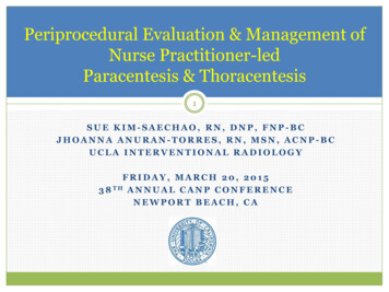
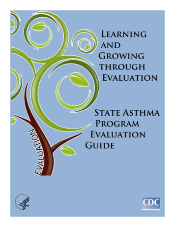
Figure 1: Treatments for Gallstone-Related DiseasesNote: These conditions are not mutually exclusive. For example, patients with cholecystitis may also have CBD stonesor cholangitis.*For University of Michigan, consult http://pancmap.med.umich.edu/.**For acute cholecystitis patients who are poor surgical candidates refer to page 13 for options.AST Aspartate Aminotransferase/ Aspartate Transaminase; BUN Blood Urea Nitrogen; CXR Chest radiograph;EKG Electrocardiogram; ERCP Endoscopic Retrograde Cholangiopancreatography; HR Heart rate; IOC Intraoperative cholangiogram; NPO Nils per os/nothing by mouth; WBC White blood cell count.4 Evaluation and Management of Gallstone Related Diseases 08/2020
Table 2. Antibiotic Guidelines for Treatment of Cholecystitis and Cholangitis in AdultsEmpiric Antibiotic TherapyCommunity-acquired, without severe sepsis/shocko 1st line: Cefuroxime1 1.5 g IV q8h /- metronidazole 500 mg PO/IV q8h (if anaerobic coverage required2)o High-risk allergy3/contraindications4 to beta-lactams: Ciprofloxacin* 400 mg IV q8h /- metronidazole 500 mg PO/IV q8h (if anaerobiccoverage is required2)Community-Acquired with severe sepsis5/shock6 OR MDR-GNR risk7o 1st line: Piperacillin-tazobactam1 4.5 g IV q6ho Low/medium-risk allergy8 to penicillins: Cefepime1 2 g IV q8h metronidazole 500 mg PO/IV q8ho Consider the addition of vancomycin to cefepime for enterococcus coverage in critically ill patients with risk factors forenterococcal infection9.o High-risk allergy3/contraindication4 to beta-lactams: Vancomycin1 aztreonam1 2 g IV q8h metronidazole 500 mg PO/IV q8hStepdown Antibiotic TherapyStep-down oral therapy can be used if the patient is tolerating oral intake, and susceptibilities (if available) do not demonstrate resistanceo Amoxicillin-clavulanic acid1 875 mg PO BID, ORo Cefuroxime1 500 mg PO BID /- metronidazole 500mg PO TID (if anaerobic coverage required2)o High-risk allergy3/contraindications4 to beta-lactams OR MDR-GNR risk7: Ciprofloxacin 750 mg PO BID /- metronidazole 500 mg POTID (if anaerobic coverage required2)Duration of Antibiotic TherapyoooooIn general: 4-7 days2After cholecystectomy: Discontinue within 24 hours unless evidence of infection outside the gallbladder wallAfter successful ERCP: 4 days post-procedurePatients with bacteremia: 7-14 days. For patients with secondary gram-negative bacteremia, a 7-day duration of IV therapy (or oralquinolone at discharge) may be appropriate for selected patients, in conjunction with ID consultation.Duration of therapy may be extended with inadequate source control or persistent clinical symptoms or signs of infection.Footnotes continued on next page5 Evaluation and Management of Gallstone Related Diseases 08/2020
Table 2. Antibiotic Guidelines for Treatment of Cholecystitis and Cholangitis in Adults (continued)1234567Adjust dose based on renal functionAnaerobic coverage (metronidazole) is not necessary for patients with community-acquired cholecystitis/cholangitis of mild-moderate severity, unless a biliaryenteric anastomosis is present.High-risk allergies include: respiratory symptoms (chest tightness, bronchospasm, wheezing, cough), angioedema (swelling, throat tightness), cardiovascularsymptoms (hypotension, dizzy/lightheadedness, syncope/passing out, arrhythmia), anaphylaxisPrevious reactions that are contraindications to further beta-lactam use (except aztreonam, which can be used unless the reaction was to ceftazidime oraztreonam) unless approved by Allergy: organ damage (kidney, liver), drug-induced immune-mediated anemia/thrombocytopenia/leukopenia, rash with mucosallesions (Stevens Johnson Syndrome/toxic epidermal necrosis), rash with pustules (acute generalized exanthematous pustulosis), rash with eosinophils and organinjury (DRESS – drug rash eosinophilia and systemic symptoms), rash with joint pain, fever, and myalgia (serum sickness)Severe Sepsis: Sepsis PLUS at least 1 organ dysfunction Sepsis: 2 SIRS criteria (heart rate greater than 90 bpm, respiratory rate greater than 20 breaths per minute, temperature less than 36oC, white bloodcount less than 4,000 cells/mm3, temperature greater than 38 C, white blood count greater than 12,000 cells/mm 3) Organ dysfunction: CV: SBP 90 mmHg or MAP 70 mmHg or require vasopressor support; Respiratory: PaO2/FiO2 250 or mechanical ventilation;Renal: decreased urine output 0.5 mg/kg/hr for 1 hour, increased SCr ( 50% from baseline); Hematologic: platelet 100,000 or increase aPTT;Metabolic: p
For most patients with acute cholecystitis, diagnosis can be based on history, physical findings, laboratory tests, and ultrasound (see Table 3 for the sonographic diagnostic criteria). In rare cases where the diagnosis remains uncertain after this evaluation, additional imaging modalities may be necessary. Treatment. The treatment of gallstone-related diseases is summarized in the Figure .File Size: 329KBPage Count: 15