Transcription
C H A P T E R11Neurodegenerative Diseasesand AutophagyAngeleen Fleming1,*, Mariella Vicinanza1,*, Maurizio Renna1,*,Claudia Puri1,*, Thomas Ricketts1,*, Jens Füllgrabe1,*,Ana Lopez1,*, Sarah M. de Jager1,*, Avraham Ashkenazi1,*,Mariana Pavel1,*, Floriana Licitra1,*, Andrea Caricasole2,*,Stephen P. Andrews2,*, John Skidmore2,* andDavid C. Rubinsztein1,31Cambridge Institute for Medical Research, University of Cambridge, Cambridge Biomedical Campus,Cambridge, United Kingdom 2Alzheimer’s Research UK Cambridge Drug Discovery Institute,University of Cambridge, Cambridge, United Kingdom 3UK Dementia Research Institute,Cambridge Biomedical Campus, Cambridge, United KingdomO U T L I N EAutophagy Cell BiologyKey Autophagy MachineryAutophagosome Membrane TraffickingEventsKey Signaling PathwaysSelective AutophagyLysosomesAutophagy in Neuronal Physiology300300Autophagy in Neurodegenerative DiseasesAlzheimer’s �s DiseasePolyglutamine DisordersAmyotrophic Lateral SclerosisHereditary Spastic ParaplegiasLafora DiseaseDynein and Dynamin MutationsDiseases Resulting from Mutations in CoreAutophagy GenesLysosomal Disorders311312314315316317318318319* Joint first authors.The Molecular and Cellular Basis of Neurodegenerative DiseasesDOI: 299 2018 Elsevier Inc. All rights reserved.
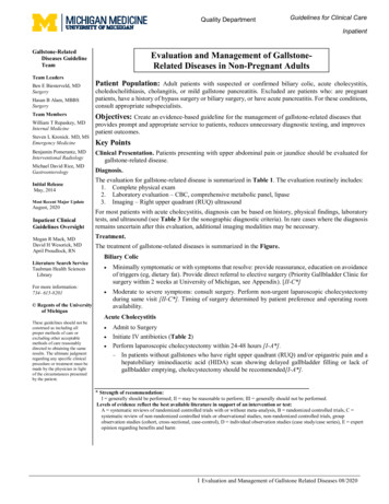
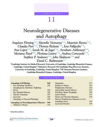
30011. NEURODEGENERATIVE DISEASES AND AUTOPHAGYAutophagy UpregulationTrehaloseRapamycinRepurposing of FDA-Approved Drugs asAutophagy 25Further Reading343324AUTOPHAGY CELL BIOLOGYKey Autophagy MachineryAutophagy (macroautophagy) is a degradation process that delivers cytoplasmic materialsto lysosomes. By doing so, autophagy sustainscellular renovation and homeostasis by recycling molecular building blocks (such as aminoacids or fatty acids) for anabolic processes. Thefirst morphologically recognizable autophagicprecursor is a flat, double-membraned, sac-likestructure (called a phagophore), whose edgeselongate and fuse while engulfing a portion ofthe cytoplasm. The resulting structure is aspherical double-membrane organelle, called theautophagosome. The formation of autophagosomes requires several steps (nucleation,elongation, and closure) governed by conservedproteins termed ATGs (AuTophaGy-relatedproteins) (Mizushima, Yoshimori, & Ohsumi,2011). Autophagy initiation and autophagosomeformation require multiple interactions betweendifferent individual proteins and protein complexes. For simplicity, these are referred to bytheir abbreviated names in the following sectionsand are described in full in Table 11.1.During autophagosome formation, theATG8 ubiquitin-like family proteins are conjugated to the lipid nes.Mammalian cells have six ATG8 orthologues;the MAP1-LC3 (LC3) and GABARAP subfamilies. Lipidated ATG8 proteins have been usedto distinguish autophagosomes from othercellular membranes (Itakura & Mizushima,2010). Measuring the LC3 lipidation, scoringthe number of LC3 vesicles, and detecting thedegradation of long-lived proteins or damagedorganelles are the mainstay methods used formonitoring autophagy (Itakura & Mizushima,2010). However, this requires careful interpretation since immune receptors engaged byphagocytosed cargoes can also enable LC3recruitment to single-membrane phagosomesin a process called LC3-associated phagocytosis (Sanjuan et al., 2007).LC3/GABARAP lipidation requires a protease and two ubiquitin-like conjugation systems(Ichimura et al., 2000; Mizushima et al., 1998) asillustrated in Fig. 11.1. The first reaction involvesthe conjugation of the proteins ATG12 to ATG5in a reaction requiring the enzymatic activities ofATG7 and ATG10. The ATG5-ATG12 conjugateforms a complex with ATG16L1. The cysteineprotease ATG4 cleaves the C-terminus of LC3exposing a glycine residue (LC3-I), which is activated by the ATG7 enzyme, initiating events forthe second conjugation reaction. In this secondreaction, the ATG12 ATG5 ATG16L1 complex,through interaction with the ATG3, acts as theE3-like ligase that determines the site of LC3 lipidation and assists the transfer of LC3-I to PE toform LC3-II (Ichimura et al., 2000). ATG8/LC3proteins may assist in the expansion and closureof autophagosomal membranes (Nakatogawa,Ichimura, & Ohsumi, 2007) as well as in autophagosome lysosome fusion and inner autophagosomal membrane degradation (Nguyen et al.,2016; Tsuboyama et al., 2016).THE MOLECULAR AND CELLULAR BASIS OF NEURODEGENERATIVE DISEASES
301AUTOPHAGY CELL BIOLOGYTABLE 11.1 List of Abbreviations of Proteins, Complexes and Cellular Structures Involved in AutophagyAbbreviationFull NameFunction of Protein or Complex in AutophagyAKTProtein kinase BSerine/threonine kinaseAMBRA-1Activating molecule in Beclin-1-regulatedautophagyPart of VPS34 complexAMPKAMP-activated protein kinaseProtein kinase complexAPPAmyloid precursor proteinMembrane protein cleaved by secretases to formAβ peptideATGsAuTophaGy-related proteinsATG3AuTophaGy-related protein 3E2-ligase-like enzymatic activityATG4AuTophaGy-related protein 4Cysteine proteaseATG7AuTophaGy-related protein 7E1-ligase-like enzymatic activityATG9AuTophaGy-related protein 9Organization of the preautophagosomalstructure/phagophore assembly siteATG10AuTophaGy-related protein 10E2-ligase-like enzymatic activityATG12AuTophaGy-related protein 12Ubiquitin-like proteinATG14AuTophaGy-related protein 14Part of VPS34 complex; regulates localization ofthe complexATG16L1 complexComplex comprising ATG5, ATG12, ATG16L1E3-ligase-like enzymatic activityBeclin-1Homologue of BEC-1 (C. elegans) and ATG6(yeast)Part of VPS34 complexCOPICoatomer protein ICoat protein complex required for ER-Golgitransport and early endosome formationCREBcAMP response element-binding proteinTranscription factorDFCP1Double FYVE-containing protein 1Interact with phospholipidsE2F1E2F transcription factor 1Transcription factorEREndoplasmic reticulumERESER-exit sitesESCRTEndosomal sorting complexes required fortransportMembrane remodelingFAM134Family with sequence similarity 134Selective autophagy cargo receptorFIP200Focal adhesion kinase family interactingprotein of 200 kDaPart of ULK1 complexFOXOForkhead boxTranscription factorFXRFarnesoid X receptorNuclear receptorFYVEZinc-finger domainBinds and inserts into PI3P membranes(Continued)THE MOLECULAR AND CELLULAR BASIS OF NEURODEGENERATIVE DISEASES
302TABLE 11.111. NEURODEGENERATIVE DISEASES AND AUTOPHAGY(Continued)AbbreviationFull NameFunction of Protein or Complex in AutophagyGABARAPγ-Aminobutyric acid receptor-associatedproteinATG8 homologueLYNUSLysosome nutrient-sensingMAMMitochondria-ER-associated membranesMAP1-LC3 or LC3Microtubule associated proteins 1A/1B lightchain 3ATG8 homologuemTORMechanistic target of rapamycinProtein kinasemTORC1mTOR complex 1; complex comprising mTOR,RAPTOR, GβLKinase complexNBR1Neighbor Of BRCA1 gene 1Selective autophagy cargo receptorNCOA4Nuclear receptor coactivator 4Selective autophagy cargo receptorNF-κBNuclear factor kappa-light-chain-enhancer ofactivated B cellsTranscription factor (protein complex)NDP52/CALCOCO2Nuclear domain 10 Protein 52/calciumbinding and coiled-coil domain 2Selective autophagy cargo receptorOPTNOptineurinSelective autophagy cargo receptorP53/TP53Tumor protein 53Transcription factor (tumor suppressor)p62/SQSTM1Ubiquitin-binding protein P62/sequestosome1Selective autophagy cargo receptorPARKINParkinson’s disease protein 2 (PARK2)E3-Ubiquitin Protein LigasePEPhosphatidylethanolaminePhospholipids found in membranesPI3PPhosphatidylinositol 3-phosphatePhospholipids found in membranesPINK1PTEN-induced putative kinase 1 (PARK6)mitochondrial serine/threonine-protein kinasePPARαPeroxisome proliferation factor-activatedreceptor αTranscription factorRAPTORRegulatory-associated protein of mTORPart of mTOR Complex 1RERecycling endosomeRPN1026S proteasome regulatory subunit RPN10Proteasome componentSNARESNAP (soluble NSF attachment protein)ReceptorMediate vesicle fusionSNX18Sorting nexin 18Membrane remodelingTAX1BP1Tax1 (human T-cell leukemia virus type I)binding protein 1Selective autophagy cargo receptorTBC1D14TBC1 domain family member 14Membrane remodelingTFEBTranscription factor EBTranscription factor(Continued)THE MOLECULAR AND CELLULAR BASIS OF NEURODEGENERATIVE DISEASES
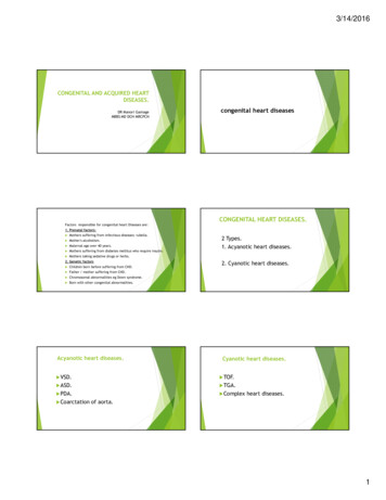
AUTOPHAGY CELL BIOLOGYTABLE 11.1303(Continued)AbbreviationFull NameFunction of Protein or Complex in AutophagyTIP6060 kDa Tat-interactive protein (Kacetyltransferase 5, KAT5)Acetyl transferaseTOLLIPToll interacting proteinSelective autophagy cargo receptorTRAF6TNF receptor-associated factor 6E3-ubiquitin protein ligaseTRIMTripartite motifPathogen recognitionUBDUbiquitin-binding domainRecognize and bind to ubiquitinULK1 and ULK2Mammalian homologs of the C. elegansuncoordinated 51 kinaseserine/threonine kinaseULK1/2-complexComplex comprising ULK1/2, ATG13,ATG101, and FIP200Regulation of autophagosome biogenesisVAMPsVesicle-associated membrane proteinsMediate vesicle fusionVPSVacuolar protein sorting-associated proteinVesicular transportVPS34 complexComplex comprising Beclin1 ATG14 VPS15 VPS34Class III phosphoinositide 3-kinase (PI3K)VTI1BVesicle transport through interaction with tSNAREs homolog 1BMediate vesicle fusionWASHWASP and Scar homologueVesicle traffickingWDR45WD repeat domain phosphoinositideinteracting protein 45Interacts with PI3WIPIsWD-repeat protein interacting withphosphoinositides (homologues of ATG18)Interact with phospholipidsZKSCAN3Zinc-finger protein with KRAB and SCANdomains 3Transcription factorAutophagy initiation and autophagosome formation requires multiple interactions between different individual proteins and proteincomplexes. For simplicity, these are referred to by their abbreviated names in the text. These proteins and complexes have many diversecellular functions other than those involved in autophagy; only their main role in autophagy is described here.ATGs are Organized in Signaling ModulesUpstream of LC3 ConjugationThe ULK1/2-complex is one of the mostupstream signaling units in autophagosome formation. This complex includes the ULK1/2homologues, ATG13, ATG101, and FIP200.AMP-activated protein kinase (AMPK) phosphorylates ATG13 and FIP200 (components ofthe ULK1/2-complex) and AMBRA-1 andBeclin-1 (components of the VPS34 complex),thereby targeting these two complexes to thepreautophagosomal membrane (see Fig. 11.2) (DiBartolomeo et al., 2010; Egan et al., 2015; Itakura& Mizushima, 2010; Jung et al., 2009; Park, Junget al., 2016; Russell et al., 2013).The generation of the lipid phosphatidylinositol 3-phosphate (PI3P) by the VPS34 complex atthe phagophore initiation site aids the recruitment of PI3P-binding ATGs, such as DFCP1 andATG18/WIPIs family proteins (Proikas-Cezanne,Takacs, Donnes, & Kohlbacher, 2015). WIPI2 iscrucial for the localization of ATG16L1 complexTHE MOLECULAR AND CELLULAR BASIS OF NEURODEGENERATIVE DISEASES
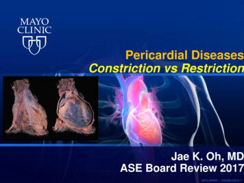
30411. NEURODEGENERATIVE DISEASES AND AUTOPHAGYLC3 conjugation 6complexAtg16Atg16Atg5Atg7LC3LC3I tructural unitsEnzymatic unitsFIGURE 11.1Conjugation steps involved in the regulation of autophagy. Two conjugation systems are requiredfor phagophore/autophagosome membrane elongation resulting in the formation of the ATG12-5-16 complex andLC3-II. Structural components are represented as squares, and enzymatic components are represented as circles.Initial conjugation of ATG12 and ATG5 is mediated by the enzymatic activity of ATG7 and ATG10. ATG5 interactswith ATG16, generating the ATG12-5-16 conjugate, which then homo-oligomerizes to form a tetrameric structurecalled ATG12-5-16 complex. LC3 is cleaved by ATG4 exposing a glycine residue and generating LC3-I, which isactivated by ATG7. The ATG12-5-16 complex through ATG3 interaction covalently conjugates LC3-I to PE to formLC3-II. WIPI 2 modulates the localization of the ATG12-5-16 complex through PI3P, facilitating the interactionof LC3-I with PE and its lipidation in the autophagosomal membrane. PE, phosphatidylethanolamine; PI3P, phosphatidylinositol 3-phosphate.and dictates where the LC3 lipidation occurs(Dooley et al., 2014). Together with ULK1, WIPI2may influence the localization of both mATG9and ATG2A/B. mATG9, the only transmembrane protein among the core ATGs, is considered one of the suppliers of lipid bilayers to theinitiation membrane during elongation (Orsiet al., 2012; Papinski et al., 2014), while ATG2A/B regulates autophagosome closure through fission/scission-type events (Knorr, Lipowsky, &Dimova, 2015; Velikkakath, Nishimura, Oita,Ishihara, & Mizushima, 2012).Autophagosome Membrane TraffickingEventsThe membrane source for autophagosomeformation remains a key open question in thefield. Membranes from different organelles arebelieved to contribute to autophagosome formation by meeting in a particular subcellularcompartment representing the autophagosomeplatform or “isolation membrane.” The isolation membrane is a compartment in proximitytomitochondria-endoplasmicreticulumTHE MOLECULAR AND CELLULAR BASIS OF NEURODEGENERATIVE DISEASES
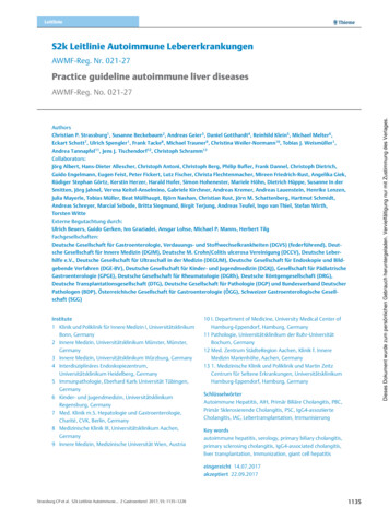
305AUTOPHAGY CELL BIOLOGYUpstream signalling in autophagosome formationStructural unitsAaEnzymatic unitsGlucoseATPCell ophoreLC3 conjugation reactions (Fig.)FIGURE 11.2 Initiation complexes controlling the initiation of autophagy. Many different cellular signals such as levelof nutrients, ATP, or cellular stress control autophagy by modulating mTORC1 activity. The activation of the Akt andthe presence of amino acids detected by Rag GTPases induce mTORC1 which in turns inhibits both Vps34 and ULK1/2complexes and hence inhibiting autophagy. Conversely, deprivation of gl
1Cambridge Institute for Medical Research, University of Cambridge, Cambridge Biomedical Campus, . Abbreviation Full Name Function of Protein or Complex in Autophagy TIP60 60 kDa Tat-interactive protein (K- acetyltransferase 5, KAT5) Acetyl transferase TOLLIP Toll interacting protein Selective autophagy cargo receptor TRAF6 TNF receptor-associated factor 6 E3-ubiquitin protein ligase TRIM .