Transcription
Congenital Diaphragmatic Hernia:Maximizing SurvivalMark F. Weems, MD,* Tim Jancelewicz, MD, MA,† Hitesh S. Sandhu, MBBS‡†‡Divisions of *Neonatal-Perinatal Medicine, Pediatric Surgery, and Critical Care, Department of Pediatrics,University of Tennessee Health Science Center, Le Bonheur Children’s Hospital, Memphis, TN.Education Gaps1. Overall mortality for patients with diaphragmatic hernia remains nearly30% but can be greatly reduced by following a standardized protocolfocused on lung-protective strategies.2. Early repair before or during extracorporeal membrane oxygenationsupport may benefit patients with severe pulmonary hypertension.AbstractCongenital diaphragmatic hernia occurs when a portion of the fetal diaphragm isabsent, allowing abdominal contents to enter the thorax, and is associated withimpaired pulmonary development. Although overall mortality is near 30%, amortality rate less than 15% may be possible by following a standardizedmultidisciplinary care plan. Fetal diagnosis and evaluation can improvecoordination of care, but there is no clear role for fetal intervention. After birth,gentle ventilation with permissive hypercapnia supports the infant whileminimizing lung injury. Appropriate cardiovascular support, treatment ofpulmonary hypertension, and extracorporeal membrane oxygenation maybenefit some patients. Timing of surgical repair depends on disease severity.All patients should have close follow-up after discharge.ObjectivesAfter completing this article, readers should be able to:1. Describe fetal development, assessment, and interventions fordiaphragmatic hernia.2. Define gentle ventilation and its role in the management of diaphragmatichernia.AUTHOR DISCLOSURE Drs Jancelewiczand Sandhu have disclosed no financialrelationships relevant to this article.Dr. Weems disclosed that he is a consultant tothe American Academy of Pediatrics, asassociate editor of Pediatrics in Review. Thiscommentary does contain a discussion of anunapproved/investigative use of acommercial product/device.3. Demonstrate the recommended preoperative stabilization of patientswith diaphragmatic hernia.4. Recognize the value of extracorporeal membrane oxygenation for patientswith diaphragmatic hernia.5. Discuss surgical planning for diaphragmatic hernia repair.Vol. 17 No. 12Downloaded from http://neoreviews.aappublications.org/ by guest on November 14, 2018DECEMBER 2016e705
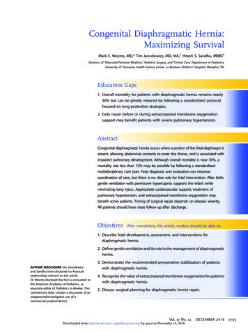
INTRODUCTIONCongenital diaphragmatic hernia (CDH) is an uncommon and complex condition that carries a high mortalityrate. Over the past 20 years, overall survival has improvedgradually from 63% to more than 70%. (1) Further advancement remains elusive because clinical trials and universally accepted standards of care for CDH management arelacking. However, implementation and adherence to a CDHprotocol may improve outcomes. (2) Management at centers reporting survival of 85% to 90% features severalcommon elements, including gentle ventilation, permissivehypercapnia, cardiovascular support, early use of extracorporeal membrane oxygenation (ECMO), and delayed surgical repair. (2)(3)(4)(5) However, it must be rememberedthat the survival rate is a function of the denominator thatis used. Case series from tertiary centers do not includethe sickest infants who die in utero, in the delivery room,or soon after admission but before transfer to a tertiarycenter. (6)Although survival rates have improved, patients withCDH are at risk for long-term morbidity. Long-term followup is recommended to monitor neurodevelopmental delay,hearing loss, pulmonary and gastrointestinal complications,and CDH recurrence. (7)EPIDEMIOLOGY AND FETAL DEVELOPMENTThe estimated prevalence of CDH varies between 2.4and 4.1 per 10,000 births. The European Surveillanceof Congenital Anomalies (EUROCAT) data from 1980to 2009 show an isolated CDH prevalence of 1.6 per10,000 births and a total (including isolated cases)nonsyndromic CDH prevalence of 2.3 per 10,000 births.These data exclude multiple births, but include allother pregnancies of more than 20 weeks of gestation.The prevalence of CDH has risen slowly over time, butthe rate of therapeutic abortion has increased, resulting in a dramatically decreased incidence among livebirths. (8)The diaphragm begins as the septum transversum and2 triangular pleuroperitoneal folds (PPFs) separate thepleural cavity from the peritoneal cavity. The mesodermalPPFs migrate across the septum transversum and give riseto muscle connective tissue fibroblasts to form the connective tissue structure of the diaphragm. Somatic muscleprecursor cells then migrate across the muscle connectivefibroblasts to complete muscularization of the diaphragmby embryonic day 16.5 in the mouse model (correlating withthe 10th week of human gestation). (9)In the case of CDH, mutated PPF-derived muscle connective tissue fibroblasts prevent muscularization in specific areas adjacent to fully muscularized regions. Therelatively weak nonmuscularized area eventually fails underpressure, allowing viscera to enter the thorax (Fig 1). Thephysical presence of abdominal contents inhibits lunggrowth and leads to pulmonary hypoplasia. (9) A 2-hit hypothesis has also been proposed, suggesting that genetic mutations leading to CDH may directly inhibit lung growth, butthis is less clearly described.ASSOCIATED ANOMALIESIsolated CDH occurs in 58% to 64% of antenatally diagnosed CDH cases. (8)(10) EUROCAT data show that chromosomal anomalies occur in 7.2%, genetic syndromesoccur in 3.5%, and nonsyndromic associated anomaliesoccur in 25% of singleton CDH cases. Among the chromosomal anomalies, the most common are trisomy 18 (4.2%),trisomy 13 (1.1%), and trisomy 21 (0.9%). (8)Fryns syndrome is the most common nontrisomy genetic syndrome associated with CDH; it is found in at least0.5% of CDH cases. Other syndromes associated withCDH include Pallister-Lillian, Wolf-Hirschorn, and Corneliade Lange. (8)Nonsyndromic anomalies are common, occurring in upto 42% of CDH pregnancies. Described anomalies includecardiac (most common), urinary, musculoskeletal, and neurologic. Genetic syndromes and other anomalies confer apoor prognosis and increased mortality. (8)(10)BENEFITS OF COORDINATED CAREIndividual cases vary widely with regard to severity of illness, size of defect, degree of pulmonary hypoplasia, andresponse to pulmonary vasodilators. As such, there aremany different opinions about appropriate CDH treatment,and management varies from center to center. Despitethese differences, several recent studies have shown the benefits of standardized care (Table 1). The details of each protocol differ, but they generally describe gentle ventilation,permissive hypercapnia, cardiovascular support, early use ofECMO, and delayed surgical repair. (2)(3)(4)(5)PRENATAL MANAGEMENTDiagnosisRecent data from the CDH Study Group revealed that 68%of 4,029 live-born infants with CDH were diagnosed antenatally. (11) Fetal ultrasonography is part of routine prenatale706 NeoReviewsDownloaded from http://neoreviews.aappublications.org/ by guest on November 14, 2018
Figure 1. Mouse model showing the development of congenital diaphragmatic hernia (CDH). A. CDH arises from early genetic mutations in a subset ofPPF-derived muscle connective tissue fibroblasts (mutant fibroblasts are yellow, wild-type fibroblasts are green). B–D. Mutant fibroblasts clonallyexpand and inhibit muscle progenitors from developing in these regions (via decreased proliferation and increased apoptosis of muscle progenitors),resulting in local regions (shown in yellow) that are amuscular but contain connective tissue fibroblasts and their associated extracellular matrix. D, E.Amuscular regions are thinner and more compliant than surrounding thicker and stiffer muscularized diaphragm and allow herniation of abdominalcontents into the thoracic cavity. E¼esophagus; NT¼neural tube; PPF¼pleuroperitoneal fold; So¼somite; ST¼septum transversum; (Reprinted withpermission from Merrell AJ, Ellis BJ, Fox ZD, Lawson JA, Weiss JA, Kardon G. Muscle connective tissue controls development of the diaphragm and is asource of congenital diaphragmatic hernias. Nat Genet. 2015;47(5):496–504.)obstetric care, but its usefulness in diagnosing CDH isvariable. Diagnostic sensitivity has regional variation anddepends on operator experience, defect laterality (left iseasier to identify than right CDH), “late” herniation ofviscera, and the presence of other anomalies. In theory, itis possible to diagnose CDH any time after completion ofdiaphragmatic development and return of the intestines tothe abdominal cavity, which occurs before the end of the firsttrimester. However, many cases may not be diagnosed untilafter 24 weeks of gestation, which could exceed the latestlegal fetal age for pregnancy termination. (6)Diagnostic features of CDH on ultrasonography includeprimary signs such as viscera in the chest and secondarysigns such as mediastinal shift, polyhydramnios, and cardiac malposition or abnormal cardiac axis. Fluid-filled viscera, most commonly the stomach, may be identified at thelevel of the 4-chamber heart view. Once CDH is identified,magnetic resonance imaging (MRI) can more reliably determine liver herniation, provide 3-dimensional (and potentially more accurate) visualization of the lungs, and detectassociated anomalies. (6) However, acquisition early in pregnancy may yield images with inadequate resolution.Once the diagnosis of CDH is made, multidisciplinaryconsultation with maternal-fetal medicine, pediatric surgery, and neonatology should be arranged to establish aplan for further evaluation, surveillance, delivery, and postnatal management.Markers of SeverityIn the fetus with CDH, the prognosis is largely determinedby gestational age, degree of lung hypoplasia, and thepresence or absence of other anomalies. In addition todetection of congenital anomalies, prenatal imaging enablesCDH severity to be estimated by directly measuring the sizeof the lungs or by assessing surrogate variables that indirectlyquantify the amount of functional lung that will be availableat birth.A large diaphragmatic defect results in more lung compression and lung hypoplasia, leading to pulmonary hypertension (PHTN) and severe disease. Prenatal identificationof liver in the chest (“liver up”) implies a large defectand thus represents a more severe subset of CDH, but itcan be difficult to distinguish between lung and liver onultrasonography. (6) In addition, some “liver up” patientsmay have good outcomes. (12) In general, liver position isnot as accurate in mortality prediction as other measures,though volume of herniated liver measured with MRI mayprovide a better survival estimate. (13)TABLE 1.Successful Diaphragmatic HerniaProtocolsAUTHORPREPROTOCOLYEAR SURVIVALPROTOCOLSURVIVALP-VALUEBagolan et al (3)200450%86%0.02Tracy et al (4)201052%85%0.006All CDH67%88%0.015CDH on ECMO20%82%0.00267%88%0.004Antonoff et al (5) 2011van den Houtet al (2)2011CDH¼congenital diaphragmatic hernia; ECMO¼extracorporealmembrane oxygenation.Vol. 17 No. 12Downloaded from http://neoreviews.aappublications.org/ by guest on November 14, 2018DECEMBER 2016e707
First reported in 1996, lung-to-head ratio (LHR) is a ratioof the contralateral area of the 2-dimensional lung to thehead circumference, as measured with ultrasonography.LHR is the most widely validated marker of CDH severity,and is most reliable between 22 and 26 weeks of gestation.Observed/expected (O/E) LHR is preferred because theLHR will change over time due to disparate growth ratesof the lung and head during gestation. (14) Ultrasonographyoperator dependence and interobserver variability canhamper the predictive value of LHR. An O/E LHR lessthan 25% portends less than 30% survival and an O/ELHR greater than 46% is associated with greater than 85%survival. (15)MRI may afford better visualization of the lung and liverthan ultrasonography, and is less dependent on maternaland fetal positioning. (16) Serial MRI can accurately monitor CDH with less operator dependence than ultrasonography. (14) Currently, MRI is the most accurate way tocalculate total lung volume, and use of O/E total lungvolume has been shown to have the best predictive valuefor mortality in a study comparing several common predictors. (16) For fetuses with an O/E total lung volume lessthan 25%, survival is less than 15%, and for those with O/Etotal lung volume greater than 35%, survival may be greaterthan 80%. (13)Fetal InterventionPrenatal therapy for CDH has included both surgical andnonsurgical approaches, with invasive measures typicallyundertaken only for those fetuses with severe CDH (lowLHR and liver herniation). Various pharmacologic treatmentsand stem cell therapy have been studied, but no definitivesurvival benefit has yet been demonstrated.Although open fetal repair of the diaphragm is possible, ithas fallen out of favor because it failed to improve mortality.Over time, prospective, randomized, controlled trials of fetal tracheal occlusion have shown increasing promise forselect populations of patients with CDH, despite the findingsof the original trial that outcomes were equivalent to thoseof standard management. (17) This procedure, now performedusing a small instrument that percutaneously delivers aremovable balloon to the fetal trachea, accelerates lunggrowth and appears to ameliorate the effects of the CDH onlung development. A randomized European study demonstrated 50% survival with tracheal occlusion compared to4.8% survival with standard management among fetuseswith severe isolated CDH. (18) An ongoing internationalrandomized trial, the Tracheal Occlusion To AccelerateLung growth (TOTAL) trial, may provide further evidencesupporting the use of percutaneous tracheal occlusion forsevere CDH. However, currently such therapy remainsexperimental and is only being undertaken in the contextof clinical trials.DELIVERY AND RESUSCITATIONPatients with CDH who are born at a high-volume centerusing a resuscitation protocol may have a survival advantage. Grushka et al report that patient risk factors wereequal in both high- and low-volume centers, but thesurvival was improved in centers delivering more than6 patients with CDH annually. (19) The mode of delivery,vaginal versus caesarian, does not seem to affect survivaland should be determined based on maternal indications.(20)There are no data to determine best practices fordelivery room resuscitation of patients with CDH. Mostpublished protocols recommend immediate intubationwith gentle ventilation, oro- or nasogastric decompression,and avoidance of mask ventilation. (4)(5)(21) The purposeof these steps is to minimize the amount of swallowedair (Fig 2) that will inflate the thoracic bowel and limitlung expansion. The 2015 update to the CDH EUROConsortium Consensus suggests that spontaneous breathing could be offered to low-risk patients, but it is unclear which population might benefit from this practice.(22)Delayed cord clamping is appropriate for most term andpreterm infants, but it cannot currently be recommendedfor patients with CDH because it would interfere with thegoal of immediate intubation.Surfactant therapy has frequently been used in these patients, but supporting data are controversial. Retrospective data from the CDH Study Group show that surfactanttherapy is associated with worse outcomes. Of 522 terminfants, those who received surfactant therapy were lesslikely to survive (57.3% vs 70.0%, P ¼ .0033), more likelyto receive ECMO (69.8% vs 50.6%, P ¼ .04), and morelikely to develop chronic lung disease (59% vs 47.6%, P ¼.0066). However, it should be recognized that this was not arandomized controlled trial, and a significant selection biasmay have skewed the findings. (23) Similarly, surfactant usein preterm infants (mean, 34 weeks) was associated with asignificantly greater odds of death than controls (odds ratio[OR] ¼ 2.17; 95% confidence interval [CI] ¼ 1.5–3.2; P .01).After controlling for gestational age and Apgar score, therewas no benefit associated with surfactant use (OR ¼ 1.58;95% CI ¼ 0.99–2.52; P ¼ .05). (24) Thus, routine use ofsurfactant therapy cannot be recommended for patientswith CDH.e708 NeoReviewsDownloaded from http://neoreviews.aappublications.org/ by guest on November 14, 2018
Figure 2. Radiographs showingdiaphragmatic hernia. A. A large amount of aircan be seen in the thoracic bowel within theleft chest and dextroposition with minimalaeration of the right lung. Umbilical arterialcatheter is in good position. The umbilicalvenous catheter tip appears to be at the levelof the diaphragm, but echocardiographyreveals that the tip does not extend beyondthe liver. B. After gastric decompression, theair in the thoracic bowel is decreased andaeration of the right lung is increased.Vascular access should be established early to providefluids, nutrition, and medications. Umbilical venous catheters (UVCs) are commonly placed in neonates, but CDHanatomy is distorted, making radiographic evaluation ofUVC position challenging (Fig 2). Alternative centralvenous access is preferred over placement of UVCs inpatients with CDH. If possible, an umbilical arterial cathetershould be placed; arterial access is helpful for measuringblood gases and monitoring blood pressure in critically illpatients with CDH.RESPIRATORY SUPPORTBefore the widespread acceptance of gentle ventilation forpatients with CDH, ventilator-induced lung injury was veryprevalent, and nearly all nonsurvivors had significant lunginjury. In what was the largest study of autopsy findingsfrom CDH nonsurvivors, 91% of infants with CDH onwhom autopsies were performed had evidence of diffusealveolar damage, 65% had evidence of air-leak, and 50%had evidence of pulmonary hemorrhage, with destructionof the alveolar-capillary interface. (25) It has been estimatedthat 25% of CDH deaths occur due to the adverse effectsof aggressive care. (26)Wung et al first championed gentle ventilation withpermissive hypercapnia for neonates with PHTN in 1985.(26) They successfully applied this strategy to patients withCDH; other studies have since supported the associationbetween gentle ventilation and improved survival. (2)(3)(4)(5)(27)(28) The specifics of gentle ventilation are looselydefined. For conventional mechanical ventilation (CMV),many recommend limiting the peak inflation pressure to25 cm H2O or less with positive end-expiratory pressure 3to 5 cm H2O. Respiratory rate is generally recommendedbetween 40 and 60 breaths per minute. Volume-targetedventilation is gaining popularity in neonatal medicine, butdata suggesting optimal tidal volume in the CDH populationare limited. Smaller-than-normal tidal volumes are oftentargeted due to presumed pulmonary hypoplasia associatedwith CDH. Te Pas et al report that patients with CDH withspontaneous respirations have a mean tidal volume of 3.8 –1.9 mL/kg. (29) Sharma et al documented a mean tidalvolume requirement of 4.6 mL/kg to achieve mean partialpressure of arterial carbon dioxide (PaCO2) of 46 mm Hg(6.1 kPa), similar to tidal volumes in infants without diaphragmatic hernia. They pointed out that the metabolic carbondioxide production in infants with CDH is no different fromthat in other infants of a similar size, and therefore it is notsurprising that they should require similar alveolar minuteventilation. (30) In our practice, we target a tidal volumeof 4 mL/kg and increase to 6 mL/kg if needed to achieve agoal partial pressure of carbon dioxide (P CO 2) of 50 to65 mm Hg (6–8.6 kPa).Many have proposed high-frequency oscillatory ventilation (HFOV) as a means of improving oxygenation with alower risk of alveolar damage, but observational data onHFOV in patients with CDH are conflicting. (31) To date, theVICI Trial is the only randomized, prospective, multicentertrial comparing CMV to HFOV. One hundred seventy-onepatients were randomized to receive either CMV or HFOV,and the remainder of each patient’s treatment followedguidelines set by the CDH EURO Consortium. (2
physical presence of abdominal contents inhibits lung growth and leadstopulmonaryhypoplasia.(9)A2-hit hypoth-esis has also been proposed, suggesting that genetic muta-tions leading to CDH may directly inhibit lung growth, but this is less clearly described. ASSOCIATED ANOMALIES Isolated CDH occurs in 58% to