Transcription
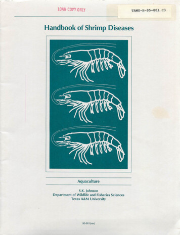
LOAN COPY ONLYTAMU-H-95-001 C3Handbook of Shrimp DiseasesAquacultureS.K. JohnsonDepartment of Wildlife and Fisheries SciencesTexas A&M University90-601 (rev)
Introduction2Shrimp Species2Shrimp Anatomy2Obvious Manifestations of Shrimp DiseaseDamaged Shells3,3Inflammation and Melanization3Emaciation and Nutritional Deficiency4Muscle NecrosisTumors and Other Tissue Problems55Surface Fouling6Cramped Shrimp6Unusual Behavior6Developmental ProblemsGrowth Problems67Color Anomalies7MicrobesVirusesBaceteria and aBody Invaders131516Surface 1818Environment20Publication of this handbook is a cooperative effort of the Texas A&M University Sea Grant College Program, theTexas A&M Department of Wildlife andFisheries Sciences and the TexasAgricultural Extension Service. Production is supported in part by InstitutionalGrant No. NA16RG0457-01 to TexasA&M University by the National SeaGrant Program, National Oceanic andAtmospheric Administration, U.S. Department of Commerce. 2.00Additional copies available from:Sea Grant College Program1716 Briarcrest Suite 603Bryan, Texas 77802TAMU-SG-90-601(r)2M August 1995NA89AA-D-SG139A/1-1
Handbook of Shrimp DiseasesS.K. JohnsonExtension Fish Disease SpecialistThis handbook is designed as an information source andfield guide for shrimp culturists, commercial fishermen, andothers interested in diseases or abnormal conditions of shrimp.It describes and illustrates common maladies, parasites andcommensals of commercially important marine shrimp. Descriptions include information on the life cycles and generalbiological characteristics of disease-producing organisms thatspend all or part of their life cycles with shrimp.Disease is one of the several causes of mortality in shrimpstocks. Death from old age is the potential fate of all shrimp,but the toll taken by predation (man being one of the majorpredators), starvation, infestation, infection and adverse environmental conditions is much more important.Although estimates of the importance of disease in naturalpopulations are generally unreliable, the influence of disease,like predation and starvation, is accepted as important in lowering numbers of natural stocks whenever they grow to excess.Disease problems are considered very important to successtail end (abdomen). The parts listed below are apparent uponoutside examination (Fig. Antennal scale6.7.8.Rostrum (horn)EyeMouthparts (several9.appendages for holdingand tearing food)Carapace (covering ofcephalothorax)10. Walking legs (pereiopods)11. Abdominal segment12. Swimmerets (pleopods)ful production in shrimp aquaculture. Because high-density,confined rearing is unnatural and may produce stress, someshrimp-associated organisms occasionally become prominentfactors in disease. Special measures are required to offset their13. Sixth abdominal segdetrimental effects.16. Gillsment14. Telson15. UropodDisease may be caused by living agents or other influencesof the general environment. Examples of influences in thegeneral environment that cause disease are lack of oxygen,poisons, low temperatures and salinity extremes. This guideconcentrates on the living agents and on visual presentation ofthe structure and effects of such agents.Shrimp SpeciesThere are many shrimp species distributed world-wide.Important shrimp of the Gulf of Mexico catch are the brownshrimp, Penaeus aztecus\ the white shrimp, Penaeus setiferus;and the pink shrimp; Penaeus duorarum.Two exotic shrimp have gained importance in Gulf Coastaquaculture operations. These are the Pacific white (white leg)shrimp, Penaeus vannamei, and the Pacific blue shrimp,Penaeus stylirostris. These two species are used likewisethroughout the Americas on both east and west coasts.In Asia, the Pacific, and to some extent the Mediterranean,the following species are used: Penaeus monodon, Penaeusmerguiensis, Penaeus chinensis, Penaeus japonicus, Penaeussemisulcatus, Penaeus indicus, Penaeus penicillatus andMetapenaeusensis. Penaeus monodon, the giant tiger (or blacktiger) shrimp is the world leader in aquaculture.Shrimp AnatomyA shrimp is covered with a protective cuticle (exoskeleton,shell) and has jointed appendages. Most organs are located inthe head end (cephalothorax) with muscles concentrated in theFig. 1. External anatomy of shrimp.(Numbers conform to list.)Inside structures include (Fig. 2)1.Esophagus2.Stomach3.4.Hemocoel (blood space)Digestive gland (hepatopancreas)5.Heart6.Intestine7.Abdominal musclesThe "skin" or hypodermisof a shrimp lies just beneaththe cuticle. It is functional insecreting the new exoskeletonthat develops to replace theold at shedding. Shedding ofthe cuticle (also known asmolting or ecdysis) occurs atintervals during a shrimp'slife and allows for change indevelopmental stage and exFig. 2. Internal anatomy ofshrimp. (Numbers conform tolist.) Jagged line representscutaway of cuticle to exposeinternal organs.pansion in size.The reproductive organs of adults are particularly noticeable. When ripe, the ovaries of females may be seen throughthe cuticle to begin in the cephalothorax and extend dorsallyinto the abdomen. Spermatophores, a pair of oval structurescontaining the sperm in adult males, are also visible throughthe cuticle when viewed from the underside near the junctureof cephalothorax and abdomen. The principle nervous struc-
ture, the ventral nerve cord, is visible along the underside ofthe body between the swimmerets.Obvious Manifestations of Shrimp DiseaseDamaged ShellsShrimp cuticle is easily damaged in aquaculture situationswhen hard structures are impacted or rubbed. (Fig. 3). Bloodruns openly (outside of vessels) under the shell of shrimps, outthrough appendages and into tiny fringe parts. When injuryoccurs to the shell, the blood quickly clots and protectsdeeperparts (Fig. 4).Shell damage may also be inflicted by the pinching or bitingof other shrimpin crowded conditions. Parts of appendagessuch as antennae may be missing. Cannibalism has an important influence on survival in some phases of shrimp culturewhere stronger individuals devour weak ones (Fig. 5).Shells may also be damaged because they become infected.A protective outer layer is part of the cuticle. If underlyingportions arc exposed opportunistic microbes will invade theshell and use it as a food base or portal for entry into deepertissue. Larger marks darken and become obvious (Fig. 6).Fig. 3. Eyes of shrimp are normally black, but rubbing of a tank wallhas caused this eye to appear whitish because of a prominent lesion.Inflammation and MelanizationDarkening of shell and deeper tissues is a frequent occurrence with shrimp and other crustaceans. In the usual case,blood cells gradually congregate in particular tissue areas (inflammation) where damage has occurred and this is followedby pigment (melanin) deposition. An infective agent, injury ora toxin may cause damage and stimulate the process (Fig. 7).Gills arc particularly prone to darkening due to their fragilenature and their function as a collecting site for elimination ofthe body's waste products (Fig. 8). Gills readily darken uponexposure to toxic metals or chemicals and as a result of infection by certain fungi (Fusarium sp.).Less common but important are dark blotches that sometimeoccur within the tails of pond shrimp. This manifestation ofnecrosis (breakdown and death) of muscle portions followed bymelanization degrades the product's market potential. It ispossible that this condition results from deep microbial invasions that run through spaces between muscle bundles but itsactual causes remain unknown (Fig. 9).Fig. 5. Cannibalism usually begins as other shrimp devour the appendages.Fig. 4. Microscopic view of a lesion on a uropod (tail part). Note creasefrom bend in part and loss of fringe setae.Fig. 6. Tail ends of two shrimp. The lower shrimp shows typical darkening of cuticle that involves microbial action. The darkening itself isconsidered a host response. The telsons of the upper shrimp areopaque because of dead inner tissue. Successful entry and tissuedestruction by bacteria was accomplished only in those parts.
Emaciation and Nutritional DeficiencyUnfed shrimp lose their normal full and robust appearanceand exhibit emaciation. The shell becomes thin and flexible asit covers underlying tissue such as tail meat that becomesgreatly resorbed for lack of nutrients. Molting is curtailed andshell and gills may darken in time (Fig. 10). Emaciation mayalso follow limited feeding behavior during chronic diseaseconditions or an exposure to unfavorable environmental conditions. Empty intestines arc easily observed through transparentcuticle and flesh.Prepared diets deficient in necessary constituents may predispose or cause disease. Vitamin C deficiency, for example,will initiate darkening of gills or certain tissues associated withFig. 7. A shrimp photographed (above) near time of back injury and(below) hours later. Injury by a toxin or disease agent will usually trigger a similar response of inflammation and melanization. kthe cuticle and eventually result in deaths.,fJ.Fig. 9. Areas of melanized necrotic tissue in tail musculature.Fig. 10. Emaciated shrimp. Gills and body fringes have become obviouslydarkened and the soft tail is covered with a thin and fragile cuticle.Fig. 8. Microscopic view of damaged and melanized gill tips.Fig. 11. Lipoid (fat) spheres in microscopic view of digestive gland tubule.
Digestive glands sometimes will become reduced in size.Among other things, this is an indication of poor nutrition.Well-fed shrimp will have an abundance of fat globules withinstorage cells of the digestive gland tubules that provide bulk tothe gland (Figs. 11 and 12).Muscle NecrosisOpaque muscles are characteristic of this condition. Whenshrimp areexposed to stressful conditions, such as low oxygenor crowding, the muscles lose their normal transparency andbecome blotched with whitish areas throughout. This mayprogress until the entire tail area takes on a whitish appearance(Fig. 13).If shrimp arc withdrawn from the adverse environmentbefore prolonged exposure, they may return to normal. Extremely affected shrimp do not recover, however, and diewithin a few minutes (Fig. 14). In moderately affected shrimp,only parts of the body return to normal; other parts, typicallythe last segments of the tail are unable to recover and are proneto bacterial infection (Fig. 15). These shrimp die within one orFig. 12. Pond-raised shrimp with full, normal and reduced, abnormaldigestive gland. Arrow points to abnormal gland.two days (Fig. 5). Shrimp muscles with this condition areknown to undergo necrosis (death or decay of tissue).Tumors and Other Tissue ProblemsConspicuous body swellings or enlargements of tissues havebeen reported in shrimp. In most cases, affected individualswerecaptured from polluted waters. Occurrenceof shrimp withevident tumors is rare in commercial catches. Miscellaneousirritations experienced by captive shrimp in tanksystems willsometimes result in focal areas of tissue overgrowth (Fig. 16).A particularly vulnerable tissue of captive juvenile and adultshrimp is found on the inner surface of the portion of carapacethat covers the gills. When microbes invade this tissue, it andFig. 13. Shrimp with necrotic muscle tissue following exposure tostressful environment. Affected tissue at arrow.the adjacent outer shell may completely disintegrate exposingthe gills. In other cases a partial loss of the tissue distally mayresult in an outward flaring of the exposed cuticle. A hemolymphoma or fluid-filled blister also forms sometime in thisFig. 14. Shrimp with advanced muscle necrosis (arrow) shown besidenormal shrimp.Fig. 15. Damage to abdomen of a shrimp as a result of Vibrio infection.Fig. 16. Tumorous growth on an adult shrimp from a tank system.
Fig. 17. Blister condition. Insetshows blister removed. The blister willdarken upon death of shrimp degrading marketability of heads-onproduct.portion of the carapace in pond shrimp (Fig. 17). Primarycauses of these manifestations are not understood.A degeneration of male reproductive tracts occasionallyoccurs in captive adults of certain penaeid species. A swellingFig. 18. Darkening of male reproductive tract of Penaeus stylirostris. A.Normal tract. B. Initial darkening. Darkening will advance until spermatophores and testes become affected. (Photos courtesy of GeorgeChamberlain.)and darkening of the tubule leading from the testes to the spcrmatophorc is readily apparent when viewed through the translucent body (Fig. 18).Surface FoulingThe surfacesof shrimps arc prone to an accumulation of various fouling organisms. Heavy infestations can interfere with mobility or breathing and influence marketability (Fig. 19).Cramped ShrimpThis is a condition described for shrimp kept in a variety ofculture situations. The tail is drawn under the body and becomes rigid to the point that it cannot be straightened (Fig. 20).The cause of cramping is unknown, but some research points tomineral imbalance.Fig. 19. Algal overgrowth on shrimp exposed to abundant light. (Photocourtesy of Steve Robertson.)Unusual BehaviorDevelopmental ProblemsDiseased shrimps often display listless behavior and ceaseto feed. In the case of water quality extremes such as low oxyDeformities are quite prevalent in some populations. Theyarise from complex interactions that involve environment, dietand gene expression. Bodies may be twisted or appendagesmisshaped or missing. Deformities arc less prevalent in wildcaught larvae than hatchery populations probably because wildshrimp have more opportunity for natural selection and exposure to normal developmental conditions (Fig. 21).gen, shrimp may surface and congregate along shores wherethey become vulnerable to bird predation. Cold water maycause shrimp to burrow and an environmental stimulation suchas low oxygen, thermal change or sudden exposures to unusualchemicals may initiate widespread molting.Fig.20. Cramped shrimp condition. Full flexure (A). Flexure maintained when pressure applied (B).
Molt arrest occurs in affected animals of some populations.Animals begin, but are unable to complete the molting process.In some cases, there is abnormal adherence to underlying skin,but most animals appear to lack the necessary stamina. Nutritional inadequacies and water quality factors have been identified as causes.Growth ProblemsGrowth problems become obvious inaquaculture stocks. Aharvested population may show a larger percentage ofrantingthan expected. Some research has connected viral disease withranting in pond stocks and it is generally held that variablegrowth may result from disease agents, genetic makeup andenvironmental influences.For unknown reasons, the shell orcuticle may become fragile in members of captive shrimp stocks.Shells are normally soft for a couple of days after molting,but shells of those suffering from soft-shell condition remainboth soft and thin and have a tendency to crack under theFig. 21. Deformed larval shrimp. Arrow points to deformed appendage.(Photo courtesy of George Chamberlain.)slightest pressure. Some evidence of cause suggests pesticidetoxicity, starvation (mentioned above) or mineral imbalance.to the cuticle or underlying skin. A genetic cause is suspected.Color AnomaliesTransformation to blue coloration from a natural brown isknown for some captive crustaceans and has been linked toShrimp of unusual color arc occasionally found among wildand farm stocks. Thestriking coloration, which may begold,blue or pink, appears throughout the tissue and is notconfinednutrition. Pond-cultured, giant tiger shrimp sometime develop acondition wheredigestive gland degeneration contributes to areddish coloration.MicrobesMicrobes are minute, living organisms, especially viruses, bacteria, rickcttsia and fungi. Sometimes protozoa arcconsidered microbes.Protozoa are microscopic, usually one-celled,animalsthat belong to the lowest division of the animal kingdom.that are capable of developing into a new individual.Bacteria are one-celled organisms that can be seen onlywith a microscope. Compared to protozoans, they arc of lesscomplex organization and normally less than 1/5,000 inch(1/2000 cm) in size.Normally, they are many times larger than bacteria. Thetypical protozoa reproduce by simple or multiple division orby budding. The more complex protozoa alternate betweenRickettsia are microbes with similarity to both virusesand bacteria and have a size that is normally somewhat in-hosts and produce cells with multiple division stages calledFungi associated with shrimp are microscopic plants thatdevelop interconnecting tubular structures. They reproduceViruses arc ultramicroscopic, infective agents capable ofmultiplying in connection with living cells. Normally, viruses are many times smaller than bacteria but may be madeclearly visible at high magnification provided by an electronby forming small cells known as spores or fruiting bodiesmicroscope.spores.between. Most think of them as small bacteria.
MicrobesVirusesOur knowledge of the diversity of shrimp viruses continues togrow. Viruses of shrimp have been assigned explicitly or tentatively to six or seven categories. Several shrimp viruses are recognized to have special economic consequence in aquaculture:BaculovirusesBaculovirus penaei — a virus common to Gulf of Mexicoshrimp. It damages tissue by entering a cell nucleus and subsequently destroys the cell as it develops (Fig. 23). An occlusionis formed (Fig. 24). This virus has become a constant problemfor many shrimp hatcheries where it damages the young larvalanimals. Occlusions of the same or closely related viruses areseen in Pacific and Atlantic Oceans of the Americas. At leastten shrimp species arc known to show disease manifestations inaquaculture settings.Monodon-typc baculovirus — one that forms sphericalocclusions (Fig. 25) and whose effects arc seen mostly in theculture of the giant tiger prawn, Penaeus monodon. Damage ofless importance has been seen in Penaeus japonicus, Penaeusmerguiensis and Penaeus plebejus.Midgut gland necrosis virus — a naked baculovirus harmfulto the Kuruma prawn, Penaeus japonicus, in Japan.Fig. 24. Occlusion bodies of Baculovirus penaei. These bodies, visibleto low power of a light microscope, are characteristic of this virus. Theocclusions and those of other baculoviruses are found mainly in thedigestive gland and digestive tract.Fig. 25. Monodon baculovirus in a tissue squash showing groups ofspherical occlusions. Light microscopy.ParvovirusesSolubility in GutIngestion ot Contaminated FoodInfection of HostFig. 23. Baculovirus life cycle. Transmission of the virus is thought tobe initiated as a susceptible shrimp ingests a viral occlusion. Virusinitially enters cell cytoplasm either by viroplexis (cell engulfs particlewith surrounding fluid) or by fusion where viral and cell membranesfuse and viral core passes into cell. Secondary infection occurs asextracellular virus continues to infect. (Redrawn by Summers andSmith, 1987. Used with permission of author and Texas AgriculturalExperiment Station, The Texas A&M University System.)Infectious hypodcrmal and hematopoietic necrosis virus —a virus affecting several commercially important shrimp and,particularly, the Pacific blue shrimp, Penaeus stylirostris.Hepatopancreatic parvo-Iikc virus — a virus causing diseasein several Asian shrimp. Transmission to Penaeus vannameidid not result in disease to that species.NodavirusTaura virus — a virus causing obvious damage to varioustissues and in the acute phase, to the hypodermis and subsequently the cuticle of Penaeus vannamei (Fig. 26). It is animportant problem for both production and marketing. Duringthe 1995 growing season, this virus caused large losses toaquaculture stocks in Texas. Damage was great in Central andSouth America beginning in 1992.
Other virusesYellow head virus — a virus causing serious disease of thegiant tiger prawn, Penaeus monodon. Large losses have beenexperienced in Asian aquaculture units. Gills and digestiveglands of infected shrimp arc pale yellow.White spot diseases — viruses of similar size and structurehave been shown to cause a similar manifestation and heavylosses to Penaeus japonicus, Penaeus monodon and Penaeuspenicillatus in Taiwan and Japan. Advanced infections showdevelopment of obvious white spots on the inside of the cuticle(Fig. 27).Several other viruses with relatively little known importancearc considered as members of the rcoviruses, rhabdoviruscs,togaviruscs.Fig. 26. Advanced stage of infection with Taura virus showing damageto cuticle. Smaller shrimp with acute infection do not show such damage but do show reddish telson and uropods.Fig. 27. Asian shrimp showing signs of white spot disease. (Photocourtesy of R. Rama Krishna.)VirusesViruses cause disease as they replicate within a host cell andthereby cause destruction or improper cell function. A virus iswithin. Other "naked" baculoviruses do not show formation ofocclusions.essentially a particle containing a core of nucleic acids, DNAor RNA. Once inside a proper host cell, the viral nucleic acidinteracts with that of a normal cell to cause reproduction of thevirus. The ability to parasitize and cause damage may be limited to a single species or closely related group of hosts, a hosttissue and usually the place within a cell in which damagetakes place.The cause and effect for all shrimp virus disease needs careful attention. Some viruses cause disease only after exposure tounusual environmental conditions. Also, impressions aboutvirus identity arc often based on results of routine examinationsthat give presumptive results. Certainly viruses cause importantdisease in particular circumstances but key understandings ofmost shrimp viruses are largely unknown: longevity withinsystems, source of infection, method of transmission, normaland unusual carriers, and potential to cause damage.ABOur ability to detect shrimp viruses is ahead of our ability toevaluate their importance or to implement controls. For viralidentification, scientists have employed the recent technologythat detects characteristic nucleic acids. This is augmented bycareful microscopical study of tissues to detect characteristicdamage to cells. Use of electron microscopy to determine sizeand shape of virus particles has also been helpful (Fig. 22).A peculiar feature of some baculoviruses of shrimp andother invertebrate animals is to occurrence of the occlusionbodies within infected cells. These are relatively large massesof consistent shape that contain virus particles embeddedFig. 22. Structure of viruses reported from shrimps. A. Baculoviridae.Size range is about 250 to 400 nanometers in length. B. Basic structure of most of the other shrimp viruses: Parvo-like viruses—20 to 24nm in diameter containing DNA; Reo-like viruses—55 to 70 nm diameter, RNA; nodavirus—30 nm diameter, RNA; toga-like virus 30 diameter, RNA, enveloped. Rhabdoviruses are elongated like baculovirusesbut a blunt end provides bullet-shapes—150 to 250 nm, RNA.
m *Fig. 28. View with light microscope of a tissue squash of infected digestive gland. Note dark necrotized tissue of tubules (arrows).Fig. 29. Transverse section of digestive gland tubules showing progression of granuloma formation. Normal tubules are to the left (N) andaffected tubules are to the right (G.).Bacteria and Rickettsialarger animals, infection becomes obvious in the digestivegland after harmful bacteria gain entry to it, presumably viaconnections to the gut.Digestive gland tissues are organized as numerous tubularstructures that ultimately feed into the digestive tract. Pondreared shrimp occasionally die in large numbers because ofdiseased digestive glands. The specialized cells that line theinside of the tubules arc particularly fragile and arc easily infected. Tubules progressively die and darken (Figs. 28 and 29).This kind of disease manifestation is seen in recent reports ofrickettsial infection. Cells of the digestive gland tubules arcseverely damaged as rickcttsiac invade and develop therein(Figs. 30 and 31).If infected by bacteria capable of using shell for nutrition,Bacterial infections of shrimp have been observed for manyyears. Scientists have noticed that bacterial infection usuallyoccurs when shrimp arc weakened. Otherwise normal shrimpalso may become infected if conditions favor presence andabundance of a particularly harmful bacterium.Shrimp body fluids are most often infected by the bacterialgroup named Vibrio. Infected shrimp show discoloration of thebody tissues in some instances, but not in others. The clottingfunction of the blood, critical in wound repair, is slowed or lostduring some infections. Members of one group of Vibrio havethe characteristic of luminescence giving heavily infected animals a "glow-in-thc-dark" appearance.Bacteria also invade the digestive tract. A typical infectionin larval animals is seen throughout the digestive system. Inthe exoskeleton will demonstrate erosive and blackened areas**.-V- - : i. .» '*' * 4«l5-Fig. 30. Histological cross section of a digestive gland tubule. Rickettsial microcolonies are shown at arrows. Rickettsiae will exhibit constantFig. 31. Electron microscope view of tissue infected with rickettsiabrownian motion and color red with Giemsa stain, but electron microsorganism (arrow). (Photo circa 1987 from Penaeus vannamei on Texascopy is needed for definite diagnosis. (Specimen courtesy of J. Brock)coast.)
(Fig. 6). These bacteria typically attack edges or tips of exoskeleton parts, but if break occurs in the exoskeleton the bacteriaare quick to enter and cause damage.Filamentous bacteria are commonly found attached to thecuticle, particularly fringe areas beset with setae (Fig. 32).When infestation is heavy, filamentous bacteria may also bepresent in large quantity on the gill filaments. Smaller, lessobvious bacteria also settle on cuticular surfaces but arc notconsidered as threatening as the filamentous type.Fig. 32. Microscopic view of filamentous bacteria on a shrimp pleopod.Microbial Disease and Digestive GlandsDigestive glands are routinely searched by pathologistsfor signs of disease. This is done after chemical preservation,microsection, slide-mounting and staining the tissue. Transverse sections of the tubules are then examined with a lightmicroscope. General damage is seen when bacteria such asVibrio species invade tubules. Rickettsiae, viruses,microsporans and haplosporans are more selective. Theyinvade cells and progressively cause damage from within.For comparative purposes, a drawing of a normal tubule iscompared with a tubule showing a variety of typical manifestations (Fig. 33).Fig. 33. A. Drawing of transverse section of digestive gland tubulewith bold lines that separate several types of disease conditions. 1.Haplosporan parasite (microsporans similar but may show fully developed spores, see Figs. 42 and 43). 2. Rickettsiae. 3. Virus infectionwith manifestation of inclusion in cytoplasm. 4. Virus infection withinclusion in nucleus. 5. Virus with occlusions in swollen nucleus. Ascells are destroyed, more general lesions are formed from viruses.Inclusions of viruses normally show distinctive shape and stainingfeatures. Particular viruses will infect particular tissue types (notalways hepatopancreas tissue) and cell locations (nucleus or cytoplasm) within preferred hosts. Cells enlarged by haplosporans andrickettsiae may be initially distinguished by comparing larger internalcomponents of the pre-spore units of haplosporans with almost submicroscopic particles of microcolonies of rickettsiae. H, earlyhaplosporan stage, H? - later stage; R - rickettsial microcolony; CI cytoplasmic inclusion of virus; Nl nuclear inclusion of virus; SN swollen nucleus with occlusions within.B. The normal tubule. Toward the digestive tract, secretory cells (1)predominate and fibrous cells (3) become more numerous. Absorptive cells (2) contain varying amounts of vacuoles according to nutritional status.10
FungusSeveral fungi are known as shrimp pathogens. Two groupscommonly infect larval shrimp, whereas another attacks thejuvenile or larger shrimp. The most common genera affectinglarval shrimp are Lagcnidium and Sirolpidium. The method ofinfection requires a thin cuticle such as that characteristic oflarval shrimp (Figs. 34 and 35).The most common genus affecting larger shrimp isFusarium. It is thought that entry into the shrimp is gained viacracks or eroded areas of the cuticle. Fusarium may be identified by the presence of canoe-shaped macroconidia that thefungus produces. Macroconidia and examples of fungal infections arc shown in Figures 36, 37 and 38.jSKbaD-Search for HostFig. 34. Transmission of Lagenidium. A. Fungus sends out dischargetube from within shrimp body. B. Vesicle forms. C. Vesicle producesmotile spores that are released. D. Motile spores contact shrimp andundergo encystment. E. Germ tube is sent into the body of the shripand fungus then spreads throughout.Fig. 35. Lagenidium infection in larval shrimp. Note extensive development of branchings of fungus throughout the body. (Photo courtesy ofFig. 36. Canoe-shaped macroconidia of Fusarium. These structuresbud off branches of the fungus and serve to transmit fungus to shrimp.Dr. Don Lightner, University of Arizona.)OXFig. 37. Shrimp photographed immediately after mole: Old appendage(arrow) is not shed due to destruction of hypodermis by active fungalinfection.Fig. 38. Microscopic view of fungus at tip of antenna.n
ProtozoaProtozoan parasites and commensals of shrimp will occuron the inside or outside of the body. Those on the outside areconsidered harmless unless present in massive or burdensomenumbers. Those on the inside can cause disease and arc representativeof several groups
Metapenaeusensis. Penaeus monodon,the giant tiger (or black tiger) shrimp is the world leader in aquaculture. Shrimp Anatomy A shrimp is covered with a protective cuticle (exoskeleton, shell) and hasjointed appendages. Most organs are located in the head end (cephalotho
