Transcription
— Archie, patientUnderstanding Brain TumorsC H A P T E R T W O / U N D E R S TA N D I N G B R A I N T U M O R SBe patient. Give yourselftime to absorb what’shappening and don’timmediately dive intothe internet.
C H A P T E R T W O / U N D E R S TA N D I N G B R A I N T U M O R SC H A P T E R 2 / U N D E R S TA N D I N G B R A I N T U M O R S . . . . . . . . . . . . . . . . . . . . . . . . . . . 5What is a brain tumor?. 7Defining brain tumors. 7Diagnosing a brain tumor. 9Scans and imaging techniques. 10 Tumor grading. . 11Tumor types. 12Diagnostic surgery. 13Pathology report. . 13The treatment team. 14Specialists. 15
2Understanding Brain TumorsThe brain, its structure, and the role that each part plays in our everyday thoughtsand behaviors is remarkable. These are only some of the reasons why a tumor in thebrain is so complex. THINGS TO REMEMBERThere are over 120 types of brain andcentral nervous system tumors. Whendiagnosed, it is important to understand: 1. The type and grade (how aggressive it is)2. Whether it is a primary or a secondarytumor 3. If it is cancerous (malignant) or not (benign)4. Where in the brain the tumor is located It helps to get a second or even thirdopinion to confirm your diagnosis andtreatment plan.Keep a list with phone numbers for allof the doctors, specialists, nurses, etc.on your team (collect business cards). You can bring someone to doctors’appointments to help you sort throughinformation, ask questions, remembernext steps, and keep notes.Keep copies of your medical records,current medication list, and diagnostic testresults. This can help you as you meet newdoctors and work with insurance.Take one step at a time. You can learnabout your diagnosis, treatment options,and what to expect after treatment, thendecide the best next-steps for you andyour family.C H A P T E R 2 / U N D E R S TA N D I N G B R A I N T U M O R S5
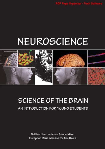
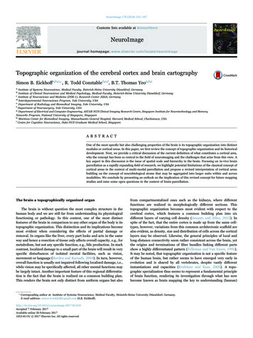
Brain and Spinal CordBRAIN STRUCTURES AND THEIR FUNCTIONSTogether, the brain and spinal cord (thecentral nervous system (CNS)) control thephysiological and psychological functionsof our body. Generally our brain includesthree major parts: The cerebrum controls thinking,learning, problem solving, emotions,speech, reading, writing, and voluntarymovement. The cerebellum controls movement,FRON TA L L OB E movement reasoning behavior memory decision making personality planning judgment initiative inhibition moodPAR I ETAL LO BE telling rightfrom left calculations sensations reading writingO C C I PI TALLO BE visionbalance, and posture. The brain stem connects the brainto the spinal cord, and controlsbreathing, heart rate, and the nervesand muscles that we use to see, hear,walk, talk, and eat.For more information aboutthe brain, view the NationalCancer Institute’s brain tumor tbrain/PatientTEMPORA LL OB E languagecomprehension behavior memory hearing emotionsPITU ITA RYGL A N D hormones growth fertilityC ER EBELLUM balance coordination fine musclecontrolBR AI N STEM breathing blood pressure heartbeat swallowingBased on an illustration from National Brain Tumor Society’s The Essential Guide to Brain Tumors6F R A N K LY S P E A K I N G A B O U T B R A I N T U M O R S
W H AT I S A B R A I N T U M O R ?DEFINING BRAIN TUMORSA brain tumor is an abnormal growthBenignspread to other areas of the body, theyof tissue in the brain or central spineThe least aggressive type of brain tumorcan spread throughout the brain or to thethat can disrupt proper brain function.is often called a benign brain tumor.spine. These tumors can be treated withDoctors refer to a tumor based on whereThey originate from cells within orsurgery, chemotherapy and radiation, butthe tumor cells began, and whethersurrounding the brain, do not containthey may recur after treatment.they are cancerous (malignant) or notcancer cells, grow slowly, and typically(benign).have clear borders that do not spreadPrimaryinto other tissue. They may become quiteWhether cancerous or benign, tumorsAll brain tumors can grow to damageareas of normal brain tissue if leftuntreated, which could be disabling andpossibly fatal.large before causing any symptoms. Ifthese tumors can be removed entirely,they tend not to return. Still, they cancause significant neurological symptomsBrain and spinal cord tumors are differentdepending on their size, and locationfor everyone. They form in different areas,near other structures in the brain. Somedevelop from different cell types, and maybenign tumors can progress to becomehave different treatment options. In thismalignant.primary brain tumors. Primary braintumors may spread to other parts ofthe brain or to the spine, but rarely toother organs.Metastatic or SecondaryMetastatic or secondary brain tumorsbook, we try to offer general guidance forbegin in another part of the body andboth low grade (benign) and high gradeMalignant(malignant) primary brain tumors forMalignant brain tumors contain canceradults.that start in cells of the brain are calledcells and often do not have clear borders.They are considered to be life-threateningbecause they grow rapidly and invadesurrounding brain tissue. Althoughmalignant brain tumors very rarelythen spread to the brain. These tumorsare more common than primary braintumors and are named by the location inwhich they begin. They are treated basedon where they originate, such as the lung,breast, colon or skin.C H A P T E R 2 / U N D E R S TA N D I N G B R A I N T U M O R S7
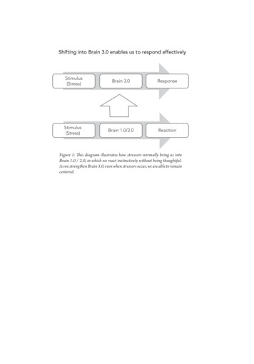
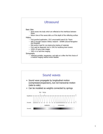
FA C T S A B O U T B R A I N T U M O R S I N T H E U N I T E D S TAT E SAn estimated 688,000 PEOPLEin the U.S. are living witha primary brain or central nervous system (CNS) tumor diagnosis:PRIMARY BRAINTUMOR TYPES138,000 WITH MALIGNANT TUMORS550,000 WITH BENIGN TUMORSAPPROXIMATELY20-40%OF ALL OTHER CANCERS LATERDEVELOP A BRAIN METASTASESIN 2012, NEW PRIMARY BRAIN TUMORDIAGNOSES INCLUDED:63%B E NI G NTUM ORS(41,980 Cases)37%This accounts for 98,000 to 170,000 newmetastatic brain tumor cases each year.M AL I G NAN TTUM iomaPituitaryNerve mbryonal, etc.)(24,300 Cases)Source: CBTRUS Statistical Report (2012)8F R A N K LY S P E A K I N G A B O U T B R A I N T U M O R S
DIAGNOSING A BRAIN TUMORWhat a crazy ride this has been. I went to bed after watching amovie and woke up in an ambulance on the way to the hospital.I didn’t know my name, didn’t recognize my wife or kids andcouldn’t understand why I was in the back of this ambulance.— Mike (from the National Brain Tumor Society’s Story Corner)Some people may have symptoms thatNeurological Examinationsuggest there is a brain tumor, othersAs part of the diagnostic testing, a doctorhave no obvious symptoms.will measure nervous system functions,Commonly, people experience longterm headaches, seizures or convulsions,difficulty thinking and speaking/findingwords, personality changes, tingling orphysical and mental alertness, andinclude the examination of normal brainfunctions from reflexes to judgment,smell and taste.stiffness in one side of the body, a loss ofIf responses are not normal, a brainbalance, vision changes, nausea, and/orscan will be ordered, or a patient will bedisorientation (see Chapter 4).referred to a neurologist or neurosurgicalIf these symptoms are occurring, a doctoroncologist for more tests.will ask questions about a person’smedical history and overall health, andprescribe a variety of diagnostic teststo determine what is causing theseproblems, and then seek remedies.C H A P T E R 2 / U N D E R S TA N D I N G B R A I N T U M O R S9
SCANS AND IMAGING TECHNIQUESMagnetic Resonance Spectroscopytissue. PET is also used during braintumor is present, and to locate exactly where(MRI Spect or MRS), measures themapping procedures.it is growing. A scan creates computerizedlevels of metabolites in the body. Animages of the brain and spinal cord byMRS can detect irregular patternspuncture), uses a special needleexamining it from different angles. Someof activity to help diagnose the typeplaced into the lower back to measurescans use a contrast agent (or a dye) toof tumor, evaluate its response topressure in the spinal canal and brainallow the doctor to see the differencetherapies, or determine aggressivenessand determine if there is an infectionbetween normal and abnormal tissue.of a tumor.or tumor cells.A scan is the first step to identify if a brainA patient may need more than one type Spinal tap (also called a lumbarPerfusion MRI examines the flow ofof scan to diagnose a tumor, dependingblood into the tissues to help assessThings to Know about Scanson its type and location.the grade/aggressiveness of tumorsAsk your neurosurgeon or nurse whatand differentiate a recurrent tumoryou have to do for a scan, where to gofrom dead tumor tissue.and how the scan works, so you can feelCommonly used scanning and imagingtechniques: 10Functional MRI (fMRI) tracks theprepared. Keep a record of your scanComputed Axial Tomography (CATuse of oxygen and blood flow inand x-ray history. This information canor CT Scan) is a computerized x-ray thatthe brain as patients perform tasks.help doctors make informed treatmentcan show a combination of soft tissue,An fMRI can identify the motor,decisions and minimize your over-bone, and blood vessels. This is oftensensory, visual and language centersexposure to radiation.the first test a person will receive in anof the brain which helps your doctoremergency room (i.e. after a seizure).carefully plan for surgery.Magnetic Resonance Imaging (MRI) Be prepared to receive multiple scansover time: first to detect the tumor; thenPositron Emission Tomographycan create clear and detailed three-to observe the site after surgery; later,(PET) scan uses a radioactivedimensional images of a brain tumor. Anwith follow-up care, to see if the tumorsubstance to visualize hypermetabolicMRI is not often used with people whoreturns.activity such as with malignant cells,have a pace maker or other metal device.or abnormalities from a tumor or scar F R A N K LY S P E A K I N G A B O U T B R A I N T U M O R S
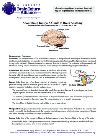
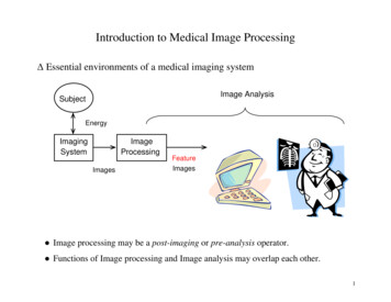
TUMOR GRADINGThe World Health Organization (WHO)Grade I Tumorhas created a standard by which all Slow-growing cells Almost normal appearance under atumors are classified. There are over120 brain tumor classifications definedby the WHO, based on the tumor cellGrade IV Tumor microscoperapidly Very abnormal appearance under amicroscope Usually not cancercomplex diagnosis. Tumors are given a Associated with long-term survivalname based on the cells where they arise, Can potentially be cured with surgerytype and location, making this a veryAbnormal cells which reproduce rapid growth and a number ranging from 1–4, usuallyForm new blood vessels to maintainAreas of dead cells (necrosis) in centerrepresented by Roman numerals I-IV.Grade II TumorThis number is called the “grade” and it Relatively slow-growing cells Slightly abnormal appearance underdetermines the grade for the entire tumora microscope(even if most of the tumor is a lower Can invade adjacent normal tissuegrade). Some tumors can change the way Can recur as a higher grade tumorthey grow and may become malignantrepresents how fast the cells can growand are likely to spread. This is criticalinformation for planning treatment andpredicting outcomes.Lower grade tumors (grades I & II)are not very aggressive and are usuallyassociated with long-term survival.Higher grade tumors (grade III & IV)treat. These are considered malignantor cancerous.cells; however, the most malignant cellover time. Your doctor can tell you if yourtumor might have this potential.Grade III Tumor Actively reproducing abnormal cells Abnormal appearance under agrow more quickly, can cause moredamage, and are often more difficult toTumors can contain several grades ofmicroscope Infiltrate adjacent normal brain tissue Tumor tends to recur, often as ahigher gradeC H A P T E R 2 / U N D E R S TA N D I N G B R A I N T U M O R S11
Tumor TypesWith over 120 tumor types, it’s challenging to diagnose and treat brain tumors. The most common primary tumor types found in adults are:GLIOMASMENINGIOMASGliomas begin from glial cells found in thesupportive tissue of the brain. There areseveral types of gliomas, categorized bywhere they are found, and where thetumor begins.Meningiomas are usually slow-growing,benign tumors that come from the outercoverings of the brain just under the skull.This type of tumor accounts for about onethird of brain tumors in adults. They mayexist for many years before being detectedand are commonly found in the cerebralhemispheres just under the skull.The following are gliomas: 12Astrocytomas begin in the supportingtissue cells (astrocytes). In adults, theyare most commonly found in thecerebrum where they cause pressure,seizures and personality changes.Astrocytomas are generally subdividedinto low (grade I & II) or high grade(grade III & IV). High grade (grade IV)are the most malignant of all braintumors, known as glioblastoma.Oligodendrogliomas also startin the supporting cells of thebrain, often found in the cerebralhemispheres (cerebrum), causingseizures, headaches, weakness,sleepiness, or changes in behavior.Oligodendrogliomas tend to respondbetter to therapies and have a betterprognosis than most other gliomas.They are grade II or III.SCHWANNOMASSchwannomas are usually benign tumorsthat arise from the supporting nerve cellscalled vestibular schwannomas or acousticneuromas. Vestibular schwannomas oftencause hearing loss, or problems withbalance or weakness on one side of theface. Surgery can be difficult becauseof where they are located. Sometimesradiation (or a combination of surgery andradiation) is used to treat these tumors.PITUITARY TUMORSThe pituitary gland is located at the baseof the brain and it produces hormonesthat control other glands in the body;specifically the thyroid, adrenal glands,ovaries and testes, glands for milkF R A N K LY S P E A K I N G A B O U T B R A I N T U M O R Sproduction in pregnant women, and thekidneys. Tumors in or around the pituitarygland can lead to problems with howthese glands function. Also, patients mayhave vision problems. Pituitary tumors arefrequently benign, and surgical removalis often the cure. Some are treated withmedication to shrink or stop the tumorfrom growing.CENTRAL NERVOUS SYSTEM(CNS) LYMPHOMACNS Lymphoma is a malignant primarybrain tumor that originates from thelymphocytes found in the brain, spinalcord, or eyes. It typically remains confinedto the CNS. Treatment commonly includeschemotherapy and/or radiation.For more information, visit theNational Cancer Institute’swebsite (www.cancer.gov); theNational Brain Tumor Society’swebsite (www.braintumor.org); theAmerican Brain Tumor Association’swebsite (www.abta.org)
D I A G N O S T I C S U R G E RYSurgeryPAT H O L O G Y R E P O R TBiopsy. A surgical procedure toA pathology report contains the analysisSurgery is used to diagnose and treatremove a small sample of tissue fromof brain tissue taken from a biopsy.brain tumors. Ideally, the brain surgeonthe tumor so the cells can be examinedSometimes the pathologist can’t make an(neurosurgeon) can completely remove aunder a microscope.exact diagnosis, so the tissue may be sentbrain tumor with surgery. If completeThere are two kinds of biopsy procedures:to another pathologist for a second opinion.removal is not possible, the surgeon will ––remove as much as possible (called aresection or debulking) withoutnegatively affecting the brain’s neurologicfunctions. If a resection is not possible,then a biopsy will be done (removing asmall piece of tumor tissue) to diagnosethe tumor type and grade so treatmentrecommendations can be made. Open Biopsy: done during acraniotomy.–– Closed Biopsy (also calledstereotactic or needle biopsy):when a needle is used to access andremove a small selection of tumortissue from an area that is difficultto reach.Craniotomy. A surgical procedure thatinvolves removing a piece of the skullto access the brain. After the tumoris resected and tested, the bone isusually put back and held in place withplates and screws. All tissue obtainedduring the procedure is evaluated by apathologist, the doctor who examinesthe tissue to identify the tumor typeand grade.The day I was told I had a 4.3 cm tumor in my head it was as ifsomeone tossed a hundred lead blankets on top of me. After itwas removed, and the diagnosis was anaplastic ependymomagrade III, that’s when it rained boulders. But I’m still here.— Mark (from the National Brain Tumor Society’s Story Corner)C H A P T E R 2 / U N D E R S TA N D I N G B R A I N T U M O R S13
T H E T R E AT M E N T T E A MIt’s likely that you will work with a largeTO PARTNER WITHYOUR TREATMENT TEAMteam of medical professionals fortreatment. Keep in mind that you can bein charge of this process – and you can Be involved in decisions.aim to like and trust the doctors you work Learn about your diagnosis and yourtreatment options by asking as manyquestions as you need to, and bylearning how to access resourcesthat may be helpful. Talk to your treatment team aboutyour worries and concerns. Try to keep all medical appointmentsand be on time. Know how to contact your treatmentteam between appointments, so youcan ask questions that need quickanswers. Ask for access to your medicalrecords: either get copies or accessyour electronic file. This can helpwhen you meet a new specialist orif you’d like another opinion.with. You have time to find a good team.A very special patient ofmine once told me “Feedyour faith and your fearswill starve to death.”— Deanna Glass-Macenka, nurse
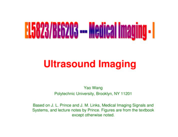
SpecialistsNEUROLOGISTA doctor specializing in disorders anddiseases affecting the brain and spinal cord(the central nervous system or CNS). Someneurologists have oncology training.It is ideal to find a neurosurgeonwith experience. Ask if at least:50%NEURO-ONCOLOGISTAn oncologist (cancer doctor) specializingin the treatment of cancers and tumorsaffecting the CNS.NEUROSURGEON (IDEALLY ANEUROSURGICAL ONCOLOGIST)A surgeon specializing in the surgicalmanagement of CNS disorders. If possible,talk to a neurosurgeon who works withbrain tumor patients 50% of the time,or more.NEURO-RADIOLOGISTA radiologist (an expert in imagingtechniques) specializing in theinterpretation of scans and images of theCNS. Some are specialists in brain tumors.NEURO-PATHOLOGISTA doctor specializing in the diagnosisof CNS disorders through microscopicexamination of biopsied tissues (tumor cells).OF T H E I R P RACT I CEI S WI T H BRAI NT UM OR PAT I E NT SNEURO-ONCOLOGY NURSECLINICAL PSYCHOLOGISTA registered nurse specializing inpatient education (including symptommanagement) and support services forbrain tumor patients.A licensed professional who can helppatients and families adjust to theeffects of illness on their lives. Neuropsychologists specialize in brain functionsand how brain damage can affect aperson’s abilities.At any point before, during or aftertreatment, the services of the followingprofessionals can be valuable:SOCIAL WORKERMedical social workers specialize incounseling and crisis intervention, and helplocate appropriate care, legal resources,and financial aid.REGISTERED DIETICIAN ORN UTRITIONISTA trained specialist with knowledge abouthow a person’s diet and daily nutritionwill impact their health. The Academy ofNutrition and Dietetics lists professionalswith oncology experience:www.eatright.org/programs/rdfinderC H A P T E R 2 / U N D E R S TA N D I N G B R A I N T U M O R S15
Cancer Support Community and the National Brain Tumor Society would like to recognize and thank all of those whocontributed to the success of this book.CONTRIBUTORS & EDITORSC O N T R I B U T I N G PA R T N E R SDESIGN & PHOTOGRAPHYDeanna Glass-Macenka, RN, BSN, CNRNJohns Hopkins HospitalAccelerate Brain Cancer CureNicola BeddowSuzanne Kleinwaks Design, LLCDesignLora Hays, L.M.F.T., R.P.T.Cancer Support Community Central IndianaCancer Support CommunityAllison Harvey, MPH, CHESRalph AlswangPhotographyAshley Varner, MSW, MBA, LCSW-CAnne Arundel Medical CenterJohns Hopkins Brain TumorEducation GroupMusella FoundationAl Musella, DPMErica Weiss, MPH, MSUPWriter/EditorNational Brian Tumor SocietyKristina KnightMichele Rhee, MBA, MPHWe’d like to extend a special thanksto focus group, interview, and surveyparticipants who shape the informationprovided in this booklet.Patrick Y. Wen, MDDana-Farber Cancer InstituteCA N C E R S U P P O RT C OM M U NITY’ S FRA N KLY SPEA KIN G A B OU T CA N CER SERIESCancer Support Community’s Frankly Speaking About Cancer: Brain Tumors program is part of a national education program thatprovides support, education, and hope to people affected by cancer and their loved ones.Frankly Speaking About Cancer booklets feature information about treatment options, how to manage side effects, the social andemotional challenges of the diagnosis, and survivorship issues.For more information about this program, the Frankly Speaking About Cancer series or Cancer Support Community, please visit ourwebsite at www.cancersupportcommunity.org or call us toll-free at 1-888-793-9355.
W W W . C A N C E R S U P P O R T C O M M U N I T Y. O R G1.888.793.9355Cancer Support Community and the National BrainTumor Society together with our partners providethis information as a service. This publication is notintended to take the place of medical care or theadvice of your doctor. We strongly suggest consultingyour doctor or another health care professional toanswer questions and learn more. 2013 Cancer Support Community. All rights reserved.C A N C E R S U P P O R T C O M M U N I T Y A N D T H E N AT I O N A L B R A I N T U M O R S O C I E T YT H A N K O U R P R O G R A M PA R T N E R S :
or CT Scan) is a computerized x-ray that can show a combination of soft tissue, bone, and blood vessels. This is often the first test a person will receive in an emergency room (i.e. after a seizure). Magnetic Res