Transcription
NeuroImage 170 (2018) 332–347Contents lists available at ScienceDirectNeuroImagejournal homepage: www.elsevier.com/locate/neuroimageTopographic organization of the cerebral cortex and brain cartographya,b,c,⁎Simon B. Eickhoffd,e,f, R. Todd Constable, B.T. Thomas YeoMARKg,h,iaInstitute of Systems Neuroscience, Medical Faculty, Heinrich-Heine University Düsseldorf, GermanyInstitute of Clinical Neuroscience and Medical Psychology, Medical Faculty, Heinrich-Heine University Düsseldorf, GermanyInstitute of Neuroscience and Medicine (INM-1), Research Center Jülich, GermanydInterdepartmental Neuroscience Program, Yale University, USAeDepartment of Radiology and Biomedical Imaging, Yale University, USAfDepartment of Neurosurgery, Yale University, USAgDepartment of Electrical and Computer Engineering, ASTAR-NUS Clinical Imaging Research Centre, Singapore Institute for Neurotechnology and MemoryNetworks Program, National University of Singapore, SingaporehMartinos Center for Biomedical Imaging, Massachusetts General Hospital, Harvard Medical School, Charlestown, USAiCentre for Cognitive Neuroscience, Duke-NUS Graduate Medical School, SingaporebcA BS T RAC TOne of the most specific but also challenging properties of the brain is its topographic organization into distinctmodules or cortical areas. In this paper, we first review the concept of topographic organization and its historicaldevelopment. Next, we provide a critical discussion of the current definition of what constitutes a cortical area,why the concept has been so central to the field of neuroimaging and the challenges that arise from this view. Akey aspect in this discussion is the issue of spatial scale and hierarchy in the brain. Focusing on in-vivo brainparcellation as a rapidly expanding field of research, we highlight potential limitations of the classical concept ofcortical areas in the context of multi-modal parcellation and propose a revised interpretation of cortical areasbuilding on the concept of neurobiological atoms that may be aggregated into larger units within and acrossmodalities. We conclude by presenting an outlook on the implication of this revised concept for future mappingstudies and raise some open questions in the context of brain parcellation.The brain a topographically organized organThe brain is without question the most complex structure in thehuman body and we are still far from understanding its physiologicalfunctioning or pathology. In this context, one of the most distinctfeatures of the brain in comparison to any other organ of the body is itstopographic organization. This distinction and its implications becomemost evident when considering the effects of partial damage orremoval. In organs like the liver, every part looks and acts in the sameway and hence a resection of tissue only affects overall capacity, e.g., formetabolism, but not any specific function, e.g., bile production. In starkcontrast, localized damage to a small part of the brain will result in veryspecific disturbances of isolated mental facilities, such as vision,movement or language (Rorden and Karnath, 2004). In turn, however,overall function is usually not impaired following localized damage, i.e.,while vision may be specifically affected, all other mental functions maybe largely intact. Another important feature of this regional differentiation is the fact that the brain is realized on a common building plan.This renders the brain not only distinct from uniform organs but also⁎from compartmentalized ones such as the kidneys, where differentfunctions are realized in morphologically different sections. Thistopographic organization becomes most evident with respect to thecerebral cortex, which features a common building plan into sixdifferent layers of varying cell density (Amunts and Zilles, 2015). Inspite of the fact, that the entire cortex is made up from the same celltypes, however, variations from this common architectonic scaffold arealso evident, as density, size and distribution of cells across the corticallayers may be observed. Likewise, the general principles of local andlong-distance connectivity seem rather consistent across the brain, yetthe origins and terminations of fiber bundles linking different partsshow a highly differentiated pattern (Felleman and Van Essen, 1991).It may be noted, that topographic organization is not a specific featureof the human brain, but rather seems to have emerged very early inevolution and is shared by all vertebrates, despite vastly differentinstantiations and capacities (Krubitzer and Kaas, 2005). A topographic specialization thus seems to represent a fundamental principleof brain function, rendering its investigation through what has nowbecome known as brain mapping the key to understanding (human)Corresponding author at: Institute of Systems Neuroscience, Medical Faculty, Heinrich-Heine University Düsseldorf, Germany.E-mail address: simon.b.eickhoff@gmail.com (S.B. 2017.02.018Accepted 7 February 2017Available online 20 February 20171053-8119/ 2017 Elsevier Inc. All rights reserved.
NeuroImage 170 (2018) 332–347S.B. Eickhoff et al.Korbinian Brodmann. In turn, von Economo and Koskinas, while alsofollowing the general concept of abrupt changes between cortical fields,were more likely to also denote transition zones and local variationswithin a given cytoarchitectonic area.Summarizing their results in a drawing showing the locationsthereof on a picture of the brain, usually an idealized surface view,they quickly established the standard language we know today. Muchlike the political organization of, e.g., Europe, the topographic organization of the human brain can be represented by a map showingindividual cortical areas of homogeneous architecture and the bordersbetween them. And although the relation between these maps and theneuropsychological symptoms arising from localized lesions was rarelydirectly assessed at that time, the prevailing notion was that thesedistinct areas should also map to distinct functions. Owing to therefinements in the methods for mapping function (based on cognitivedeficits following brain lesions) and architecture (based on histologicalexamination), the next decades provided increasingly complex brainmaps, defining smaller and smaller sub-areas.In spite of this general trend, which may be considered ongoing ifnot accentuated with the advent of neuroimaging (Raichle, 2009), it isnoteworthy that the compartmentalization of the brain has beenchallenged throughout its entire history. For example, shortly afterthe localization of language-relevant areas by Broca and Wernicke,Ludwig Lichtheim already proposed a model that highlighted the roleof connections and interactions in the context of language production.In turn, while researchers such as Edith Gerhard and the RussianSchool provided increasingly detailed anatomical maps of the humanbrain, Gerhardt von Bonin and Percival Bailey proposed a map thatconsidered almost the entire cortex as largely uniform, advocating aholistic view of brain organization (Bailey and von Bonin, 1951). Yet,the prevailing view was that of topographic organization suggested byanatomical brain maps, localized lesions and physical white-matterdissection in post-mortem brains, even though the relationships amongthem remained opaque.brain organization.Historical development and conceptual changesOne of the most curious facets in the history of human brainmapping is the fact that virtually all of its key concepts were anticipatedby the 18th century phrenologists long before the emergence ofneuroscience as the current biomedical discipline (Finger, 2004).Evidently, their simplistic view of mental organs or facilities and theidea that these could be assessed by examining craniological features isso remote from the current scientific paradigm, that “neo-phrenology”has become a derogative term for over-simplified localization ofcognitive functions. It is worth noting, though, that the works by theprotagonists of that field, such as Johann Kaspar Lavater (1741–1801)and Franz Joseph Gall (1758–1828), actually spelled out most of thefundamental ideas that form the basis of human brain mapping and itsclinical applications. Most importantly, they postulated that the brainwas the physical basis of the mind and not a homogeneous organ. Theyhypothesized that the brain consisted of a topographically organizedmosaic of individual modules supporting different functions andshowing inter-individual variability that related to phenotypical features.The first step towards establishing the topographic specificity ofmental functions compatible with current scientific standards wastaken in 1861 when Paul Broca described his famous patient sufferingfrom non-fluent aphasia, but no other deficits. This condition haddeveloped following a lesion in the left inferior frontal cortex. Broca'shypothesis that this specific deficit was caused by isolated damage to aspecific part of the brain received heavy criticism from his peerssupporting a holistic view on brain function (equipotentiality, pioneered by Lashley and Flourens). In the decades that followed,however, inferring the localization of cognitive functions throughobservation of the effects of localized lesions not only established itselfas the primary approach towards understanding the organization of thehuman brain, but also shaped emerging concepts on brain organization. Most importantly, it fostered the notion of topographic specialization and structure-function relationships that has remained largelyintact until the most recent focus on brain connectomics (Sporns,2011).One of the key developments that supported this localizing,topographic view of (human) brain organization was the emergenceof microscopical anatomy in the late 19th and early 20th century. Usinghistological analysis of brain sections stained for cell bodies or myelin,researchers like Paul Flechsig, Cécile and Oskar Vogt, von Economoand Koskinas, and Korbinian Brodmann (for a detailed review, cf. Zillesand Palomero-Gallagher, 2011) provided detailed descriptions of themicroarchitecture of the human cerebral cortex. Histological analysis ofstained, serially sectioned post-mortem brains provided the firstopportunity to investigate the heterogeneity of microstructural featuresacross the whole brain and hence provide complete maps of itsdifferentiation. More specifically, cytoarchitectonic analysis is basedon sections stained for cell bodies and assesses the relative width andcell-density of the six canonical layers of the (neo-) cortex, thedistribution of different cell types and the size and arrangement of(in particular) pyramidal cells. In turn, myeloarchitectonic investigations are performed on sections stained for myelin and focus on thedistribution of radial and tangential fibers within the cortex as well asthe presence and location of myelin-dense bands. In these “classical”brain mapping studies, it was noted, that the cyto- or myeloarchitectonic characteristics of the cortex usually don’t vary smoothly acrossthe brain but tend to change abruptly at particular locations, whichwere considered to represent the borders between different corticalareas. This concept was most clearly advocated by the Vogts’ based onthe myeloarchitectonic work, who posited “haarscharfe Grenzen”(borders sharp as a hair) in the cerebral cortex, a feature that is alsoclearly expressed in the cytoarchitectonic work of their studentNew perspectives studying non-human primatesWhile many early anatomical studies were already comparative innature and features such as the cortical column were initially describedin the cat (Mountcastle, 1957), views on human brain organizationwere most profoundly influenced by the strong growth of physiologicalstudies in non-human primates in the 1970s. Such studies entailed twosubstantial advantages that were critical for the conceptual development of brain mapping. First, studies in non-human primates enabledthe use of invasive methods that to date must be considered the goldstandard for the investigation of brain function, connectivity andstructure. Invasive electrophysiological recordings provided for thefirst time spatially detailed information about neuronal responseproperties in the intact brain under a wide variety of experimentalcontexts (Hubel and Wiesel, 1968). In turn, size and location ofnaturally occurring lesions in the human brain are strongly influencedby the vascular architecture and their effects can only be assessedfollowing recovery of the patient, i.e., after substantial plasticity andadaptation has already happened (Mah et al., 2014). Single unitrecordings and the measurement of local field potentials in monkeysthus provided an unprecedented view on local differentiation of brainfunction, confirming and extending previous theories on topographicorganization (Levy and Goldman-Rakic, 2000). In addition, anteroand retrograde tracers that are absorbed by the neurons at the injectionsite and then moved towards either the soma (retrograde tracers) or theaxon terminals (anterograde tracers) via axonal transport provideddetailed information on the long-range connections between differentbrain regions (Rockland and Pandya, 1979). These studies revealedthat long-range connections follow a precise topography and specifically link individual areas across the brain. Finally, given that theanimals needed to be sacrificed to verify the location of the recording333
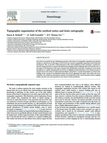
NeuroImage 170 (2018) 332–347S.B. Eickhoff et al.cortical area inherently requires that within any single modality, i.e.,structure, function and connectivity, there are clearly demarcatedborders, rather than gradients or transition zones. In turn, within aparticular area, this feature should be homogeneous. While both ofthese fundamental assumptions are rooted in the classical anatomicalworks by Brodmann or the Vogts, who advocated the concept ofhaarscharfe grenzen, i.e., borders sharp as a hair, they are actuallyfar from trivial or undisputed. On the one hand, there is ampleevidence that structural and functional features may be organized ingradients, in particular in the posterior parietal and prefrontal cortex(cf. Carmichael and Price, 1994 and references therein). On the otherhand, other aspects of cortical organization such as the internaldifferentiation of early visual areas into blobs, stripes and oculardominance columns (reviewed in Yoshioka et al. (1996)) likewisechallenge the assumption of homogeneous areas with sharp transitionsof neurobiological features at their borders.The second fundamental assumption is that a cortical area shouldbe differentiated from its neighbors by all three aspects, i.e., structure,function and connectivity, with the respective borders from eachcriterion being in close agreement with each other. There is ampleevidence from non-human primates demonstrating such agreementmade possible through microscopic spatial resolution and the possibility to perform multi-modal comparisons of all three aspects in thesame individual as described above. Nevertheless, there are alsonumerous instances in which a sharp change in one property is notmirrored in other aspects of cortical organization. One particularlyinteresting example of this situation, which is not confounded bydifferent spatial scales, inter-subject registration or blurring is provided by in-vitro multi-receptor autoradiography (Amunts and Zilles,2015). In this work, the binding sites for various receptors for classicalneurotransmitters are marked on neighboring 20 μm thick sections ofpost-mortem brains using tritiated ligands. Looking at the early visualcortex, i.e., the part of the brain from which the concept of a corticalarea was primarily derived, it may be noted, that in virtually allinstances borders, evidenced by changes in mean receptor concentration or laminar pattern of receptor density, are closely matching thecytoarchitectonic borders between visual areas (Eickhoff et al., 2008a,2008b). However, at the same time any particular border is usuallyonly revealed by a subset of the investigated receptors (Fig. 1). Thisduality of a close match between revealed borders but frequently absenttransitions in some features has recently been mirrored by anintegrated analysis of cortical myelination, functional activation properties and resting-state connectivity.sites and to reveal the transported tracers, histological investigationsare readily performed using the same cyto- and myelo-architectonicapproaches that formed the basis of classical human brain maps (VanEssen et al., 1981).The above naturally leads to the second critical advantage of suchstudies over the then (and one could argue currently) availablemethods for studying the human brain. In non-human primates,structure, function and connectivity of the brain may not only beassessed with a very high precision and specificity, but moreover allthree aspects may be investigated within the same animal and hencedirectly compared to each other. That is, all three aspects of corticaldifferentiation were not only assessed with high fidelity but couldmoreover be spatially related to each at the millimeter-scale within thesame individual. Throughout many of the seminal publications over thenext decades, one of the most consistent observations was a goodcongruence between microanatomical borders, distinctions in connectivity patterns and variations in functional response properties. That is,if injection sites were at different sides of a cytoarchitectonic border,they usually differed in their projection patterns, if two locationsshowed different response properties, they were distinct in myeloarchitectonic features and so on (cf. Burton et al., 1995; Krubitzeret al., 1995).This within-subject agreement of topographic differentiation acrossdifferent features (at least within the sensory and motor cortices whichwere predominantly investigated) confirmed, albeit in different species,the prevailing notion that histologically defined areas in the humanbrain should correspond to functional modules whose damage wouldthen lead to particular neuropsychological symptoms. The observationsoutlined above thus culminated in what is now considered the“classical” definition of a cortical area.The concept of cortical areasIn non-human primates, a cortical area is conventionally defined asa region of the cortex that may be distinguished from its neighbors byseveral criteria (for reviews see Felleman and Van Essen, 1991; Orbanet al., 2004; Krubitzer and Kaas, 2005; Eickhoff and Grefkes, 2011).First, a cortical area should show different global functional propertiesthan the surrounding cortex. Second, it should be microstructurallydistinct, i.e., differ in cyto- and myeloarchitecture. Third, it shouldfeature a different pattern of inter-regional connectivity than itsneighbors. Finally, given that most of the experimental work that hasled to this conceptual definition of cortical areas has been performed insensory cortices, in particular the visual system, the presence of acomplete representational, e.g., retinotopic, map has likewise enteredthe definition of a cortical area. This definition not only reflects thehistorical development of concepts on topographic organization asoutlined above, but has moreover been among the concepts moststrongly shaping the emerging field of neuroimaging. Several aspects ofthe outlined definition are particularly noteworthy in the context of thisreview, most of which are attributable to the fact that the concept ofcortical areas was primarily defined based on investigations of sensorycortices, in particular the visual system. Maybe the most obvious is theimportance of topographic maps, which is indeed a hallmark of sensoryareas to the extent that a double representation of a particular part ofthe body or visual field provides one of the strongest indications of thepresence of an additional cortical area (Krubitzer et al., 1995; cf. Zekiet al., 1998). When moving away from early uni-modal cortices,however, such topical organizations become increasingly difficult todefine, even though there is evidence for, e.g., retinotopic features inassociation cortex (Selemon and Goldman-Rakic, 1985). Consequently,this criterion has been down-weighted when considering areas outsideuni-modal sensory cortices.Two implications of the classical definition of a cortical area hadformative influences on the fields of neuroimaging and in-vivo brainparcellation (cf. Amunts et al., 2014). First, the classical definition of aTheoretical and practical relevance of the “cortical area”conceptIn spite of the conceptual limitations noted above, which werealready known by the time that neuroimaging emerged and humanbrain mapping began to grow into the large and highly dynamic field itis now, the concept of a cortical area has a tremendous impact on ourfield. In fact, we would argue that the idea of a cortical area as a fieldthat is distinguished from its neighbors by (sharp) changes in structure,function and connectivity, must be considered as the conceptualbackdrop of most research in human brain mapping. But why has thisconcept caught on so well in neuroimaging and to date remained one ofthe core concepts for the analysis and description of brain organization?The key appeal from both the theoretical and computational side isthe idea of modular brain organization encoded by the term “corticalarea”, rather than the concept itself. The most important aspect here isthat such modules or “cortical” areas may be regarded as units ofcoordinated information processing that act as a unified whole in termsof the sustained functions and in terms of interactions with othermodules (Eickhoff and Grefkes, 2011; Fox and Friston, 2012; Decoet al., 2015). This view not only accommodates the notion that the334
NeuroImage 170 (2018) 332–347S.B. Eickhoff et al.Fig. 1. Autoradiographic visualization of the distribution of binding sites for three different neurotransmitter receptors as well as a corresponding cell body stained section. Corticalborders within the early visual cortex as evident by each feature are marked in the image and areas are labeled in the cytoarchitectonic section. Two important aspects become evidentfrom this histological data. First, not every feature or modality reveals all cortical borders. Second, in case that multiple features indicate a given border, the locations of these borders areusually in very good spatial agreement.for most standard computers (Loewe et al., 2016). Likewise, anymultivariate analysis of voxel-wise data, in particular across a largenumber of subjects, will be inevitably challenging. Moreover, even ifcomputationally tractable, analysis of neuroimaging data at the resolution of individual voxels is complicated by smoothing effects both fromMR physics sources as well as from the reliance on the spatially broadBOLD effect (Ugurbil, 2016).Using cortical areas as a neurobiologically meaningful dimensionality reduction thus entails many advantages. First, working at the levelof a cortical area, rather than at the voxel level greatly ameliorates thenumber of features and hence provides a much more efficient framework for, in particular large-sample, analysis. More importantly,however, it provides an a priori, biologically motivated approach tofeature reduction, providing a potential solution to the important butultimately open question of how to define nodes for connectivityanalyses (cf. Zalesky et al., 2010; Caspers et al., 2013; Bellec et al.,2015; Thirion et al., 2014). That is, if a parcellation of the cortex is inline with the conceptual definition of a cortical area, these modulesshould each be homogeneous in, but distinct from, each other in termsof all measured or derived properties, e.g., volumetric measures orconnectivity-profiles. Consequently, a parcellation into cortical areasbrain is topographically organized into distinct units, but also acknowledges the importance of specific interactions between them through thecriterion of distinct connectivity (patterns). The idea of a “cortical area”thus provides the currently most widely accepted view of the brain as ahighly-specifically interconnected network between specialized modules (Bullmore and Sporns, 2012; Park and Friston, 2013; Amuntset al., 2014).Besides this conceptual relevance of defining what could beconsidered as the smallest meaningful building block (cf. next section),it is worth reviewing the concept of cortical areas or modules as theunits of investigation. In particular, the resolution of current neuroimaging data (Uğurbil et al., 2013) is well coarser than the size of acortical column (Horton and Adams, 2005 but cf. Yacoub et al., 2008),but finer than the macroscopic neurobiological organization of thebrain. Hence, the nominal dimensionality of 200,000–400,000 greymatter voxels (at a resolution of 1.5–2 mm isotropic voxel size anddepending on the amount of included brainstem) is almost certainlynot the one that would best represent the phenomenon of interest, i.e.,cortical differentiation. Rather, it poses a substantial computationalchallenge, e.g., when trying to compute whole brain dense (voxel xvoxel) connectomes, leading to matrices that require too much memory335
NeuroImage 170 (2018) 332–347S.B. Eickhoff et al.based on Brodmann's schematic drawing and was only meant forillustrative purposes, multiple (again extrapolated) versions of theTalairach atlas (e.g., Lancaster et al., 2000) are still widely used inuse today. Thus, while many much more detailed classical and modernhistological maps of the brain are available, the subjective guess ofwhere the areas of the coarsest available atlas should be located in 3Dspace is widely considered as the standard reference of the brain.Obviously, however, these areas are much coarser than any functionaldifferentiation, leading to two scenarios. In some cases, a host ofdifferent functions is assigned to the same Brodmann area (BA), asevident, e.g., for BA 9 or BA 39. In other cases, the broad labels of theTalairach / Brodmann atlas are unsystematically mixed with labelsoriginating from primate research, which may be far from congruent,e.g., V3 (BA 19, cf. Rottschy et al., 2007) or PMd (BA 6, cf. Genon et al.,2016). While they may provide some common language of where afinding is located, it becomes clear that, like cortical columns, suchlarge anatomical regions do not seem to reflect the true modularorganization of the brain.These considerations evidently lead to the crucial question of the“right” granularity from the neurobiological point of view. Here it isimportant to remember, that the brain is a hierarchically organizedsystem in which individual elements (at the lowest level most likely thecortical column) are working together in a coordinated manner toimplement a particular process. Neighboring sets of columns perform asimilar process that is used in concert. The ensuing ensemble is closelylinked with others that usually cooperate in most settings andultimately with yet other regions that may be considered part of alimited number of canonical large scale networks (Scannell et al., 1999;Ross et al., 2000; Sporns et al., 2004; Fox et al., 2005; Kaiser, 2011;Yeo et al., 2011; Power et al., 2011; Fox and Friston, 2012).Consequently, neither the fact that there are several million corticalcolumns nor the answer that there are 5–15 large scale networksprovides incorrect or biologically meaningless answers to how manymodules comprise the brain.Nevertheless, in many cases the actual unit of interest should bewhat we will call neurobiological atoms. We would conceptualize themas modules that perform (only) one specific operation (independentlyof how this can be mapped to psychological terms) and behave as a unitin all relevant neurobiological features (that may yet need to bedefined), while being distinct from all other units, even those withwhich they cooperate. We note, that this largely reproduces thedefinition of a cortical area, but deviates from the notion of spatialhomogeneity and functional specificity. This reformulation has particular relevance in topographically organized regions, given that, e.g.,the hand representation of the primary motor cortex is actually verydistinct from that of the face, rendering them potentially differentatoms as demonstrated in recent mapping studies.One question that remains open for the time being, though, is thescale of these neurobiological atoms and the extent to which this scaleis homogeneous across the brain. Addressing this question in a mannerthat is unbiased by the effective resolution of the applied imaging dataor constrained by the computational approach remains one of thebiggest challenges in understanding the topographic organization ofthe human brain as a very specifically connected network of specializedmodules.promises to be optimal for the investigation of any sort of data. Incontrast, any data driven dimensionality reduction (Craddock et al.,2013; Eickhoff et al., 2015) should be modality and potentially datasetspecific. Moreover, given that cortical areas are supposed to reflectmodules sustaining specific operations, there would be a natural linkbetween the units of analysis and physiological as well as pathologicalfunction. Last but not least, the analysis of the structure, function andconnectivity of cortical areas, rather than voxels or data-driven lowerdimensional representations, can provide a standard space that allowscomparisons between studies while still allowing voxel-wise analyses,which could be allocated to the respective modules. In summary, it mayhence not surprise that the concept of “cortical areas” has been aprimary focus of neuroimaging throughout its existence.The issue of scale and granularityImportantly, it may be argued that cortical areas in the manner thatthey are usually defined by the aforementioned criteria in humans andexperimental animals seem to provide a very useful intermediate levelof detail. This may be best exemplified by juxtaposing a
Feb 07, 2017 · Historical development and conceptual changes . observation of the effects of localized lesions not only established itself as the primary approach towards understanding the organization of the human brain, but also shaped emerging concepts on brain organiza- . brain, G