Transcription
Pediatric Cardiac Acute Care HandbookEdition 2 (2018-2019)Compiled by:Alaina K. Kipps, MD, MS; Inger Olson, MD; Neha Purkey, MD; Charitha Reddy, MDI. General Principles of Cardiologya. Cardiac Anatomy .1b. History and Physical .6c. Cardiac Catheterization . .10d. Echocardiography .14II. Common Complaints in Cardiologya. Murmurs .25b. Chest Pain .28c. Syncope .30d. Preventative Cardiology .32III. EKG Interpretation and Common Arrhythmiasa. EKG Reading . .34b. Arrhythmia Algorithm .42c. Common Arrhythmias .45IV. Congenital Heart Diseasea. Neonatal Presentation of CHD .64V. Acyanotic Lesionsa. ASD . .68b. VSD . .71c. AVSD . .74d. PDA . .77e. Ebstein Anomaly . .81f. Bicuspid Aortic Valve . . .84g. Aortic Stenosis . . .86h. Pulmonary Stenosis . .89i. Coarctation . . . .92j. Interrupted Aortic Arch . .95
VI. Cyanotic Lesionsa. DTGA . .97b. TOF . . .100c. TOF/PA/MAPCAs . .103d. Truncus . . 107e. DORV . 109f. HLHS .112g. PA/IVS . .117h. Tricuspid Atresia . .119i. TAPVC . . .122VII. Surgical Repairs . .126VIII. Cardiomyopathies .136IX. Congestive Heart Failure . .141X. Pulmonary Hypertension . .143XI. Acquired Heart Disease . 148XII. Common Associationsa. Genetic Syndromes . . .161XIII. Appendix .173
Cardiac Anatomy: Learning the Language In congenital heart disease, the heart can be located anywhere in the chest, and thecomponents of the heart can be arranged in a number of different ways.“Right” and “left” do not refer to the side of to the body, but to specific anatomic criteria thatidentify different components of the heart.Cardiac Position Levocardia: heart is in the left chest, apex points leftward (normal position) Mesocardia: heart in the midline, apex points inferiorly Dextrocardia: heart in the right chest, apex points rightward Dextroposition: heart in the right chest, with apex pointing leftward Dextrorotation: heart in the left chest, apex rotated rightward Ectopia cordis: heart partially or completely outside of the chest/sternum1
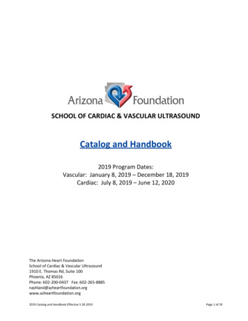
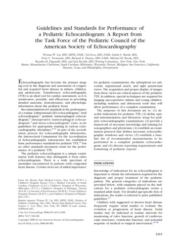
The Main Chambers of the HeartNetter’s Correlative Imaging: Cardiothoracic AnatomyRight Atrium: receives the SVC, IVC and coronary sinus, limbus of fossa ovalis present Broad-based, triangular appendage with pectinate muscles that extend into the right atrial body Crista terminalis presentLeft Atrium: receives the pulmonary veins (but this is not a defining feature) Narrow-based, thin, finger-like appendage with pectinate muscles that are confined to theappendage Visible attachments of septum primum to septum secundumRight Ventricle: Coarse trabeculae with a prominent septal band, parietal band and moderator band Septophilic attachments of the tricuspid valve (attaches to the septum and free wall) Well-developed infundibulum (or conus) – the muscle underneath the semilunar valve, whichresults in lack of fibrous continuity between the tricuspid and semilunar valves The tricuspid valve always belongs to the right ventricleLeft Ventricle: Smooth septal surface with fine trabeculae Septophobic attachments of the mitral valve (attaches only to the free wall) No infundibulum under the semilunar valve, so there is fibrous continuity between the mitraland semilunar valves2
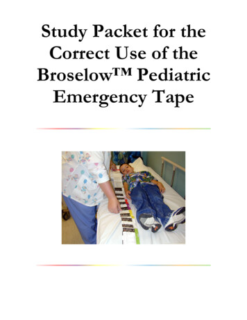
Segmental or “Van Praaghian” Approach to Cardiac Anatomy { , , }1st letter { , X,X} Visceral and atrial situs Position of the abdominal organs (liver, spleen and stomach) and atria Solitus (S): normal anatomy with liver on right, spleen and stomach on left, right atrium on right,left atrium on left Inversus (I): mirror image of normal: liver on left, spleen and stomach on right, right atrium onleft, left atrium on right Ambiguous (A): indeterminate situs: midline liver, cannot differentiate between right and leftatria, usually associated with heterotaxy or “atrial isomerism”www.pedscards.com2nd letter, {X, , X}: Ventricular Looping D-loop (D): normal anatomy with right ventricle to the right of the left ventricle L-loop (L): left ventricle to the right of the right ventricle The “hand rule” demonstrates the chirality of the ventricles. A D-looped right ventricle is a “righthanded” ventricle because only the right hand will fit inside the ventricle so the thumb is in theinflow, fingers are in the outflow, and the palm faces the septum. An L-looped right ventricle is“left handed” because the left hand will fit in the right ventricle.3
Right hand fits in the right ventricle D-loopwww.pedscards.com3rd letter, {X,X, }: Great Arteries the relative position of the semilunar valves to each other Solitus (S): normal anatomy with aortic valve posterior and to the right of the pulmonary valve Inversus (I): mirror image with aortic valve posterior and to the left of the pulmonary valve D-malposition (D): aortic valve is anterior and to the right of the pulmonary valve L-malposition (L): aortic valve is anterior and to the left of the pulmonary valve Anterior (A): aortic valve is directly anterior to the pulmonary valve Posterior (P): aortic valve is directly posterior to the pulmonary valvewww.pedscards.comIf the anatomy of a segment cannot be determined, then an “X” is used for that segment.The connecting segments and ventriculo-arterial connections are described separately.The Andersonian Approach to Cardiac AnatomyA different approach to describing cardiac anatomy was championed by Dr. Bob Anderson. If the rightatrium connects to the right ventricle, and the left atrium connects to the left ventricle, this is describedas atrioventricular concordance. If the pulmonary artery arises from the right ventricle and the aortaarises from the left ventricle, this is described as ventriculoarterial concordance. If the right atrium4
connects to the left ventricle, this is termed atrioventricular discordance. If the pulmonary artery arisesfrom the left ventricle, this is termed ventriculoarterial discordance.Questions What are the anatomic features of the tricuspid and mitral valves? What are the anatomic features of the semilunar valves? What is the anatomic relationship of the systemic arteries? Describe normal branching of the aortic arch.ResourcesAnderson RH, Becker AE, Freedom RM, et al. Sequential segmental analysis of congenital heart disease.Pediatric Cardiology 1984; 5(4): 281-287.Edwards WD, Maleszewski JJ. Cardiac Anatomy and Examination of Cardiac Specimens. In: Allen HD,Driscoll DJ, Shaddy RE, Feltes TF, 8th, editors. Moss and Adams’ Heart Disease in Infants, Children, andAdolescents: Including the Fetus and Young Adult. Philadelphia: Lippincott Williams & Wilkins; 2013.p.1-31.Van Praagh R. Terminology of congenital heart disease: Glossary and commentary. Circulation 1977;56:139-143.5
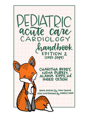
The Cardiac History & Physical ExamQuestions to Ask How is the child growing? How much activity can the child tolerate? Can they keep up with peers? How was the mother’s pregnancy? Did the mother have diabetes or other medical problems?Did she take any drugs or prescription medications during the pregnancy? Is there a family history of congenital heart disease, sudden death or early cardiac death? Does the child have a syndrome that is associated with cardiac disease? Does the child have any symptoms: poor growth, poor feeding, developmental delay,diaphoresis, poor exercise tolerance, cyanosis, syncope, palpitations, or edema?Vitals Signs Are Vital! Tachycardia is a clue that the child requires increased cardiac output Tachypnea can be a consequence of increased pulmonary blood flow or congestion.o Quiet/comfortable or with respiratory distress. Pulse oximetry will identify if an infant is cyanoticInspection Look for a precordial bulge or hyperdynamic precordium Look for extracardiac signs: dysmorphic facial features, clubbing of the digits, edema, respiratorydistress (grunting, retractions)Palpation Arterial exam:o Pulses: rate, rhythm, volume, character. Assess for brachial-femoral delay. Femoral pulses should be part of every exam, particularly in neonateso Assess distal perfusion by the warmth of the digits and capillary refill time. Venous exam:o Assess position, size, consistency of the livero Assess for jugular venous distention in an older child Precordial exam:o Entire palm and hand should be placed on chest to assess for thrills or heaveso Assess the position and character of the apical impulseAuscultation S1: the first heart soundo From closure of the mitral and tricuspid valves, can occasionally be audible as a split S1o Best heard with the diaphragm of the stethoscope S2: the second heart soundo From closure of the semilunar valveso Usually consists of a louder, earlier sound from aortic valve closure (A2) followed by aquieter sound from pulmonary valve closure (P2).o Splitting of S2 during inspiration or “physiologic splitting” is a normal finding related toincreased right heart filling and decreased left heart filling during inspiration, causing6
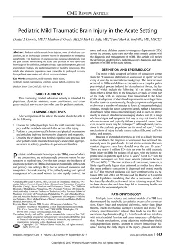
the right ventricle to take longer to empty, and the pulmonary valve to take longer toclose. To say the “S2 is normal” is to say you heard this variable splitting!!o A loud S2 is likely from early closure of the pulmonary valve and may suggest pulmonaryartery hypertension.S3 gallop: “Kentucky”o Heard early in diastole during rapid fillingo May be normal in older children and competitive athletesS4 gallop: “Tennessee”o Heard when the myocardium is poorly compliant, from rapid filling of the ventricleduring atrial contraction (late in diastole)o Always pathologic in children, usually associated with CHFEjection Clicks: early systolic, high-frequency sound associated with abnormal semilunar valvesMidsystolic click: midsystolic, high-frequency sound associated with mitral valve prolapse4thAdapted from: a/Wiggers Diagram.png7
Murmurs: caused by turbulent blood flow creating audible sound waves in the range of 20Hz to2000Hzo Described by: Timing: When in the cardiac cycle does the murmur occur? What is the murmur’s relationship to S1 and S2? Intensity or loudness: Depends on the size of the orifice or vessel through which blood flows,the pressure difference or gradient across the site, and the blood flowor volume across the site For systolic murmurs:o Grade 1: heard only with intense concentrationo Grade 2: faint, but heard immediatelyo Grade 3: easily heard, of intermediate intensityo Grade 4: easily heard, associated with a thrill (a palpablevibration on the chest wall)o Grade 5: very loud with a thrill, audible with only the edge ofthe stethoscope on the chest wallo Grade 6: audible with the stethoscope off the chest wall Duration of the murmur Configuration: the dynamic shape of the murmur Pitch: the frequency range of the murmur Quality: the presence of harmonics or overtones Location on the chest wall: Point of maximal intensity: Where is the sound loudest? Extent of radiation: Over what area is the sound audible?o Systolic Murmurs occur between S1 and S2 Holosystolic murmurs: obscure S1 and terminate at S2 Ejection murmurs: crescendo-decrescendo or diamond-shaped Early systolic murmurs: start abruptly at S1, but taper and disappear before S2– associated exclusively with small muscular VSDs Mid- to late systolic murmurs: begin midway through systole, often heard withmidsystolic clicks and the mitral regurgitation heard with mitral valve prolapseo Diastolic Murmurs occur between S2 and S1 Early diastolic murmurs: decrescendo murmurs, arise from aortic or pulmonaryvalve regurgitation Mid-diastolic murmurs: diamond shaped murmurs, associated with increasedflow across a normal mitral or tricuspid valve, or normal flow across a stenoticmitral or tricuspid valve Late diastolic murmurs: crescendo murmurs, created by stenotic or narrowedmitral or tricuspid valves, the noise is associated with atrial contraction ALWAYS pathologic!o Continuous Murmurs occur throughout the cardiac cycle Flow through vessels beyond the semilunar valves is not confined to systole ordiastole Murmur is heard from S1 and extends beyond S2 into diastole These can be innocent (e.g. venous hum).8
References and Advanced ReadingPelech A. Evaluation of the Pediatric Patient with a Cardiac Murmur. Pediatr Clin North Am 1999;46(2):167-88.Pelech A. Physiology of Cardiac Auscultation. Pediatr Clin North Am 2004; 51(6):1515-35, vii-viii.Gersh BJ. “Auscultation of heart sounds.” UpToDateonline.comFurther Questions: Name common causes of holosystolic murmurs? Systolic ejection murmurs? Diastolic murmurs? What is pulsus paradoxus? How do you measure it? What is the differential diagnosis? What is a widened pulse pressure? What is the differential diagnosis? How do you distinguish between central cyanosis and acrocyanosis? Name common causes of widely split or fixed splitting of S2.9
Introduction to Cardiac CathWhat information can be obtained in the cath lab? Measure pressures in the cardiac chambers Sample blood to measure oxygen saturations in each chamber Examine anatomy by injecting contrast Use the numbers obtained to calculate cardiac output, shunts and vascular resistanceIndications for Cath Confirm or complete the anatomic diagnosis Obtain hemodynamic information Clinical signs and symptoms do not fit with a patient’s diagnosis Clinical course is not progressing as expected Specific interventionsCALCULATIONS:Oxygen Capacity: maximal amount of oxygen that can be taken up by hemoglobin in the bloodO2 capacity (mL/L) 1.36 x Hgb (gm/dL) x 10Oxygen Content: Amount of oxygen present in a blood sample (includes amount bound to hemoglobinand amount dissolved in plasma). At normal body temperature (37 C):O2 content (mL/L) O2 bound to hemoglobin O2 dissolved in plasma (O2 capacity x O2 saturation/100) (0.03 x PO2)In room air, amount of dissolved O2 in the sample is ignored as it represents 1.5% of total body oxygen, so we simplify to:O2 content (mL/L) (1.36 x Hgb x 10) x O2 saturation/100Fick Equation: uses the speed of oxygen usage to estimate blood flowUptake of a substance Flow x [concentration of substance in – concentration of substance out]QP pulmonary blood flowQS systemic blood flow Cardiac IndexQP (L/min) Oxygen consumption (mL/min)Pulmonary venous O2 content – Pulmonary arterial O2 contentQS (L/min) Oxygen consumption (mL/min)Systemic arterial O2 content – Mixed venous O2 contentQp:QS* Systemic arterial O2 saturation – Mixed venous O2 saturationPulmonary venous O2 saturation – Pulmonary arterial O2 saturation*assuming samples were obtained in room airOxygen consumption is an estimated value based on the patient’s age, gender and heart rate10
Ohm’s Law:RP pulmonary vascular resistanceRS systemic vascular resistanceResistance RP RS Change in PressureFlowPressure in Pulmonary Artery – Pressure in Left AtriumQPPressure in Aorta – Pressure in Right AtriumQSMean pressures are used for all calculations11
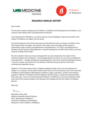
PCWNORMAL VALUES:Cardiac Index 3-5, PVR 2Common Post-Cath Complications 1% risk of major complications (death, myocardial infarction, stroke) Inducible arrhythmias: atrial tachyarrhythmias, ventricular tachycardia, ventricular fibrillation,bradycardia, AV conduction abnormalities Perforation of the heart or great vessels Allergic reaction to local anesthetic or iodinated contrast Valvar damage, particularly during endomyocardial biopsies Radiation exposure Infection Local vascular complications (most common): acute thrombosis, dissection, hemorrhage,hematoma, arteriovenous fistula, pseudoaneurysm12
Questions: What information is obtained during a pre-Glenn cath? What information is obtained during a cath in a patient with Tetralogy of Fallot, PulmonaryAtresia and MAPCAs?Case A: 3 month old patient, intubated in 21% FiO2. Patient’s hemoglobin in 14.7g/dL. Assume theoxygen consumption is 150mL/min and pulmonary venous saturations are 100%. Calculate the QP.o Calculate QS.o What is the PVR?o What is the likely lesion and why?SiteSVCRARVPAPVLVAoPressures (mm Hg)mean 480/320/8, mean 12mean 580/582/45Oxygen Saturation60%62%63%63%96%75%75%Case B: 1 day old patient, intubated in 50% FiO2. O2 capacity is 200mL. Assume the oxygenconsumption is 120mL/min/m2 and pulmonary venous saturations are 100%.o Calculate the QP.o Calculate QS.o What is the QP:QS?o What is the likely lesion and why?SiteSVCRARVPAPVLVAoPressures (mm Hg)mean 675/560/40, mean 46mean 560/675/55, mean 62Oxygen Saturation40%62%65%92%100%96%65%Oxygen Content (mL/L)80124130184200192130ReferencesTaggart NW, Cabalka AK. Cardiac Catheterization and Angiography. In: Allen HD, Driscoll DJ, Shaddy RE,Feltes TF, 8th, editors. Moss and Adams’ Heart Disease in Infants, Children, and Adolescents: Includingthe Fetus and Young Adult. Philadelphia: Lippincott Williams & Wilkins; 2013. p.258-287.Carrozza JP. Complications of diagnostic cardiac catheterization. Uptodate.com13
14
15
16
17
18
19
20
21
22
23
24
MurmursSee Cardiac History & Physical Exam for introductory discussion of murmurs.Innocent Murmurs of ChildhoodAgeStill’sPulmonaryflow murmurPeripheralpulmonicstenosisVenous ortic systolicmurmurMammaryArtery Soufflé2-6 years,may beaudiblefrominfancy toadulthoodAll ages0-6months 3-8yearsTiming &ConfigurationEarly -3Low tomediumVibratory“twang” or“musical”LLSB, extendsto apexVentricular falsetendons2nd and 3rdintercostalspacesAudible flowacrosspulmonaryoutflow tractAcute take offof the branchPAs in neonates when supineEarly- eginning inmid-systoleGrade2-3Continuousmurmur, in diastoleGrade1-3Grade1-2Rough,dissonantLow tomedium when supineWhining,roaring scendoGrade1-3Low womenEjectionGrade1-3Low tomediumSystolicmurmur,extends intodiastoleGrade1-3High when supine with headturned awayfrom examiner withcompression ofjugular veinDisappearswithhyperextensionof shouldersVaries fromday to dayLUSB,radiates tobilateralaxillae andbackLow anteriorneck, extendstoinfraclaviculararea, R L withrespiratoryinfectionsTurbulence atconfluence ofjugular andsubclavian veinsas they enterSVC, orangulation ofIJV as it coursesover transverseprocess of atlasAboveclavicles,radiates toneckMajorbrachiocephalicvessels arisingfrom aortaRUSB with anxiety,anemia,hyperthyroidismor fearBlood flow inarteries andveins leading toand frombreastsAnteriorchest wallover breast25
Evaluation of a Murmur Take a good history, including symptoms with exercise and family history“The Seven S’s” of Innocent Murmurs Sensitive – varying intensity with changes in posture and respiration, loudest when supine Short duration – not holosystolic Single – no associated clicks or gallops Small – limited to a small area, non-radiating Soft – low amplitude Sweet – not harsh Systolic – occurs only during in systole26
Red Flags History suggestive of cardiac disease Presence of a holosystolic or diastolic murmur Grade 3 or higher murmur Abnormal S2 or audible click Systolic murmur that intensifies with standing Neonates and very young infants (murmurs are more likely to represent congenital heartdisease, though young infants can have innocent murmurs too!)Questions When should you refer to a patient with a murmur to Cardiology? How can you distinguish a pulmonary flow murmur from the murmur caused by an ASD?Or from the murmur caused by pulmonary valve stenosis?References:Cassidy SC, Allen HD, Phillips JR. History and Physical Examination. In: Allen HD, Driscoll DJ, Shaddy RE,Feltes TF, 8th, editors. Moss and Adams’ Heart Disease in Infants, Children, and Adolescents: Includingthe Fetus and Young Adult. Philadelphia: Lippincott Williams & Wilkins; 2013. p.91-92.Frank JE, Jacobe KM. Evaluation and management of heart murmurs in children. Am Fam Physician2011; 84(7):793-800.Pelech A. Evaluation of the Pediatric Patient with a Cardiac Murmur. Pediatr Clin North Am 1999;46(2):167-88.27
Chest PainChest pain is generally a benign condition in pediatrics (94-99% of the time!)It is a common chief complaint and there is a large differential diagnosis (see flow chart). Idiopathic andchest wall pain are the most common etiologies, followed by GI pain (e.g. GERD) and asthma.Cardiac Causes: usually from compromised coronary supply and/or a mismatch of oxygen supply anddemand to cardiac tissue Left ventricular outflow tract obstructiono Hypertrophic cardiomyopathyo Aortic stenosiso Coarctation of the aorta Coronary artery abnormalitieso Abnormal coronary originso Sequelae of Kawasaki Diseaseo Slit-like coronary orifice or acute angle of take-off of the coronary arteryo Myocardial bridge (muscle covering part of coronary vessel) Classic angina in patients with severe hyperlipidemia or a history of Kawasaki Disease Variant angina (coronary vasospasm) after recreational drug use (cocaine, amphetamines, bathsalts, marijuana, synthetic cannabinoids) Pericarditis Myocarditis Dilated cardiomyopathy Tachyarrhythmias Aortic root dissection Ruptured sinus of Valsalva aneurysm Pulmonary HypertensionCardiac Chest Pain Red Flags Patient describes chest pain that is “deep,” “crushing,” or “substernal” with radiation to theneck, shoulder or arm. Chest pain associated with vomiting, diaphoresis, change in mental status, dizziness, dyspnea Chest pain with exertion Decreased exercise tolerance Exertional syncope or dizziness Palpitations Heart failure symptoms History of congenital heart disease, heart transplant, Kawasaki disease, substance abuse Family history of cardiomyopathy, cardiac arrhythmia, sudden death in parents or siblingsbefore the age of 50, connective tissue disorders, or hypercoagulable statesDiagnostic Work-up History, history, history (and family history!) Vital signs EKG Consider a chest X-ray Consider a trial of NSAIDs if musculoskeletal cause is suspected Cardiology consultation for echo or exercise stress testing28
Questions When should a patient be referred to Cardiology clinic? To the ED? When should a patient be restricted from exercise? For each of the cardiac causes listed above, make a list of other signs or symptoms suggestive ofthe diagnosis.ReferencesJohnson JN, Driscoll DJ. Chest Pain in Children and Adolescents. In: Allen HD, Driscoll DJ, Shaddy RE,Feltes TF, 8th, editors. Moss and Adams’ Heart Disease in Infants, Children, and Adolescents: Includingthe Fetus and Young Adult. Philadelphia: Lippincott Williams & Wilkins; 2013. p.1509-1513.Geggel RL, Endom EE. Causes of nontraumatic chest pain in children and adolescents.Uptodateonline.comGeggel RL, Endom EE. Nontraumatic chest pain in children and adolescents: Approach and initialmanagement. Uptodateonline.comKocis KC. Chest Pain in Pediatrics. Pediatric Clinics of North America. 1999; 46(2): 189-203.29
Syncope 95% of all syncopal episodes in otherwise healthy adolescents are of benign etiologyNon-Cardiac Causes Neurocardiogenic syncope Breath holding spells POTS Seizures PsychogenicCardiac Causes Arrhythmiao Long QT Syndromeo Heart blocko SVTo VT Structural cardiac disease (left ventricular outflow tract obstruction) Vascular diseaseRed Flags for Cardiac Disease No prodrome (dizziness, nausea, pallor) prior to loss of consciousness Syncope in response to loud noise, surprise or extreme emotional stress Syncope during exercise Syncope while supine Family history of sudden death in a close family member before age 30 LVH on the EKGEvaluation History, history, history! Vitals, including testing orthostatics EKG Consider a cardiac event monitor Consider a neurologic work-up with an EEGA Word on Postural Orthostatic Tachycardia Syndrome (POTS) Increase in heart rate by more than 30bpm within 10 minutes, or overall heart rate increase toabove 120bpm in adultso Children may have hypotension, but adults typically do noto Exercise intoleranceo Lightheadednesso Palpitations Typically seen in patients aged 15-50 years of age, 75-80% are female Etiologyo Possibly related to central hypovolemia with excessively decreased venous return tothe heart while upright, an abnormal inflammatory response or autonomic systemdysfunctiono Often symptoms begin after a viral illness, pregnancy, major surgery or trauma30
o Association between POTS and joint hypermobility (such as Ehlers-Danlos Syndrome)Treatmento Increase water intake to 8-10 16oz glasses of water per dayo Increase sodium intake to 200mEq per dayo Cross the legs and fold the arms, especially while standing, to maintain blood pressureo Increase aerobic exercise and resistance trainingo Consider drug therapy: fludrocortisone, alpha agonists, beta blockers, SSRIsQuestions What questions should you ask when evaluating a patient with syncope? When would you refer a patient with syncope to the ED? How would you test for orthostasis in the clinic or ED? How would you follow a patient with syncope as an outpatient? When would you refer toCardiology?ReferencesAckerman MJ. Cardiac Channelopathies, Syncope and Sudden Death. In: Allen HD, Driscoll DJ, Shaddy RE,Feltes TF, 8th, editors. Moss and Adams’ Heart Disease in Infants, Children, and Adolescents: Includingthe Fetus and Young Adult. Philadelphia: Lippincott Williams & Wilkins; 2013. p.397-398.Lewis DA, Dhala A. Syncope in the pediatric patient. Pediatric Clinics North America 1999; 46(2):205-219.McLeod KA. Syncope in Childhood. Arch Dis Child 2003; 88:350-353.Medow MS, Stewart J. The Postural Tachycardia Syndrome. Cardiology in Review 2007; 15(2):67-75.31
Preventive CardiologyRisk factors associated with atherosclerotic changes & coronary artery disease: Overweight (BMI 85th percentile for age and gender) or obese (BMI 95th percentile) Hypertension: BP consistently 95th percentile for age, height and gender Dyslipidemia: higher levels of total serum and LDL, lower levels of HDL, higher levels oftriglycerideso Look for xanthomas (firm, fatty deposits) under the skino Consider inherited disorders of LDL metabolism (Familial Hypercholesterolemia, FamilialCombined Hyperlipidemia, Hyperapobetalipoproteinemia, PolygenicHypercholesterolemia, Small Dense LDL) Cigarette smoke exposure, including second-hand smoke Physical inactivity Obstructive Sleep Apnea Positive family historyo Heart attack, treated angina, interventions for coronary artery disease, stroke, orsudden cardiac disease in a male parent or sibling before age 55, or a female parent orsibling before age 65High risk conditions associated with coronary artery disease before age 30: Diabetes (type 1 or 2) Chronic kidney disease Heart transplant recipients Kawasaki disease with persistent coronary aneurysmsModerate risk conditions associated with evidence of accelerated atherosclerosis before age 30: Kawasaki disease with regressed coronary aneurysms Chronic inflammatory disease HIV infection Nephrotic syndrome Major depressive disorder and bipolar disorderRoutine screening recommended at health supervision visits by the AAP, the AHA and the NHLBI: Obtain history of diet, physical activity and possible smoke exposure Review the family history for premature coronary artery disease Identify conditions associated with higher risk of early atherosclerosis Measure blood pressure for every well-child visit over 3 years of ageo Use tables for age, height and gender to establish patient’s percentileo Confirm appropriate size blood pressure cuffo Normal: 90th percentileo Prehypertension: 90th percentile or 120/80 AND 95th percentileo Stage 1 Hypertension: 95th percentile to 99th percentile 5mm Hgo Stage 2 Hypertension: 99th percentile 5mm Hg Measure height and weight and calculate BMI32
Perform lipid screening at ages 9-11 AND ages 17-21 or every 3 years in children with a familyhistory of early cardiovascular disease or a parent with high cholesterolObtain a fasting blood sugar every 2 years in patients age 10 or older with a BMI above the 85thpercentile and 2 additional risk factors for diabetes (family history of type 2 diabetes, ethnicitywith a high risk for diabetes, signs of insulin resistance, or conditions with a high risk fordiabetes such as Polycystic Ovary Syndrome)Questions Define the categories of hypertension and review proper technique for diagnosing hypertension What other studies or physical exam maneuvers should the pediatrician do to evaluate forsecondary hypertension? When should you refer to cardiology?ReferencesDe Ferranti SD, Newburger JW. “Risk factors and development of atherosclerosis in childhood.”UptoDateOnline.comDe Ferranti SD, Newburger JW. “Pediatric prevention of adult cardiovascular disease: promoting ahealthy lifestyle and identifying at-risk children.” UptoDateOnline.comDe Ferranti SD, Newburger JW. “Overvie
Pelech A. Evaluation of the Pediatric Patient with a Cardiac Murmur. Pediatr Clin North Am 1999; 46(2):167-88. Pelech A. Physiology of Cardiac Auscultation. Pediatr Clin North Am 2004; 51(6):1515-35, vii-viii. Gersh BJ. “Auscultation of heart sounds.” UpToDateonline.com . Further