Transcription
The Malaysian Journal of Analytical Sciences, Vol 15 No 1 (2011): 54 - 60A SIMPLE AND COST EFFECTIVE ISOLATION AND PURIFICATIONPROTOCOL OF MITRAGYNINE FROM MITRAGYNA SPECIOSA KORTH(KETUM) LEAVES(Satu Protokol Pengasingan Mitragina daripada Daun Mitragyna speciosa Korth (Ketum) yangMudah dan Kos Efektif)Goh Teik Beng1* , Mohamad Razak Hamdan1, Mohammad Jamshed Siddiqui2, Mohd Nizam Mordi1and Sharif Mahsufi Mansor11Centre of Drug Research,School of Pharmaceutical Science,Universiti Sains Malaysia, Minden 11800, Penang, Malaysia.2*Corresponding author: gtb.dd04@student.usm.myAbstractThe objective of the present study was to develop a simple and cost effective method for the isolation of mitragynine fromMitragyna speciosa Korth leaves. The results of our study showed that around 0.088 % (w/w) pure mitragynine was obtained bythis newly developed method with a purity of 99.0 % (w/w) based on GC-MS analysis. Moreover, the recovery of the puremitragynine from the chloroform extract was also more than 95.0 %. Advantages of this new method were simpler, faster andmore economical. In conclusion this simple and cost effective method helps to isolate mitragynine with higher purity incomparison with the published methods.Keywords: Mitragynine speciosa Korth, mitragynine, mitragynine extractionAbstrakObjektif kajian ini adalah untuk membangunkan satu kaedah yang mudah dan kos efektif untuk pengasingan mitragininadaripada daun Mitragyna speciosa Korth. Keputusan kajian menunjukkan bahawa lebih kurang 0.088 % (w/w) mitraginina tulentelah diperolehi dengan menggunakan kaedah baru yang dibangunkan ini dengan ketulenan 99.0 % (w/w) berdasarkan analysisGC-MS. Malahan, hasil pemerolehan semula mitraginina tulen daripada ekstrak kloroform juga melebihi 95.0 %. Faedahkaedah baru ini adalah lebih mudah, cepat dan ekonomikal. Kesimpulannya, kaedah yang lebih mudah dan kos efektif inimembantu mengasingkan mitraginina dengan ketulenan yang lebih tinggi berbanding dengan kaedah yang telah pun diterbitkan.Kata kunci: Mitragynine speciosa Korth, mitragina, pengekstrakan mitraginaIntroductionMitragynine is the main alkaloid present in Mitragyna speciosa leaf with opioid agonistic activity [1,2,3,4]. Themitragynine content in M. speciosa leaves varied from place to place depending on the geographical location andseason [4]. The mitragynine content of Thai M. speciosa was 66% of the total alkaloids while the Malaysian speciescontained only 12% of the total alkaloids [5]. This necessitates finding a simple, fast and reproducible method toobtain mitragynine from Malaysian species of M. speciosa.Currently, several methods are available for the extraction and isolation of mitragynine from Mitragyna speciosaleaf. These extraction and isolation methods published have their own advantages and disadvantages. Most of thesemethods commonly employs soxhlet extraction method using methanol as solvent to obtain the crude extract of M.speciosa leaves [6,7,8]. However, these methods were very slow and require days to obtain crude extract of M.Speciosa. The extract obtained by these methods were sticky, agglutinated and might need sonication to facilitatethe extraction of mitragynine from the crude extracts. Moreover, the process required to obtain pure mitragynine54
Goh et al: A SIMPLE AND COST EFFECTIVE ISOLATION AND PURIFICATION PROTOCOL OFMITRAGYNINE FROM MITRAGYNA SPECIOSA KORTH (KETUM) LEAVESfrom the crude extract was too lengthy and laborious which involves acidification, neutralisation followed by liquidliquid extraction and finally column chromatography with a series of mobile phase with increasing polarity [6,7,8].Hence, our main objective of this study is to find a simple and rapid method with maximum output of puremitragynine from dried leaves of M. speciosa as it is not commercially available at cheaper price.ExperimentalChemicals and solventsPicric acid, potassium bromide (KBr) and acetanilide were obtained from Sigma-Aldrich, St. Louis, USA. Puremitragynine standard was obtained from IMR (Institute of Medical Research, Kuala Lumpur, Malaysia).Commercial pure mitragynine standard (ASB-00013890-025) was obtained from Chromadex, Irvin, USA. Solventshexane, petroleum ether, methanol, acetone, chloroform, ethyl acetate and HCL used were of analytical gradepurchased from Fischer Scientific, Loughborough, U.K.Plant material identificationLeaves of M. speciosa were collected from Perlis, Malaysia and were identified by a botanist in the School ofBiological Science, Universiti Sains Malaysia (USM). A voucher specimen (No 11074) was deposited at theHerbarium of the School of Biological Science, USM for further references.Extraction and isolation of mitragynineFresh Mitragyna speciosa Korth leaves (1.0 kg) were washed with water to remove dirt and adhering material, ovendried at 45-50 C for three days and milled into fine powder using a blender to get the dry powder (297.0 g).Soxhlet extraction was carried out using 297.0 g powdered leaves with 4 L petroleum ether (40-60 oC) for 8 hour,then petroleum ether solution was discarded and the defatted powdered leaves were dried and the extraction wasrepeated with 4 L chloroform for 8 hour. The chloroform solution obtained was filtered, concentrated, evaporated todryness under the reduced pressure using rotary evaporator and was kept in a refrigerator (-200C). The dried crudechloroform extract was subjected for flash chromatography according to the method of Still et al.[10]. Crudefraction containing mitragynine was obtained by eluting with hexane and ethyl acetate (80:20 v/v) and this fraction(100 mL) was subjected to liquid-liquid fractionation using petroleum ether (100 mL X 3 times) for furtherpurification. The petroleum ether layer was discarded and the remaining solution was concentrated under thereduced pressure using a Buchi R215 Rotavapor (Flawil, Switzerland) to obtain crude mitragynine.Purification of mitragynine by crystallizationApproximately 0.80 g of the isolated crude mitragynine was dissolved in minimum quantity of methanol and thissolution was added to a 5 mL saturated methanolic picric acid solution. Side of test tube was scratched and thissolution was kept in refrigerator for 20 minutes to obtain orange colored mitragynine picrate crystals. These crystalswere filtered, washed with methanol followed by acetone and the melting point of these crystals was determined.The mitragynine picrate crystals were converted to mitragynine free base by dissolving them in hot saturatedacetone solution and by adding this solution to an excess of dilute ammonia solution lead to the liberation of freebase which was extracted with diethyl ether. This solution was dried to obtain a pale brown, amorphous compound.This compound along with IMR pure standard was subjected for TLC for the comparison of hR f value.Qualitative characterisation of mitragynine free baseVarious analytical methods such as UV-Visible, IR, NMR, mass spectroscopy, CHN analysis, HPTLC, meltingpoint and determination of dissociation constant were carried out to characterize the isolated pure mitragynine freebase. UV-Visible spectroscopy was carried out using Shimadzu UV 160-A double beam spectrophotometer (Kyoto,Japan), the UV spectra of 0.015 ppm pure mitragynine in methanol was obtained against a blank containingmethanol. FTIR spectra were recorded using KBr pellets by a Thermal Scientific Nicolet 6700 spectrophotometerwith Omnic software (Franklin, USA). 1H-NMR and 13C –NMR were performed using 400 MHz and 100 MHzBruker spectrometer (Karlsruhe, Germany), respectively. GC-MS spectra were taken using HP 6890A GC system(Santa Clara, USA) with HP-5MS Polyimide coated capillary column (30 m x 0.25 mm i.d. x 0.1 μm), heated from100oC to 280oC at 10oC / min, 10 Kpa helium at 1.00 mL/min flow rate and an injection volume of I μL with splitratio of 5 : 1. CHN elemental analysis was conducted using Perkin Elmer CHN Analyzer Model 2400-2(Massachusetts, USA). HPTLC study was performed on a 0.50 mm thick Silica gel 60 G 254 (10 cm x 20 cm) using a55
The Malaysian Journal of Analytical Sciences, Vol 15 No 1 (2011): 54 - 60mobile phase hexane:ethyl acetate (80:20 v/v). TLC plates were then visualized under UV light (254 and 366 nm).Melting point was measured by Koffler Block Electrothermal Melting Point (London, UK) with Acetanilide (113114oC) as calibration standard. pKa of isolated and purified mitragynine free base was determined by titrationmethod using 0.0001 N HCL with Cyberlab 14 L pH meter (Millbury, USA) in pH modes. In this procedure, 50 mgpure mitragynine in 30 mL methanol was used. The long term stability of the stock solution of mitragynine (1000ng mL-1) refrigerated at -20 oC was tested after storage period of 60 days. The stock was diluted to 1 ng mL -1 withpure methanol in three replicates prior to the analysis. The result was expressed as the difference from time t 0day. The percentage difference from time t 0 day not exceeded 15% indicating that the stock solution ofmitragynine was stable [11].Statistical analysisResults were expressed as the mean S.E.M of three independent experiments. Student’s t-test was used forstatistical analysis; P values 0.05 were considered to be significant [12]. Statistical calculations were carried outwith the Minitab 15.0 English for windows software package.Results and DiscussionExtraction is an important step involved in the isolation of bioactive compounds from the plants with medicinalvalue. In our study we followed simple soxhlet extraction method to defat Mitragyna speciosa Korth leaves usingpetroleum ether followed by extraction with chloroform which yielded 7.22 g of crude chloroform extract. It wasevident from Table 1 that chloroform extract obtained was pure and rich in mitragynine content (70.0 %) than themethanol extract of the first stage from the published method [6,7,8] with a mitragynine content of only 19.0%.Approximately 0.80 g of crude mitragynine was obtained by flash chromatography of the crude extract followed byliquid-liquid fractionation and the mitragynine in this extract appeared as one single peak in GC-MS with relativepurity of 98.0 %. This newly developed method even employed only one step flash chromatography to obtain 98.0% pure mitragynine (Table. 1) whereas the published methods followed a series of mobile phase to obtain the sameresult [7]. Thus, this newly developed method required less solvent, incurred less steps and more economical.Table 1: Comparison of GC-MS purity of mitragynine extracts at various stage between our methodand published methodsStage ofExtractionMethanolAcid rmFirst columnSecond columnPetroleum etherwashingCrystallizationGC-MS Purity of MitragynineUsing Our MethodUsing Published Method [6, 7, 8]*19.070.092.098.099.0*43.0*76.096.0-The data is the percentage area of GC-MS based on ca.300.0 g M. speciosa dried leaves at each stage* Results obtained in our lab using published method [6,7,8]Results of crystallisation showed that orange colored crystals of mitragynine picrate salt (ca. 0.60 g.) obtained fromcrude mitragynine have a melting point of 220-225 C. Precipitation of these orange colored crystals leads to theformation of a pale brown, amorphous mass of mitragynine free base (ca. 0.26 g). It was very soluble in methanoland gives a very strongly fluorescent solution. The result of TLC study shown that the hR f (81.9 cm) value of theisolated and purified free base of mitragynine was in accordance with that of IMR pure mitragynine standard56
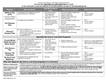
Goh et al: A SIMPLE AND COST EFFECTIVE ISOLATION AND PURIFICATION PROTOCOL OFMITRAGYNINE FROM MITRAGYNA SPECIOSA KORTH (KETUM) LEAVES(Table 5). Moreover, the purity of the mitragynine obtained by this newly developed method was approximately99.0 %. In addition, the purified mitragynine showed a single chromatographic peak upon spectroscopic analysis byGC-MS. The spectrum of purified mitragynine (C23H30N2O4, exact molecular mass 398.2207) was confirmed withthe IMR pure standard and to the NIST 98 mass spectral library. Mitragynine obtained from this newly developedmethod was 99% pure, when peak area of mitragynine was compared to other minor impurities in the GC-MSanalysis (Table 2), and was used as the analytical standard to build the calibration curve (Figure 1) for quantitativeanalysis.Figure 1: Pure mitragynine calibration curveThe pure mitragynine standard calibration curve was linear with the equation of y 105.7x – 8.2 over the range of0.5-50 ng (n 5, r2 0.999 0.002) (Figure 1).The mean coefficients of variation for within day assay precision was2.33 0.25 % at a concentration range of 0.5-50 ng. For the day to day variation, the mean coefficient of variationwas 4.90 0.60 % for the same concentration range. The detection limit (LOD) and quantification limit (LOQ) ofassay for mitragynine was 0.50 ng and 2.00 ng respectively. The concentrations giving signal-to-noise ratios of 3:1and 10:1 were considered to be the LOD and the LOQ respectively.It is clear from Table 2 that the pure mitragynine obtained (99.0 %) by this newly developed method was stable forsix months and its strength was 100 % comparable with that of IMR pure mitragynine standard (Table. 2). Therecovery of pure mitragynine from chloroform extract of M. speciosa leaves from Perlis, Malaysia by this methodwas more than 95.0 % at each stage (Table. 3).57
The Malaysian Journal of Analytical Sciences, Vol 15 No 1 (2011): 54 - 60Table 2: Comparison of purity and stability of mitragynine obtained by our method and standard mitragynine(Chromadex, Irvin, USA)Characteristic ofPure MitragyninePurity (%)*Purity strength (%)Stability, 1000 ngmL-1Difference from t 0 (ngmL-1),after 6 months at -20oCIsolatedmitragynine99.0100.00.40Standard mitragynine96.098.00.30*Purity strength (%) was in comparison with IMR standardTable 3: Recovery (%) of mitragynine from chloroform extract of M. speciosa leaves from Perlis, Malaysiaby our method.Type of extractActual yield (g)Theoretical yield (g)ChloroformColumnchromatographyPetroleum etherWashingCrystalization7.22 0.331.54 0.091.55Recovery from chloroform extract(%)99.35 0.100.78 0.060.8097.50 0.120.26 0.010.2796.29 0.15Values are the means of three replicates standard error of the mean (S.E.M.).From Table 3, it was confirmed that our method is feasible to isolate pure mitragynine. The pure mitragynine (99.0%) yield obtained by this method was 0.26 0.01 g from ca.300.0 g of M. speciosa dry leaves (Table. 4) and thecontent of mitragynine we obtained from the M. speciosa leaves of Perlis, Malaysia was 0.088 % w/w (0.88mg/g of dried leaves). This was within the range of 0.08-0.10% as reported in the literature for leaves extracts of M.speciosa [6, 9].Table 4: Yields (%) and GC-MS purity (%) of mitragynine obtained from M. speciosa leaves of Perlisby our method at each stage.Stage ofextractionChloroformColumnChromatographyHexane washingCrystallizationActual yield (g)Actual yield (%)GC-MS Purity (%)7.22 0.33a1.54 0.09b2.600.5670.0 0.20e92.0 0.54f0.78 0.06c0.26 0.01d0.260.08898.0 0.32g99.0 0.25hValues are the means of three replicates standard error of the mean (S.E.M). The different alphabet of a,b,c and d represents thevalues different significantly for the yield obtained whereas the different alphabet e,f,g and h represents the values differentsignificantly for the GC-MS purity at P 0.05 based on Student t-Test Minitab (Illinois USA) (a,bP 0.05 and e,fP 0.05 incomparison between chloroform extraction and column chromatography, b,cP 0.05 and f,gP 0.05 in comparison between columnchromatography and hexane washing, c,dP 0.05 and g,hP 0.05 in comparison between hexane washing and crystallization forboth yields and GC-MS purity respectively.58
Goh et al: A SIMPLE AND COST EFFECTIVE ISOLATION AND PURIFICATION PROTOCOL OFMITRAGYNINE FROM MITRAGYNA SPECIOSA KORTH (KETUM) LEAVESThe details about the HPTLC results of isolated and pure mitragynine are given in Table 5. The hR f of isolatedmitragynine (81.9) was comparable with the hRf of IMR standard (82.0) which shows that the isolated compound ismitragynine. Figure 2a, 2b shows the HPTLC profile of fractions collected (1-8) from flash chromatography as wellas IMR mitragynine standard under UV 365 and 254 nm. It is noted from the Figure 2a and 2b that fractions 1-3does not contain mitragynine whereas fractions 4-8 containing mitragynine. The hRf of mitragynine (81.6) present inthe fractions 4-8 were in comparison with the hRf of IMR standard (82.0).Table 5: HPTLC fingerprint of isolated mitragynine and the standard mitragynine (IMR)ParametersQuantity sample appliedDetectorStationary PhaseMobile PhaseApplied Concentration, mg/mlElution Length, cmhRf, cmVisual appearance of Band :White lightUV 365UV 254IodineDragendorff reagentTLC conditions / resultsIsolated mitragynineStandard mitragynine (IMR)2 ul2 ul12 W VL. 6.1C UV detector12 W VL. 6.1C UV detector6 W : 254 nm6 W : 254 nm6 W : 365 nm6 W : 365 nmSilica Gel 60G254Silica Gel 60G254Hexane : Ethyl acetate (80 : 20)Hexane : Ethyl acetate (80 : 20)0.030.038.0-10.08.0-10.081.982.0Reddish brownGreenish brownBrownishYellowish brownOrangeReddish brownGreenish brownBrownishYellowish brownOrangeFigures 2a and 2b: HPTLC profile of different fractions (1-8) of flash chromatography and standard mitragynine (a)under UV 365 and UV 254 nm.59
The Malaysian Journal of Analytical Sciences, Vol 15 No 1 (2011): 54 - 60Mitragynine free base: Yellow amorphous solid. m.p.: 103-1050C. pKa 5.5. UV/Vis λmax (MeOH) nm (log ε):225 (4.76),290 (3.94). IR (KBr): 1567.2, 1704.5, 3439.3 cm-1. hRf : 0.82 (C6H14: EtOOCMe, 80:20). GCMS (EI, 70eV): m/z (%) 398 [M ] (92), 214. (100),383 (44), 200 (26). CHN : Calcd for C 23H30N2O4: C, 69.35; H, 7.54;N,7.04. Found C, 69.35; H , 7.50; N, 7.00. 1H NMR (400 MHz, MeOD): δ 7.68 (1H, br-s, N-H), 7.53 (1H, s, H-17),6.91 (1H, dd, J 7.9, 7.9 Hz, H-11), 6.89 (1H, d, J 7.9 Hz, H-12), 6.41 (1H, d,J 7.9, H-10), 3.84 (3H, s, 9-OCH3),3.78 (3H, s, 17-OCH3), 3.68 (3H, s, 22-OCH3), 3.15 (1H, br-d, J 9.8 Hz, H3), 3.07 (1H, m, H-6), 3.04-3.07 (2H,overlapped, H-15, H-21), 2.90-3.00 (2H, overlapped, H-5 and H-6), 2.40-2.55 (3H, overlapped, H-5, H-14, H-21),1.75-1.80 (2H, overlapped, H-14, H-19), 1.64(1H, br-d,J 11.3, H-20), 1.30(1H, m, H-19), 0.87 (3H, dd, H-18).13CNMR (100 MHz, MeOD): 169.36 (C-22), 160.65 (C-17), 154.49 (C-9), 137.33 (C-13), 133.69 (C-2), 121.91 (C-11),117.72 (C-8), 111.55 (C-16), 107.91 (C-7), 104.26 (C-10), 99.80 (C-12), 61.65 (17-OCH3), 61.20 (C-3), 57.77 (C21), 53.85 (C-5), 51.45 (22-COOCH3),55.40 (9-OCH3), 39.95 (C-20), 31.99 (C-15), 29.78 (C-14), 22.78 (C-6),14.22 (C-19), 12.95 (C-18).ConclusionIn conclusion, we have developed an alternative method to isolate and purify the mitragynine from M. speciosaleaves. The main advantage of this method was simple, fast and cost effective with high reproducibility for theextraction of mitragynine. Morover this method give improved yield of mitragynine (0.088 % w/w) with purity of99.0 % based on GCMS analysis which was better than the purity of the mitragynine obtained from the otherpublished methods.AcknowledgementsThis work was supported by USM Research University Grant and R&D Initiative Fund, Ministry of Science,Technology and Innovation, Malaysia (MOSTI).ReferencesTakayama, H., Ishikawa, H., Kurihara, M., Kitajima, M., Aimi, N., Ponglux, D., Koyama, F., Matsumoto, K.,Moriyama,T., Yamamoto, L.T., Watanabe, K., Horie, K. 2002. Studies on the synthesis and opioid agonisticactivities ofmitragynine-related índole alkaloids: Discovery of opioid agonists structurally different from otheropioid ligands.Journal of Medicinal Chemistry, 45: 1949-1956.2. Grewal, K.S. 1932. Observations on the pharmacology of mitragynine. Journal of Pharmacology andExperimental Therapeutics, 46: 251-271.3. Suwanlert, L.S. 1975. A study of Kratom eaters in Thailand. Bulletine on Narcotics, 27:
methanol. FTIR spectra were recorded using KBr pellets by a Thermal Scientific Nicolet 6700 spectrophotometer with Omnic software (Franklin, USA). 1H-NMR and 13C –NMR were performed using 400 MHz and 100 MHz Bruker spectrometer (Karlsruhe, Germany), respec