Transcription
DISSERTATIONTitel der DissertationVAF347, a novel low molecular weight immunomodulator, inhibitsInterleukin-6 production in the human monocytic cell line Monomac1 viaactivation of the aryl hydrocarbon receptorangestrebter akademischer GradDoktor der Naturwissenschaften (Dr. rer. nat.)Verfasser:Mag. Hermann NovakMatrikel-Nummer:9920080Dissertationsgebiet (lt.Studienblatt):Genetik - MikrobiologieBetreuerin:Ao. Univ.-Prof. Dipl.-Biol. Dr. Angela WitteWien, am 20. September 2008
Für ihre Hilfe und persönliche Unterstützung möchte ich mich besondersbei folgenden Personen bedanken:Den Supervisoren:Univ.-Doz. Dr. Maximilian WoisetschlägerAo. Univ.-Prof. Dipl. –Biol. Dr. Angela WitteDer Labormannschaft:Ing. Nathalie HarrerMag. Petra GruberWolfgang NerudaMitarbeitern anderer Labors:Dr. Nicole UrtzEdith PurschRobert CsongaMonique Bongersund nicht zuletzt:meiner Familie, vor allem NoraFranziska WohlkönigBernhard Mayer2
Table of ContentsTable of ContentsAbstract7Kurzfassung8Abbreviations91. Introduction101.1 The immune system: cytokines and their function101.1.1 The discovery of Interleukin-6101.1.2 Structure and expression of Interleukin-6101.1.3 The Interleukin-6 promoter111.1.4 Cellular signaling and regulation of Interleukin-6131.1.5 Biological effects of Interleukin-6151.1.6 Interleukin-6 in rheumatoid arthritis151.2 Dendritic cells and their functions161.3 VAF347, a novel low molecular weight immunomodulator171.3.1 Identifying the molecular target(s) of VAF347181.4 The aryl hydrocarbon receptor (AhR)191.4.1 Function and structure of the AhR191.4.2 Ligands of AhR201.4.3 The AhR signaling pathway211.4.4 Molecular mechanisms of AhR functions in the regulation of cytochrome23P450 genes1.4.5 AhR-null mice241.4.6 Roles of AhR in cell physiology241.5 Aim252. Materials and Methods262.1 Standard Methods262.1.1 Qiagen Plasmid Miniprep262.1.2 Qiagen Plasmid Maxiprep262.1.3 DNA-Concentration Measurement272.1.4 Agarose gel electrophoresis272.1.5 QIAquick Gel Extraction Kit272.2 Generation of templates usable for RT-PCR282.2.1 Absolutely RNA RT-PCR Miniprep Kit (Stratagene)282.2.2 RNA-Concentration Measurement293
Table of Contents2.2.3 iScript cDNA Synthesis292.3 Quantitative Reverse Transcription Polymerase Chain reaction (RT-PCR)292.4 Cloning strategy of promoter reporter constructs, expression and silencing vectors312.4.1 Cloning of an Interleukin-6 promoter reporter construct312.4.2.A. Amplification of the wild type Interleukin-6 Promoter312.4.2.B. Restriction digestion of PCR products312.4.2.C. Ligation of PCR products and vector322.4.2.D. Transformation322.4.3 Cloning of a retroviral trans-dominant negative AhR expression construct332.4.3.A. Amplification of trans-dominant negative AhR332.4.3.B. Restriction digestion of AhR515 PCR product and vector332.4.4. Cloning of a retroviral AhR gene silencing construct LMP-shAhR342.4.5. Cloning of a retroviral NF-IL6 expression construct342.4.6. Cloning of a retroviral NF-κB p65 expression construct352.4.7. Cloning of the lentiviral PU.1 gene silencing construct GIPZ-sh-PU.1352.5 ELISA for human Interleukin-6362.6 Western blot analysis362.6.1 Preparation of whole cell lysates362.6.2 Tris-Glycine gel electrophoresis, blotting to nitrocellulose membrane and blocking 372.6.3 Western Blot of the aryl hydrocarbon receptor372.6.4 Western Blot of STAT5 and STAT6372.6.5 Western Blot of the p44/p42 MAP kinases382.6.6 Western Blot of NF-IL6382.6.7 Western Blot of NF-κB p65382.6.8 Western Blot of PU.1392.6.9 Substrate for HRP and western blot developing392.7 Cells, cell culture conditions and inducers2.7.1 Thawing and growing of cells from liquid nitrogen392.7.2 Freezing of cells for liquid nitrogen storage392.7.3 Monomac1 cells402.7.4 PhoenixA cells402.7.5 PhoenixGP cells402.7.6 Cell inducers, compounds and inhibitors402.8 Induction of Monomac1 cells2.8.1 Induction of Monomac1 cells for RT-PCR and IL-6 ELISA4394141
Table of Contents2.8.2 Induction of Monomac1 cells for analysis of intracellular signaling events2.9 Transient transfection of MM1 using the Amaxa electroporation device2.9.1 Luciferase activity measurement2.10 Viral infection of Monomac1 cells424242432.10.1 Transfection of packaging cell lines with retroviral constructs432.10.2 Transfection of a packaging cell line with lentiviral constructs432.10.3 Retroviral infection of Momomac1 cells432.10.4 Lentiviral infection of Momomac1 cells442.11 Chromatin Immunoprecipitation442.11.1 Induction of Monomac1 cells442.11.2 Crosslinking and lysis442.11.3 Random DNA fragmentation452.11.4 Immunoprecipitation and washing452.11.5 Elution and Proteinase K digest462.11.6 Phenol/chloroform extraction and PCR462.12 Statistical analysis473. Results483.1 IL-4 GM-CSF induce Interleukin-6 expression in human Monomac1 cells483.2 VAF347 inhibits IL-4 GM-CSF induced IL-6 production in Monomac1 cells493.3 VAF347 induces CYP1a1 expression in Monomac1523.4 TCDD and omeprazole have a similar activity profile in MM1 compared to VAF347533.5 PMA induces IL-6 expression in Monomac1 cells563.6 Functional AhR is necessary to mediate the biological phenotype of VAF347583.6.1 A trans-dominant negative AhR protein abolishes VAF347 activity593.6.2 Silencing of the AhR gene abolishes VAF347 activity633.7 Signaling events in Monomac1 after cytokine stimulation643.7.1 IL-4 GM-CSF lead to activation of members of the JAK/STAT family653.7.2 IL-4 GM-CSF lead to activation of proteins of the MAP Kinase cascade673.7.3 Goe6979, a potent JAK-2 inhibitor, inhibits IL-6 expression in Monomac1713.7.4 NF-κB inhibitors do not alter IL-6 expression in Monomac1733.8 IL-6 promoter studies743.8.1 VAF347 inhibits the induction of an IL-6 promoter reporter construct743.8.2 VAF347 blocks the binding of NF-IL6 to the IL-6 promoter763.8.3 VAF347 does not inhibit the expression of IL-6 relevant transcription factors773.9 Interference of VAF347 with IL-6 relevant transcription factors785
Table of Contents3.9.1 Over-expression of NF-IL6 partially abolishes the inhibitory effect of VAF347793.9.2 Over-expression of NF-κB p65 does not alter the inhibitory effect of VAF347823.9.3 Silencing of the PU.1 gene abolishes the inhibitory effect of VAF347834. Discussion875. References94Curriculum Vitae114Lebenslauf1156
AbstractAbstractInterleukin (IL) -6 is a pleiotropic cytokine that is involved in the regulation of immune reactions,hematopoiesis, bone metabolism and inflammation. Deregulation of IL-6 has been shown to beinvolved in several inflammatory and autoimmune disorders, including rheumatoid arthritis,Castleman’s disease and Crohn’s disease. Therefore, interception of the IL-6 signaling pathwayrepresents an interesting therapeutic target for the treatment of these diseases.VAF347 is a novel low molecular weight immunomodulator which has been shown to selectivelyblock IgE synthesis in human B cells and to inhibit IL-6 production in human monocyte-deriveddendritic cells. This study was designed to identify the molecular target(s) of VAF347 that lead toinhibition of IL-6 synthesis. Similar to primary cells, VAF347 inhibits IL-4 and GM-CSF as well asPMA induced IL-6 production in the human monocytic cell line Monomac1. We demonstrate, thatthe aryl hydrocarbon receptor (AhR) is the functionally relevant target for inhibition of IL-6response by VAF347. The AhR is responsible for mediating the biological activity of VAF347 inMonomac1, since 1) other AhR ligands display an identical activity profile, 2) ectopic overexpression of a trans-dominant negative AhR protein abolishes VAF347 activity, and 3) silencing ofAhR gene expression renders Monomac1 resistant to VAF347.Furthermore, VAF347 inhibits the activation of an IL-6 promoter reporter gene construct and blocksthe binding of the transcription factor NF-IL6, which is thought to be one of the main factorsinvolved in IL-6 regulation, to the IL-6 promoter. Over-expression of wild type NF-IL6 partiallyabolishes VAF347 activity. These findings showed that VAF347 mediates its inhibitory effect at thetranscriptional level by inhibiting the interaction of NF-IL6 with its binding site on the IL-6promoter.Collectively, these data identify the AhR as the functional target of VAF347. Given the fact thatIL-6 deregulation is involved in the pathology of several inflammatory and autoimmune diseases,targeting the AhR may provide a new tool for the treatment of these diseases.7
KurzfassungKurzfassungInterleukin (IL) -6 ist ein pleiotropes Zytokin das sowohl an der Regulation von Immunreaktionen,der Hämatopoese, dem Metabolismus des Knochens als auch an Entzündungsreaktionen eimehrerenEntzündungs-undAutoimmunerkrankungen gefunden worden, wie etwa bei Gelenkrheumatismus, Morbus Castlemanoder Morbus Crohn. Deshalb stellt die Unterbrechung des IL-6 Signalweges ein interessantestherapeutisches Angriffsziel für die Behandlung dieser Krankheiten dar.Es wurde bereits gezeigt, dass VAF347 als ein neuer niedermolekularer Immunmodulator sowohlspezifisch die Immunglobulin-E Synthese von humanen B-Zellen, als auch die IL-6 Produktion vonhumanen Dendritischen Zellen monozytären Ursprungs inhibiert.Diese Studie wurde gestaltet, um das (die) molekulare(n) Ziel(e) von VAF347, das zur Inhibierungder IL-6 Produktion führt, zu identifizieren. Ähnlich wie bei primären Zellen inhibiert VAF347 dievon IL-4 und GM-CSF als auch die von PMA induzierte IL-6 Produktion in der humanenmonozytären Zelllinie Monomac1. Es wird gezeigt, dass der Aryl Hydrocarbon Rezeptor (AhR) dasfunktionell bedeutende Target für die Inhibition der IL-6 Produktion durch VAF347 ist. Der AhRist für die biologische Aktivität von VAF347 in Monomac1 Zellen verantwortlich, da 1) andereAhR Liganden ein ähnliches Aktivitätsprofil zeigen, 2) ektopische Überexpression einestransdominant negativen AhR Proteins die biologische Aktivität von VAF347 aufhebt und 3) dieStilllegung des AhR Gens Monomac1 Zellen resistent gegen VAF347 macht.Darüber hinaus inhibiert VAF347 die Expression eines IL-6 Promoter Reporter-Gen Konstrukts undblockiert die Bindung des Transkriptionsfaktors NF-IL6, welcher als einer der Hauptfaktoren fürdie Regulation der IL-6 Expression angesehen wird, an den IL-6 Promoter. Die Überexpression desWildtyp NF-IL6 Proteins hebt teilweise die Aktivität von VAF347 auf. Diese Ergebnissedemonstrieren, dass VAF347 seinen inhibitorischen Effekt auf transkriptioneller Ebene ausübt, dadie Interaktion von NF-IL6 mit dessen Bindungsstelle im IL-6 Promoter verhindert wurde.Zusammenfassend identifizieren diese Daten AhR als das funktionell entscheidende Target vonVAF347. Da die Deregulation von IL-6 am Krankheitsbild mehrerer inflammatorischer- undAutoimmunerkrankungen beteiligt ist, könnte das Targeting von AhR eine neue Möglichkeit zurBehandlung dieser Krankheiten darstellen.8
AbbreviationsAbbreviationsaaamino acid(s)AhRaryl hydrocarbon receptorAhRRaryl hydrocarbon receptor repressorARNTaryl hydrocarbon receptor nuclear translocatorAmpampicillinbpbase pair(s)ChIPchromatin immunoprecipitationCYP1A1cytochrome P450 1A1DC(s)dendritic cell(s)DNAdeoxyribonucleic aciddsdouble strandedEtOH96% EthanolGFPgreen fluorescent proteinGM-CSFgranulocyte macrophage colony-stimulating factorHRPhorse radish s)Janus Kinase(s)kDakilo DaltonMAPKmitogen-activated protein kinaseMM1Monomac1PBSphosphate buffered salinePKCprotein kinase CPMAphorbol 12-myristate 13-acetateRNAribonucleic acidRTroom temperaturesssingle strandedSTATsignal transducer and activator of inTH17Interleukin-17 producing T cellTregregulatory T cell9
Introduction1. Introduction1.1 The immune system: cytokines and their functionThe development of an effective immune response involves the interaction of multiple cell typesincluding myeloid cells and lymphocytes. The complex interactions among these cells are in partmediated by cytokines. Cytokines are relatively small regulatory proteins that are secreted fromcells of innate and adaptive immunity as well as various other cell types in response to a number ofstimuli. On one hand, a subgroup of these proteins stimulate the growth and differentiation ofvarious cells during immune homeostasis, while on the other hand, cytokines facilitate theactivation and differentiation of effector cells both during the innate and adaptive phases of immuneresponses against foreign antigens such as bacteria, viruses or self-antigens.Binding of cytokines to specific receptors on the membrane of target cells leads to the activation ofdiverse signal-transduction pathways which ultimately lead to altered gene expression.In recent years cytokines and their receptors have become important targets for specific antagonistsin various immune and inflammatory diseases. One of these cytokines that is considered as such atarget, due to its pro-inflammatory functions is the cytokine Interleukin-6 (IL-6). IL-6 is apleiotropic cytokine that is involved in the regulation of immune reactions, hematopoiesis, bonemetabolism and inflammatory state. The overproduction of IL-6 has been implicated in thepathology of several inflammatory and autoimmune disorders, including rheumatoid arthritis,Crohn’s disease, Castleman’s disease and systemic-onset juvenile idiopathic arthritis. Thereforeinterception of the IL-6 signaling pathway has become an interesting target for the treatment ofthese diseases.1.1.1 The discovery of Interleukin-6The history of nomenclature reflects the diverse biological functions of this cytokine: IL-6 wasinitially identified as a T cell-derived B-cell differentiation factor that induces B-cells todifferentiate into antibody-producing cells (1), and was therefore first termed B-cell stimulatoryfactor-2 (BSF-2). The complementary DNA of BSF-2 was cloned in 1986 (2). Following itsdiscovery, BSF-2 was found to be identical to the 26 kDa protein (3), an interferon-β2 protein (4), ahybridoma/plasmacytoma growth factor (5,6) and a hepatocyte-stimulating factor (7). Later thesenames were unified as IL-6.1.1.2 Structure and expression of Interleukin-6The human IL-6 gene contains four introns and four exons and maps to chromosome 7p21 (8). Witha length of 212 amino acids (aa), human IL-6 has a molecular mass of 21 to 28 kilo Dalton (kDa)10
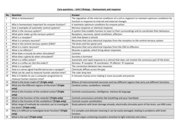
Introductionand contains two N-glycosylation sites (2). Four cysteine residues are conserved in the centralportions of human and murine IL-6, which are not necessary for IL-6 activity (9). Human IL-6shows a sequence homology of 65% at the DNA level and 42% at the protein level compared withmurine IL-6 (10).IL-6 is not constitutively expressed in the human body, but is induced by a large number of multiplestimuli including bacterial and viral infections, microbial components such as lipopolysaccharide(LPS) and different cytokines (11). IL-1β, TNF-α and IFN-γ have been shown to induce IL-6expression in many cell types including T-cells, B-cells, monocytes, endothelial cells, fibroblastsand several kinds of tumor cells.1.1.3 The Interleukin-6 promoterA series of transcription factors have been described to contribute to the complex regulation of theIL-6 gene. Their interactions may vary depending on the extracellular stimuli as well as the celltype. So far, several functional cis-regulatory elements in the human IL-6 promoter have beenidentified, including nuclear factor-kappa B (NF-κB) (position -62 to –49 relative to thetranslational start codon), nuclear factor-IL6 (NF-IL6) (-144 to -132), cAMP response elementbinding protein (CREB) (-152 to -145) and activator protein 1 (AP-1) (-271 to –263) binding sites(Figure 1.1.3.1).NF-κB is a member of the Rel family of transcription factors that form homo- or heterodimersthrough the Rel homology domain (12). In resting cells, NF-κB is located in the cytoplasm bound toa member of the family of inhibitors of κB (IκBs). Upon activation of cells by diverse stimuli, IκBsbecome phosphorylated and are rapidly degraded. The degraded IκBs can no longer bind to NF-κBwhich is therefore released and translocated to the nucleus where it binds to its specific DNArecognition site and contributes to the transcriptional activation of multiple genes.NF-IL6, also known as CCAAT/enhancer binding protein beta (C/EBP β), belongs to the family ofC/EBP transcription factors which are characterized by a basic leucine-zipper structure which isresponsible for homo- or heterodimerization (13). In primary cells, NF-IL6 is usually expressed atlow levels and is strongly induced upon stimulation of cells by TNF-α, IL-1β or LPS. In contrast,NF-IL6 is constitutively expressed in many cell lines. Phosphorylation of NF-IL6 has been shownto increase its transcriptional activity (14).11
TGTCT-152-145 -144-132TGCGATGCTAAAGGACGTCA AAATGTGGGATTTTCCCATGAGTCTCAATATTAGAGTCTCAATATA boxNF-κB-17-10 1CCCCCAATAAATATAGGACTGGAGATGTATA boxFigure 1.1.3.1: The human IL-6 promoter. A part of the IL-6 promoter is shown in whichcore sequences for each transcription factor binding site are indicated in bold. The startcodon ATG for the IL-6 gene is boxed, the TATA box is bolded. The numbers on top ofcorresponding nucleotides give the position relative to the start codon ATG.The AP-1 transcription factors are homo- or heterodimers of basic region-leucine zipper proteinsbelonging to the Jun and Fos subfamilies (15). Since Fos proteins do not form stable dimers, theyneed to form dimers with Jun proteins to bind DNA. The members of the Jun protein family varyconsiderably in their DNA binding affinities and transactivation capacities, with c-Jun showing thehighest activation potential (16).Studies in various cell types have shown that induction of IL-6 gene transcription is complex andinvolves the coordinated regulation of factors that associate with the evolutionary conservedNF-κB, NF-IL6 and AP-1 sites in the mouse and human IL-6 promoter (17-21).12
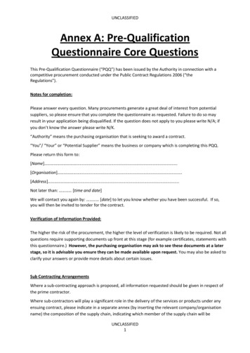
Introduction1.1.4 Cellular signaling and regulation of Interleukin-6IL-6 has a unique receptor system (22). The IL-6 receptor consists of two functional membraneproteins: an 80 kDa IL-6 binding chain (IL-6R, IL-6R α-chain, CD126) (23) and a 130 kDasignal transducer (gp130, IL-6R β-chain CD130) involved in IL-6 signal transduction (24,25). The80 kDa IL-6 receptor can exist in transmembrane form or in soluble form. The transmembrane formhas a short intracytoplasmatic region consisting of 82 aa, that lacks tyrosine-kinase domains anddoes not have an enzymatic role in IL-6 signaling.Binding of IL-6 to the membranous IL-6 receptor brings the intracellular segments of two IL-6Rchains in close proximity, thus permitting the association of the intracellular IL-6R segments withthe 130 kDa glycoprotein (Figure 1.1.4.1). This is thought to induce the formation of a multimericcomplex consisting of two IL-6 molecules bound to two IL-6R proteins and two gp130 moleculesbound to the intracytoplasmatic segments of the two IL-6R proteins.On the other hand, the soluble form of IL-6R, which is produced by cleavage of the membranousIL-6R or by alternative splicing, can bind IL-6. A complex of two IL-6 molecules bound to twosoluble IL-6R proteins forms a complex with two gp130 proteins. This complex is able to transmitsignals in a process called trans-signaling (26).Dimerization of gp130 proteins transduces a signal to activate members of the JAK tyrosine kinases(Janus family tyrosine kinases). JAKs represent a family of four non-receptor tyrosine kinases,comprising the members JAK1, JAK2, JAK3 and Tyk2, which selectively phosphorylate the signaltransducer and activator of transcription (STAT) proteins thus leading to their activation. In the caseof IL-6R signaling, JAKs recrui
2.4.7. Cloning of the lentiviral PU.1 gene silencing construct GIPZ-sh-PU.1 35 2.5 ELISA for human Interleukin-6 36 2.6 Western blot analysis 36 2.6.1 Preparation of whole cell lysates 36 2.6.2 Tris-Glycine gel electrophor