Transcription
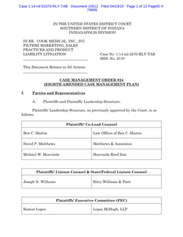
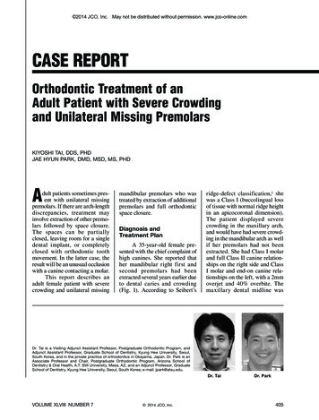
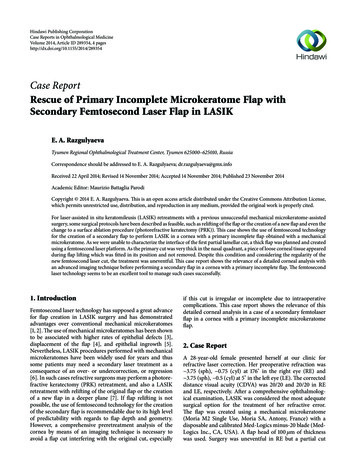
Hindawi Publishing CorporationCase Reports in MedicineVolume 2013, Article ID 292961, 4 pageshttp://dx.doi.org/10.1155/2013/292961Case ReportTelescoping Intestine in an AdultKhaldoon Shaheen,1 Naseem Eisa,1 Abdul Hamid Alraiyes,2M. Chadi Alraies,1 and Srinivas Merugu31Department of Hospital Medicine, Institute of Medicine, Cleveland Clinic, Cleveland, OH 44195, USADepartment of Pulmonary, Critical Care and Environmental Medicine, Tulane University Health Sciences Center,New Orleans, LA 70118, USA3Department of Medicine, Case Western Reserve University St. Vincent Charity Medical Center, Cleveland, OH 44115, USA2Correspondence should be addressed to Khaldoon Shaheen; khaldoonshaheen@yahoo.comReceived 24 January 2013; Accepted 20 June 2013Academic Editor: Gerald S. SupinskiCopyright 2013 Khaldoon Shaheen et al. This is an open access article distributed under the Creative Commons AttributionLicense, which permits unrestricted use, distribution, and reproduction in any medium, provided the original work is properlycited.Protrusion of a bowel segment into another (intussusception) produces severe abdominal pain and culminates in intestinalobstruction. In adults, intestinal obstruction due to intussusception is relatively rare phenomenon, as it accounts for minority ofintestinal obstructions in this population demographic. Organic lesion is usually identifiable as the cause of adult intussusceptions,neoplasms account for the majority. Therefore, surgical resection without reduction is almost always necessary and is advocated asthe best treatment of adult intussusception. Here, we describe a rare case of a 44-year-old male with a diffuse large B-cell lymphomainvolving the terminal ileum, which had caused ileocolic intussusception and subsequently developed intestinal obstructionrequiring surgical intervention. This case emphasizes the importance of recognizing intussusception as the initial presentationfor bowel malignancy.1. IntroductionProtrusion of a bowel segment into another produces severeabdominal pain and culminates in intestinal obstruction.In adults, intestinal obstruction due to intussusception isrelatively rare phenomenon, as it accounts for minority ofintestinal obstructions in this population demographics. Inthis case, we provide diagnostic, therapeutic, and prognostickeys to bowel intussusception in adults.2. The CaseA 44-year-old construction worker man, with no past medical problems, was admitted with two-month history ofrecurrent and progressively worsening abdominal pain andbleeding per rectum. Pain was colicky in nature and welllocalized to lower abdomen causing significant distress andinsomnia. He denied similar symptoms in the past and henever had a colonoscopy. His condition-associated with lossof weight of about 20 pounds. Patient denied hematuria,frequency, and dysuria. There was no history of fever. He hasno medications and denied using NSAIDs. His social historywas significant for smoking (20 pack-year-histories) andoccasional alcohol use. He denied any illicit drug abuse. Histemperature was 36.4 C, blood pressure 103/63 mmHg, heartrate 96/minute, respiratory rate 18/minute, and SaO2 of 97%on room air. He was in mild distress secondary to abdominalpain. Abdominal examination was significant for mild tenderness in the suprapubic region and left iliac fossa with norebound tenderness. Rectal exam was positive for fecal occultblood. Remainder of the physical exam was unremarkable.Laboratory investigation was significant for WBC 13.400cells/mm3 with normal differential, hemoglobin 7.4 g/dL,hematocrit 23.1%, and platelet count 701,000 cells/mm3 .Abdominal X-ray (Figure 1) showed dilated small bowel loopsand multiple air-fluid levels suggesting distal small bowelobstruction. This was followed with computed tomography(CT) of the abdomen which revealed ileocolic intussusception (Figure 2). Surgery was consulted and patient wastaken to operating room where a right hemicolectomy and
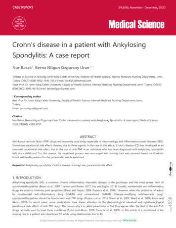
2Case Reports in Medicine(a)(b)Figure 1: X-ray abdomen. (a) Supine abdominal radiograph shows dilated small bowel loops and absence or paucity of gas in the large boweldue to distal small bowel obstruction. (b) Upright abdominal radiograph shows multiple air-fluid levels in upper and central abdomen thatrepresent dilated proximal and central fluid-filled small bowel due to distal small bowel obstruction (arrows).(a)(b)Figure 2: A computed tomography (CT) scan of the abdomen. (a) Axial CT scan at level of iliac crests shows a longitudinal sectional image ofthe intussusception. A portion of the terminal ileum is inside the cecum and far from the invaginated ileal mesentery that separates the walls ofthe two bowel segments (arrow). (b) Cross-sectional image of the midportion of intussusception (arrow) illustrates small bowel invaginationthrough the ascending colon just above the cecum (target sign). These findings are consistent with ileocolic intussusception.extended ileac resection was performed. Surgery revealeda thickened mass involving the terminal ileum responsiblefor the ileocolic intussusception. Histopathology of the massshowed a diffuse large B-cell lymphoma involving the terminal ileum (Figure 3). No systemic lymphadenopathy wasfound suggesting a primary gastrointestinal non-Hodgkin’slymphoma. HIV test was negative. His postoperative coursewas uneventful, and later he was discharged home in stablecondition.3. DiscussionIntussusception is an invagination or “telescoping” of a proximal segment of bowel into the lumen of a distal segmentleading to obstruction and compromise of mesenteric bloodflow, with resultant ischemia of the bowel wall [1]. Anyintraluminal lesion (leading point) is able to trigger an intraluminal invagination finally causing an intussusception.Subsequent peristaltic bowel activity produces an area ofsequence constriction and relaxation, thus telescoping theleading point through the distal bowel lumen.Intussusception is commonly seen in children andreported as the second most common abdominal emergencyin children, trailing only appendicitis. Adult intussusception,however, accounts only for 5% of all cases of intussusceptionand 1% of all cases of bowel obstruction in adults [2]. Themean age for intussusception is 50 years of age. Incidence isabout the same in males and females [2].Intussusceptions can be categorized into four types:enteroenteric, colocolic, enterocolic, and ileocecal. Enterocolic intussusception is the most common type [2]. Incontrast to the pediatric population, where intussusception
Case Reports in Medicine(a)(c)3(b)(d)Figure 3: Diffuse large B-cell lymphoma (DLBCL): the tumor composed of diffuse infiltration of large lymphoid cells in all wall layers of theaffected terminal ileum. (a) Staining with hematoxylin-eosin, magnification 40. (b) Staining with hematoxylin-eosin, magnification 200.(c) Staining with hematoxylin-eosin, magnification 400. (d) Immunoperoxidase staining for CD20, magnification 400.is usually idiopathic or secondary to viral illness, an organiclesion (leading point) is usually the identifiable culprit inadults. The vast majority (90%) of cases is due to neoplasmand the remainders (10%) are idiopathic. Tumors are 80%benign: that is, adhesions, lymphoid hyperplasia, trauma,lipomas, leiomyomas, Meckel’s diverticulum, gastrointestinalstromal tumors, hemangiomas, or Peutz-Jegher adenoma.Only few cases (less than 20%) of small intestinal intussusceptions are malignant. In contrast, the colon is more likely(60%) to have malignant lesion as the cause of intussusception; two-thirds are due to primary colon adenocarcinomaand one-third is due to malignant lymphoma—the twoconstitute the most common malignant lesions in the colon[3].An increased incidence of intussusception has beenreported in patients with acquired immune deficiency syndrome (AIDS). This is due to the high incidence of infectiousand neoplastic conditions like lymphoid hyperplasia, Kaposi’ssarcoma, and non-Hodgkin’s lymphoma. In HIV patients,lymphoma accounts for about 10% of all malignancies. It isthus not surprising that this population, more so than otherpopulation groups, is more frequently referred to surgery forabdominal complaints [4]. In our case, a 53-year-old malewith diffuse large B-cell lymphoma involving the terminalileum and had caused an ileocolic intussusception. Ourliterature search revealed that few cases of B-cell lymphoma,presenting as intussusceptions of terminal ileum, in HIVseronegative adults, have ever been reported [5–8].Intussusception in adults often present as chronic, intermittent abdominal pain associated with intermittent partialbowel obstruction which can cause nausea, vomiting, melena,weight loss, fever, and constipation [9]. Abdominal massesare palpable in 24%–42% of patients, and identification of ashifting mass or one that is palpable only when symptomsare present is suggestive of intussusception [3]. The relativelylow incidence and varied presentation, both, account forthe difficulty in making diagnosis of intussusception beforesurgery. Reijnen et al. reported a preoperative diagnostic rateof 50% [10], while Eisen et al. reported a lower rate of 40.7%[11].A number of different radiologic methods have been described as useful in the diagnosis of intussusception. Plain abdominal films are typically the first diagnostic tool [12]; suchfilms usually demonstrate signs of intestinal obstruction andsuggest location of obstruction. Upper gastrointestinal seriesmay show a “stacked coins” or “coiled spring” appearance[11]. Barium enema examination may be useful in patientswith colonic or ileocolic intussusception in which a “cupshaped” filling defect is a characteristic finding [11]. Ultrasonography is another useful measurement in the diagnosisof intussusceptions. The characteristic sonographic findingsare the “target” or “doughnut” signs on the transverse viewand the “pseudo-kidney” sign or “hay-fork” sign in thelongitudinal view [13]. Certainly, an experienced radiologistis required to confirm such findings. Abdominal CT has beenreported to be the most useful tool for diagnosis of intestinal
4intussusception and is superior to other contrast studies,ultrasonography or endoscopy [14]. A “target sign” may beseen on CT on perpendicular view (Figure 2(b)), while theintussusception will appear as a sausage-shaped mass whenthe CT beam is parallel to the longitudinal axis (Figure 2(a)).The distended loop of bowel (intussuscipiens) has a thickenedwall because it represents two layers of bowel.While pediatric intussusception is usually due to a benignetiology and can usually be managed with nonoperativereduction (use of barium or air-contrast enemas), surgicalresection without reduction is almost always necessary andis advocated as the best treatment of adult intussusception,given the high percentage of associated malignancy [14].Nevertheless, some authors have recommended a selectiveapproach to resection, particularly for small bowel intussusceptions, as the lower malignancy rate for small bowelintussusception makes the argument for initial resection lessconvincing. Furthermore, the choice of using a laparoscopicor open approach for small bowel resection depends on theclinical condition of the patient, the location and extent ofintussusception, the possibility of underlying disease, and theavailability of experienced surgeons [15].The overall five-year survival for a primary gastrointestinal lymphoma is 38%. Curative resections yielded a survivalof 60% regardless of site while palliative resections offeredonly a 17% chance of cure. As expected, survival was inverselyproportional to extent of nodal spread. Postoperative radiotherapy is recommended for residual disease [16].Case Reports in Medicine[6][7][8][9][10][11][12][13][14][15]4. ConclusionIntussusception in adults is a rare entity and diagnosis maybe challenging because of nonspecific symptoms. Cliniciansshould be familiar with the various treatment options,because the real cause of the intussusception often is accurately diagnosed by laparotomy. CT is the most useful imaging modality in the diagnosis of intussusception. Treatmentusually requires resection of the involved bowel segment.Reduction can be attempted in small-bowel intussusception ifthe segment involved is viable or malignancy is not suspected;however, a more careful approach is recommended in colonicintussusception because of a significantly higher chance ofmalignancy.References[1] A. Marinis, A. Yiallourou, L. Samanides et al., “Intussusceptionof the bowel in adults: a review,” World Journal of Gastroenterology, vol. 15, no. 4, pp. 407–411, 2009.[2] F. P. Agha, “Intussception in adults,” American Journal of Roentgenology, vol. 146, no. 3, pp. 527–532, 1986.[3] O. Başar, B. Odemiş, I. Ertuğrul et al., “Ileo-ileal invaginationcaused by lymphoma,” Chinese Medical Journal, vol. 120, no. 12,pp. 1119–1120, 2007.[4] M. Corti, M. F. Villafañe, O. Palmieri et al., “Ileocolic intussusception due to a large B cell lymphoma in a patient with AIDS,”Acta Gastroenterol Latinoam, vol. 38, no. 1, pp. 51–55, 2008.[5] N. Takiguchi, H. Sarashina, N. Saitoh et al., “Ileocolic intussusception in adult due to malignant lymphoma in the cecum with[16]intramural metastasis,” Journal of Gastroenterology, vol. 31, no.4, pp. 603–606, 1996.K. Contreary, F. C. Nance, and W. F. Becker, “Primary lymphoma of the gastrointestinal tract,” Annals of Surgery, vol. 191,no. 5, pp. 593–598, 1980.K. Murata, H. Kase, and H. Kanemoto, “Primary malignantlymphoma of the ileum presenting as intussusception,” InternalMedicine, vol. 48, no. 17, pp. 1559–1560, 2009.M. Matsushita, K. Hajiro, T. Kajiyama et al., “Malignant lymphoma in the ileocecal region causing intussusception,” Journalof Gastroenterology, vol. 29, no. 2, pp. 203–207, 1994.G. Gayer, R. Zissin, S. Apter, M. Papa, and M. Hertz, “Adultintussusception—a CT diagnosis,” British Journal of Radiology,vol. 75, no. 890, pp. 185–190, 2002.H. A. M. Reijnen, H. J. M. Joosten, and H. H. M. De Boer,“Diagnosis and treatment of adult intussusception,” AmericanJournal of Surgery, vol. 158, no. 1, pp. 25–28, 1989.L. K. Eisen, J. D. Cunningham, and A. H. Aufses Jr., “Intussusception in adults: institutional review,” Journal of the AmericanCollege of Surgeons, vol. 188, no. 4, pp. 390–395, 1999.J. R. Cogley, S. C. O’Connor, R. Houshyar et al., “Emergent pediatric US: what every Radiologist should know,” Radiographics,vol. 32, no. 3, pp. 651–665, 2012.D. M. Nagorney, M. G. Sarr, and D. C. McIlrath, “Surgicalmanagement of intussusception in the adult,” Annals of Surgery,vol. 193, no. 2, pp. 230–236, 1981.T. Azar and D. L. Berger, “Adult intussusception,” Annals ofSurgery, vol. 226, no. 2, pp. 134–138, 1997.V. Alonso, E. M. Targarona, G. E. Bendahan et al., “Laparoscopictreatment for intussusception of the small intestine in the adult,”Surgical Laparoscopy, Endoscopy and Percutaneous Techniques,vol. 13, no. 6, pp. 394–396, 2003.K. Contreary, F. C. Nance, and W. F. Becker, “Primary lymphoma of the gastrointestinal tract,” Annals of Surgery, vol. 191,no. 5, pp. 593–598, 1980.
MEDIATORSofINFLAMMATIONThe ScientificWorld JournalHindawi Publishing Corporationhttp://www.hindawi.comVolume 2014GastroenterologyResearch and PracticeHindawi Publishing Corporationhttp://www.hindawi.comVolume 2014Journal ofHindawi Publishing Corporationhttp://www.hindawi.comDiabetes ResearchVolume 2014Hindawi Publishing Corporationhttp://www.hindawi.comVolume 2014Hindawi Publishing Corporationhttp://www.hindawi.comVolume 2014International Journal ofJournal ofEndocrinologyImmunology ResearchHindawi Publishing Corporationhttp://www.hindawi.comDisease MarkersHindawi Publishing Corporationhttp://www.hindawi.comVolume 2014Volume 2014Submit your manuscripts athttp://www.hindawi.comBioMedResearch InternationalPPAR ResearchHindawi Publishing Corporationhttp://www.hindawi.comHindawi Publishing Corporationhttp://www.hindawi.comVolume 2014Volume 2014Journal ofObesityJournal ofOphthalmologyHindawi Publishing Corporationhttp://www.hindawi.comVolume 2014Evidence-BasedComplementary andAlternative MedicineStem CellsInternationalHindawi Publishing Corporationhttp://www.hindawi.comVolume 2014Hindawi Publishing Corporationhttp://www.hindawi.comVolume 2014Journal ofOncologyHindawi Publishing Corporationhttp://www.hindawi.comVolume 2014Hindawi Publishing Corporationhttp://www.hindawi.comVolume 2014Parkinson’sDiseaseComputational andMathematical Methodsin MedicineHindawi Publishing Corporationhttp://www.hindawi.comVolume 2014AIDSBehaviouralNeurologyHindawi Publishing Corporationhttp://www.hindawi.comResearch and TreatmentVolume 2014Hindawi Publishing Corporationhttp://www.hindawi.comVolume 2014Hindawi Publishing Corporationhttp://www.hindawi.comVolume 2014Oxidative Medicine andCellular LongevityHindawi Publishing Corporationhttp://www.hindawi.comVolume 2014
Department of Pulmonary, Critical Care and Environmental Medicine, Tulane University Health Sciences Center, New Orleans, LA , USA Department of Medicine, Case Western Reserve University St. Vincent Charity Medical Center, Cleveland, OH , USA . G.Gayer,R.Zissin,S.Apter,M.Papa,andM.Hertz,