Transcription
Hindawi Publishing CorporationCase Reports in Ophthalmological MedicineVolume 2014, Article ID 289354, 4 pageshttp://dx.doi.org/10.1155/2014/289354Case ReportRescue of Primary Incomplete Microkeratome Flap withSecondary Femtosecond Laser Flap in LASIKE. A. RazgulyaevaTyumen Regional Ophthalmological Treatment Center, Tyumen 625000–625010, RussiaCorrespondence should be addressed to E. A. Razgulyaeva; dr.razgulyaeva@gmx.infoReceived 22 April 2014; Revised 14 November 2014; Accepted 14 November 2014; Published 23 November 2014Academic Editor: Maurizio Battaglia ParodiCopyright 2014 E. A. Razgulyaeva. This is an open access article distributed under the Creative Commons Attribution License,which permits unrestricted use, distribution, and reproduction in any medium, provided the original work is properly cited.For laser-assisted in situ keratomileusis (LASIK) retreatments with a previous unsuccessful mechanical microkeratome-assistedsurgery, some surgical protocols have been described as feasible, such as relifting of the flap or the creation of a new flap and even thechange to a surface ablation procedure (photorefractive keratectomy (PRK)). This case shows the use of femtosecond technologyfor the creation of a secondary flap to perform LASIK in a cornea with a primary incomplete flap obtained with a mechanicalmicrokeratome. As we were unable to characterize the interface of the first partial lamellar cut, a thick flap was planned and createdusing a femtosecond laser platform. As the primary cut was very thick in the nasal quadrant, a piece of loose corneal tissue appearedduring flap lifting which was fitted in its position and not removed. Despite this condition and considering the regularity of thenew femtosecond laser cut, the treatment was uneventful. This case report shows the relevance of a detailed corneal analysis withan advanced imaging technique before performing a secondary flap in a cornea with a primary incomplete flap. The femtosecondlaser technology seems to be an excellent tool to manage such cases successfully.1. IntroductionFemtosecond laser technology has supposed a great advancefor flap creation in LASIK surgery and has demonstratedadvantages over conventional mechanical microkeratomes[1, 2]. The use of mechanical microkeratomes has been shownto be associated with higher rates of epithelial defects [3],displacement of the flap [4], and epithelial ingrowth [5].Nevertheless, LASIK procedures performed with mechanicalmicrokeratomes have been widely used for years and thussome patients may need a secondary laser treatment as aconsequence of an over- or undercorrection, or regression[6]. In such cases refractive surgeons may perform a photorefractive keratectomy (PRK) retreatment, and also a LASIKretreatment with relifting of the original flap or the creationof a new flap in a deeper plane [7]. If flap relifting is notpossible, the use of femtosecond technology for the creationof the secondary flap is recommendable due to its high levelof predictability with regards to flap depth and geometry.However, a comprehensive preretreatment analysis of thecornea by means of an imaging technique is necessary toavoid a flap cut interfering with the original cut, especiallyif this cut is irregular or incomplete due to intraoperativecomplications. This case report shows the relevance of thisdetailed corneal analysis in a case of a secondary femtolaserflap in a cornea with a primary incomplete microkeratomeflap.2. Case ReportA 28-year-old female presented herself at our clinic forrefractive laser correction. Her preoperative refraction was 3.75 (sph), 0.75 (cyl) at 176 in the right eye (RE) and 3.75 (sph), 0.5 (cyl) at 5 in the left eye (LE). The correcteddistance visual acuity (CDVA) was 20/20 and 20/20 in REand LE, respectively. After a comprehensive ophthalmological examination, LASIK was considered the most adequatesurgical option for the treatment of her refractive error.The flap was created using a mechanical microkeratome(Moria M2 Single Use, Moria SA, Antony, France) with adisposable and calibrated Med-Logics minus-20 blade (MedLogics Inc., CA, USA). A flap head of 100 𝜇m of thicknesswas used. Surgery was uneventful in RE but a partial cut
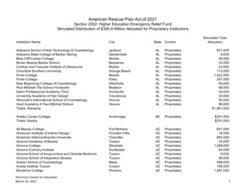
2Case Reports in Ophthalmological MedicineFigure 1: Slit-lamp visualization of both flap edges.Figure 3: In the lower quadrant, the cut done primarily with themicrokeratome opened up from inside after colliding with the newfemtosecond flap, leading to an epithelial defect that was visualizedwith the OCT system.Figure 2: Delimitation (black arrows) of the loose corneal tissue thatwas present in the nasal quadrant and evidenced after lifting the flap.with the microkeratome was performed in LE due to a lossof suction, and consequently the laser treatment was aborted.Three months after this episode, when the refraction andthe topography were stable, an analysis of the LE cornea wasperformed with the optical coherence tomography (OCT)system Cirrus HD-OCT (Carl Zeiss Meditec, Germany) inorder to determine the position and trajectory of the irregularcut, but the interface of the incomplete primary flap was notvisible, even with high magnification. Considering that theprimary intended flap had a theoretical central thickness of100 𝜇m, a new cut using the IntraLase femtosecond laser FS60 (Abbott Medical Optics, Inc.) was performed using thefollowing settings: diameter of 9 mm and flap thickness of140 𝜇m. This planning was aimed at avoiding an interferencewith the primary incomplete cut. Although the planning was140 𝜇m, according to the OCT image, the flap was approximately of 100–110 𝜇m in the center. There was no subepithelialopacity that may have predisposed to an incomplete lamellarpass due to the vertical femtosecond gas breakthrough.At the immediate postoperative visit, the slit-lamp examination allowed the visualization of both flap edges (Figure 1).Intraoperatively, a piece of loose corneal tissue was observedin the nasal quadrant after lifting the flap which was thoughtto be the result of the presence of a primary cut of morethan 140 𝜇m at this position. This piece of corneal tissuewas fitted in its position and not removed. Its extensioncould be also delimited at the slit lamp (Figure 2). In thelower quadrant, the primary microkeratome cut opened upfrom inside after colliding with the new femtosecond flap,Figure 4: OCT image of both flaps in the temporal quadrantshowing the abrupt end of the primary cut done with the mechanicalmicrokeratome.leading to an epithelial defect (Figure 3). In the temporalquadrant, both flaps were clearly positioned at different levelsand the abrupt end of the primary mechanical cut was visiblein the OCT analysis (Figure 4). Despite this condition andconsidering the regularity of the new femtosecond cut, lasertreatment was uneventfully applied. The day after surgery,the patient achieved an uncorrected distance visual acuity(UDVA) of 0.9 (decimal notation).3. DiscussionAs shown in this case report, femtosecond laser-assistedLASIK is a safe option to manage unsuccessful previousLASIK procedures with mechanical microkeratomes. If theprimary flap has been uneventfully done and relifting withmanual dissection is not possible because of its strongadhesion, the creation of a new vertical side cut with theuse of the femtosecond laser creates a net interface thatdiminishes the mechanical trauma to the epithelium (Güellet al. [8]) and prevents epithelial ingrowth due to the verticalside cut configuration of the femtosecond flap compared tothe sloping edge of the mechanical-based flap [9]. Vaddavalliet al. found that femtosecond retreatment using a side cutonly algorithm is effective in the treatment of residual errorsafter mechanical microkeratome-based LASIK, reducing the
Case Reports in Ophthalmological Medicinerates of epithelial ingrowth [10]. The technique from Güellet al. [8] was shown to be also useful if the primary cut isirregular or incomplete, as in our case.The preoperative assessment of the primary flap throughan imaging technique, such as OCT, is essential, especiallyin the peripheral area, where both primary and secondaryflaps are expected to interfere due to the wide variability ofthickness of a flap obtained with a mechanical microkeratome[11–13]. Anterior segment OCT (AS-OCT) is an imagingtechnology that would provide enough information about thecharacteristics of the primary flap in cases like that presentedhere [14–16], but in some cases the difficulty in detecting theinterface can be significant. This difficulty is especially relevant when a posterior segment OCT is used for the analysisof the cornea because of the unavailability of an AS-OCT atthe clinical setting, as happened in our case. This inability ofcharacterizing the interface prior to the second cut can leadto complications such as the presence of some loose tissue,scars, or loss of a sliver of tissue. Pietilä et al. used the FEMTOLDV femtosecond laser (Ziemer Ophthalmic Systems, Port,Switzerland) to perform a new thin flap (90 𝜇m) in 81eyes previously operated with a mechanical microkeratome.They found an improved predictability in flap thicknesswith their cutting and a low rate of complications such asadhesions related to scars of the primary flap, with no furtherprevention of the excimer laser ablation treatment. However,these authors did not recommend the creation of a newfemtosecond flap in cases of primary free cap or visible scarsin the old flap [17]. Garcia-Gonzalez and Teus created a newfemtosecond mini-flap, smaller in diameter compared to theprevious one, in 10 eyes of 7 patients to avoid the adhesionof the primary flap edges, and without interferences withthe peripheral interface, preventing the risk of dislocation ofthe primary flap [18]. In our case, the lack of an accurateOCT image delimiting the previous partial flap led us toprogram a new thick femtosecond flap, assuming a higherrisk of interference between the primary and secondary flapin the peripheral cornea and consequently to the generationof loose tissue at this area. Fortunately, this complication waseasily managed with no further consequences. In conclusion,femtosecond laser-assisted LASIK retreatment in eyes withprevious unsuccessful surgery with the creation of an incomplete or irregular flap using a mechanical microkeratomeseems to be a safe and highly predictable option. It shouldbe considered that an incomplete microkeratome flap resultsin an irregular topographic pattern [19, 20] that shouldbe treated by means of topography or wavefront-guidedprocedures in order to minimize the higher order aberrationsand corneal irregularity induced with the incomplete cut [21–24]. In such cases, it is highly recommended to perform acomplete and detailed analysis of the primary flap throughAS-OCT imaging technology [25, 26] prior to the retreatmentin order to define the most appropriate planning for thelamellar cut. Likewise, AS-OCT is able to evaluate epithelialremodeling which can be considered another importantbiological metric to take into account when evaluating thisor any similar complicated refractive surgery cases [25].3Conflict of InterestsThe author has no proprietary or commercial interest in themedical devices that are involved in this paper.References[1] S. Chen, Y. Feng, A. Stojanovic, M. R. Jankov II, and Q. Wang,“Intralase femtosecond laser vs mechanical microkeratomesin LASIK for myopia: a systematic review and meta-analysis,”Journal of Refractive Surgery, vol. 28, no. 1, pp. 15–24, 2012.[2] A. A. Farjo, A. Sugar, S. C. Schallhorn et al., “Femtosecond lasersfor LASIK flap creation: a report by the American academyof ophthalmology,” Ophthalmology, vol. 120, no. 3, pp. e5–e20,2013.[3] G. M. Kezirian and K. G. Stonecipher, “Comparison of theIntraLase femtosecond laser and mechanical keratomes forlaser in situ keratomileusis,” Journal of Cataract and RefractiveSurgery, vol. 30, no. 4, pp. 804–811, 2004.[4] G. Clare, T. C. B. Moore, C. Grills, A. Leccisotti, J. E. Moore,and S. Schallhorn, “Early flap displacement after LASIK,” Ophthalmology, vol. 118, no. 9, pp. 1760–1765, 2011.[5] E. Letko, M. O. Price, and F. W. Price Jr., “Influence of originalflap creation method on incidence of epithelial ingrowth afterLASIK retreatment,” Journal of Refractive Surgery, vol. 25, no.11, pp. 1039–1041, 2009.[6] P. S. Hersh, K. L. Fry, and D. S. Bishop, “Incidence andassociations of retreatment after LASIK,” Ophthalmology, vol.110, no. 4, pp. 748–754, 2003.[7] E. A. Davis, D. R. Hardten, M. Lindstrom, T. W. Samuelson, andR. L. Lindstrom, “LASIK enhancements: a comparison of liftingto recutting the flap,” Ophthalmology, vol. 109, no. 12, pp. 2308–2313, 2002.[8] J. L. Güell, D. Elies, O. Gris, F. Manero, and M. Morral,“Femtosecond laser-assisted enhancements after laser in situkeratomileusis,” Journal of Cataract and Refractive Surgery, vol.37, no. 11, pp. 1928–1931, 2011.[9] P. K. Vaddavalli and S. H. Yoo, “Femtosecond laser in-situkeratomileusis flap configurations,” Current Opinion in Ophthalmology, vol. 22, no. 4, pp. 245–250, 2011.[10] P. K. Vaddavalli, V. F. Diakonis, A. P. Canto et al., “Complications of femtosecond laser-assisted re-treatment for residualrefractive errors after LASIK,” Journal of Refractive Surgery, vol.29, no. 8, pp. 577–580, 2013.[11] D. Z. Reinstein, H. F. S. Sutton, S. Srivannaboon, R. H. Silverman, T. J. Archer, and D. J. Coleman, “Evaluating microkeratome efficacy by 3D corneal lamellar flap thickness accuracyand reproducibility using Artemis VHF digital ultrasound arcscanning,” Journal of Refractive Surgery, vol. 22, no. 5, pp. 431–440, 2006.[12] J. L. Alió and D. P. Piñero, “Very high-frequency digital ultrasound measurement of the LASIK flap thickness profile usingthe IntraLase femtosecond laser and M2 and Carriazo-Pendularmicrokeratomes,” Journal of Refractive Surgery, vol. 24, no. 1, pp.12–23, 2008.[13] Y. Zhang, Y.-G. Chen, and Y.-J. Xia, “Comparison of cornealflap morphology using AS-OCT in LASIK with the waveLightFS200 femtosecond laser versus a mechanical microkeratome,”Journal of Refractive Surgery, vol. 29, no. 5, pp. 320–324, 2013.[14] J. E. Stahl, D. S. Durrie, F. J. Schwendeman, and A. J. Boghossian,“Anterior segment OCT analysis of thin IntraLase femtosecond
ase Reports in Ophthalmological Medicineflaps,” Journal of Refractive Surgery, vol. 23, no. 6, pp. 555–558,2007.A. C. K. Cheng, T. Ho, S. Lau, A. L. Wong, C. Leung, and D. S.C. Lam, “Measurement of LASIK flap thickness with anteriorsegment optical coherence tomography,” Journal of RefractiveSurgery, vol. 24, no. 9, pp. 879–884, 2008.A. B. Cummings, B. K. Cummings, and G. E. Kelly, “Predictability of corneal flap thickness in laser in situ keratomileusis usinga 200 kHz femtosecond laser,” Journal of Cataract & RefractiveSurgery, vol. 39, no. 3, pp. 378–385, 2013.J. Pietilä, A. Huhtala, P. Mäkinen, and H. Uusitalo, “Laser insitu keratomileusis enhancements with the Ziemer FEMTOLDV femtosecond laser following previous LASIK treatments,”Graefe’s Archive for Clinical and Experimental Ophthalmology,vol. 251, no. 2, pp. 597–602, 2013.M. Garcia-Gonzalez and M. A. Teus, “Creation of a newfemtosecond laser-assisted mini-flap to enhance late regressionafter LASIK,” Journal of Refractive Surgery, vol. 29, no. 8, pp.564–568, 2013.V. J. Katsanevaki, N. S. Tsiklis, N. I. Astyrakakis, and I. G.Pallikaris, “Intraoperative management of partial flap duringLASIK: a small case series report,” Ophthalmology, vol. 112, no.10, pp. 1710.e1–1710.e5, 2005.I. G. Pallikaris, V. J. Katsanevaki, and S. I. Panagopoulou, “Laserin situ keratomileusis intraoperative complications using onetype of microkeratome,” Ophthalmology, vol. 109, no. 1, pp. 57–63, 2002.X. Chen, A. Stojanovic, W. Zhou, T. P. Utheim, F. Stojanovic,and Q. Wang, “Transepithelial, topography-guided ablation inthe treatment of visual disturbances in LASIK flap or interfacecomplications,” Journal of Refractive Surgery, vol. 28, no. 2, pp.120–126, 2012.J. L. Alió, D. P. Piñero, and A. B. Plaza Puche, “Corneal wavefront-guided photorefractive keratectomy in patients withirregular corneas after corneal refractive surgery,” Journal ofCataract and Refractive Surgery, vol. 34, no. 10, pp. 1727–1735,2008.J. L. Alió, D. P. Piñero, and A. B. Plaza Puche, “Corneal wavefront-guided enhancement for high levels of corneal comaaberration after laser in situ keratomileusis,” Journal of Cataractand Refractive Surgery, vol. 34, no. 2, pp. 222–231, 2008.M. R. Jankov II, S. I. Panagopoulou, N. S. Tsiklis, G. C.Hajitanasis, I. M. Aslanides, and I. G. Pallikaris, “Topographyguided treatment of irregular astigmatism with the wavelightexcimer laser,” Journal of Refractive Surgery, vol. 22, no. 4, pp.335–344, 2006.K. Maia Rocha and R. R. Krueger, “Spectral-domain opticalcoherence tomography epithelial and flap thickness mappingin femtosecond laser-assisted in situ keratomileusis,” AmericanJournal of Ophthalmology, vol. 158, pp. 293.e1–301.e1, 2014.J. Zhang, Y. H. Zhou, L. Tian, and C. B. Zhai, “Comparison ofZiemer FEMTO LDV “Classic” and “Crystal Line” femtosecondlaser flap quality by Fourier-domain optical coherence tomography,” International Journal of Ophthalmology, vol. 6, no. 5, pp.611–617, 2013.
MEDIATORSofINFLAMMATIONThe ScientificWorld JournalHindawi Publishing Corporationhttp://www.hindawi.comVolume 2014GastroenterologyResearch and PracticeHindawi Publishing Corporationhttp://www.hindawi.comVolume 2014Journal ofHindawi Publishing Corporationhttp://www.hindawi.comDiabetes ResearchVolume 2014Hindawi Publishing Corporationhttp://www.hindawi.comVolume 2014Hindawi Publishing Corporationhttp://www.hindawi.comVolume 2014International Journal ofJournal ofEndocrinologyImmunology ResearchHindawi Publishing Corporationhttp://www.hindawi.comDisease MarkersHindawi Publishing Corporationhttp://www.hindawi.comVolume 2014Volume 2014Submit your manuscripts athttp://www.hindawi.comBioMedResearch InternationalPPAR ResearchHindawi Publishing Corporationhttp://www.hindawi.comHindawi Publishing Corporationhttp://www.hindawi.comVolume 2014Volume 2014Journal ofObesityJournal ofOphthalmologyHindawi Publishing Corporationhttp://www.hindawi.comVolume 2014Evidence-BasedComplementary andAlternative MedicineStem CellsInternationalHindawi Publishing Corporationhttp://www.hindawi.comVolume 2014Hindawi Publishing Corporationhttp://www.hindawi.comVolume 2014Journal ofOncologyHindawi Publishing Corporationhttp://www.hindawi.comVolume 2014Hindawi Publishing Corporationhttp://www.hindawi.comVolume 2014Parkinson’sDiseaseComputational andMathematical Methodsin MedicineHindawi Publishing Corporationhttp://www.hindawi.comVolume 2014AIDSBehaviouralNeurologyHindawi Publishing Corporationhttp://www.hindawi.comResearch and TreatmentVolume 2014Hindawi Publishing Corporationhttp://www.hindawi.comVolume 2014Hindawi Publishing Corporationhttp://www.hindawi.comVolume 2014Oxidative Medicine andCellular LongevityHindawi Publishing Corporationhttp://www.hindawi.comVolume 2014
Case Report Rescue of Primary Incomplete Microkeratome Flap with Secondary Femtosecond Laser Flap in LASIK E.A.Razgulyaeva Tyumen Re