Transcription
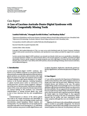
Hindawi Publishing CorporationCase Reports in DentistryVolume 2016, Article ID 8563961, 4 pageshttp://dx.doi.org/10.1155/2016/8563961Case ReportA Case of Lacrimo-Auriculo-Dento-Digital Syndrome withMultiple Congenitally Missing TeethLumbini Pathivada,1 Munagala Karthik Krishna,2 and Mandeep Rallan11Department of Paedodontics and Preventive Dentistry, Teerthanker Mahaveer Dental College and Research Centre, Moradabad, IndiaDepartment of Periodontology, Teerthanker Mahaveer Dental College and Research Centre, Moradabad, India2Correspondence should be addressed to Lumbini Pathivada; drlumbinip@gmail.comReceived 31 July 2016; Accepted 6 September 2016Academic Editor: Zühre AkarslanCopyright 2016 Lumbini Pathivada et al. This is an open access article distributed under the Creative Commons AttributionLicense, which permits unrestricted use, distribution, and reproduction in any medium, provided the original work is properlycited.Lacrimo-auriculo-dento-digital (LADD) syndrome is an extremely rare disorder which may occur sporadically or inheritably asan autosomal dominant condition. It is characterized by defects in the lacrimal apparatus, ear problems, and dental and digitalabnormalities. However, specific symptoms vary greatly among the cases with a high degree of overlap with other similar geneticdisorders. Here, we describe a 7-year-old boy with LADD syndrome, clinical and radiological findings, dental treatment undertaken,and its differential diagnosis.1. IntroductionLacrimo-auriculo-dento-digital (LADD) syndrome, alsoknown as Levy-Hollister syndrome, is a rare genetic disordercharacterized by anomalies affecting the lacrimal and salivaryglands and ducts, ears, dentition, and extremities with a highdegree of variation in the signs and symptoms and an overlapof similar findings in other genetic disorders [1–3]. Malformations in the lacrimal apparatus, including hypoplasticor aplastic lacrimal puncta and/or an obstruction of thenasolacrimal duct, excessive tearing (epiphora), inflammation of the tear sac (dacryocystitis), and dryness and inflammation of the cornea and conjunctiva (keratoconjunctivitis),are common findings in this syndrome. Less commonly,underdeveloped or missing lacrimal glands may resultin lack of tears (alacrima) and dry eyes (xerophthalmia)[4–6].Underdevelopment or absence of the salivary glandsmay result in dry mouth (xerostomia) and vulnerability tosevere dental caries. Other oral findings include small teeth(microdontia), enamel hypoplasia, belated eruption, andmissing teeth [7–9]. Cup-shaped, low-set ears maybe presentassociated with mild to severe hearing loss [1, 2]. Individualswith this syndrome may present variable digital features suchas aplasia, hypoplasia, duplication, clinodactyly, partial syndactyly, digitalization, and abnormal placement of fingers [9–16].2. Case ReportA 7-year-old boy reported to the Department of Pedodonticsand Preventive Dentistry with a chief complaint of dryness ofmouth since birth and decayed teeth. Additional concernsincluded dryness and itchiness in the eyes. The patientweighed 28 kgs and the measured height was 115.2 cm. He hadabundant scalp hair but normal hair density was observed onthe body and limbs. His ears appeared to be normally locatedbut were small and cup-shaped. Shape of the head was abnormal in that the parietal aspect appeared prominent althoughthe frontal region was normal (Figure 1). Eyes appeareddry with lack of tearing at the time of examination. Patientreported that his mother and sister experienced similarocular symptoms although they were not available for examination.Palpation of soft tissues in the submandibular and parotidregions revealed no perceptible enlargement. On intraoralexamination, reduced saliva with multiple carious lesions andenamel dysplasia were observed. Several primary teeth were
2Case Reports in DentistryFigure 3: Panoramic radiograph showing lack of primary dentitiontooth buds.Figure 1: Presence of cup-shaped ear and prominent parietal aspectof head.Figure 4: MRI showing aplasia of lacrimal and salivary glands.protocol. Evaluation after 3 years showed eruption of lowerpermanent canines and right second premolar (Figure 6).3. DiscussionFigure 2: Intraoral view showing multiple carious lesions, reducedsaliva, and missing primary dentition.absent as per their chronological age of eruption (Figure 2).A panoramic radiograph confirmed this finding, althoughtooth buds of unerupted permanent teeth were evident(Figure 3). MRI scan revealed aplasia of bilateral parotidand submandibular salivary glands and hypertrophied minorsalivary glands along oropharyngeal wall. Lacrimal apparatusagenesis was also evident (Figure 4).Dental treatment involved scaling, restorations in relationto primary maxillary canines and second molars, and extraction of root stumps of mandibular canines. Band adaptationwas done in relation to permanent molars and alginateimpressions were taken for the purpose of fabricating spacemaintainers. Patient was instructed in oral hygiene measures, prescribed fluoride mouthrinse and salivary substitute,and recalled. During the second visit, a lingual arch spacemaintainer was given in the lower arch, and topical fluoridevarnish and resin-based pit and fissure sealants were applied(Figure 5). Patient refused an upper arch space maintainerdue to apparent discomfort and was placed on a regular recallIn this case report, we have described a 7-year-old boywith reduced lacrimal secretions, cup-shaped ears, and dental anomalies, characteristic of LADD syndrome. However,the child lacked significant digital malformations. Reducedtears and saliva production were probably as a consequenceof aplasia of lacrimal and salivary glands, respectively, asconfirmed by imaging methods. The proband demonstratedbilateral cup-shaped ears that were positioned normally andno concomitant hearing loss.A significant finding in the present case was the absence ofseveral primary teeth. Maintenance of arch space in suchcases presents a clinical challenge due to increased caries riskand discomfort in wearing appliances because of diminishedsalivary flow. However, in the present case, prescriptionof salivary substitute may have played a role in enablingthe patient to wear the appliance comfortably due to itslubricating properties which proved to be beneficial in thatthe patient could wear the appliance for a period sufficientto prevent pathological migration and provided space foreruption of permanent teeth.LADD syndrome is an autosomal dominant disordercaused due to mutations in one of at least three genes, thefibroblast growth factor receptor 2 (FGFR2), fibroblastgrowth factor receptor 3 (FGFR3), and fibroblast growth
Case Reports in Dentistry3associated systemic manifestations. Multiple missing primarydentition is an unusual association that requires comprehensive dental therapy.Competing InterestsThe authors claim to have no financial interest in anycompany or any of the products mentioned in this article.ReferencesFigure 5: Posttreatment view with restored caries and placement ofspace maintainer in lower arch.Figure 6: Three-year follow-up view with erupted permanentdentition.factor 10 (FGF10) [11–13]. Several other conditions have common, overlapping clinical features and similar genetic etiology but are yet distinct from LADD syndrome. Aplasia of thelacrimal and salivary glands (ALSG) presents with symptomsincluding xerophthalmia, xerostomia, scarring of the conjunctiva, dental erosion, periodontal disease, and increasedrisk of dental caries. ALSG is inherited as an autosomaldominant disorder caused due to mutation in the FGF10 gene[14]. Ectrodactyly-ectodermal dysplasia-cleft lip/palate (EECsyndrome) is another autosomally dominant syndrome andcharacterized by digital malformations, cleft palate, andcleft lip. EEC individuals may present with features thatoverlap with the LADD phenotype, including abnormalities of lacrimal ducts, chronic conjunctivitis, hypodontia,and/or microdontia [15]. Labyrinthine aplasia, microtia, andmicrodontia (LAMM) syndrome presents with Michel aplasia (complete bony and membranous aplasia of the inner ear)in association with microdontia and microtia. LAMM is characterized by an autosomal recessive pattern of inheritanceinvolving mutations in the FGF3 gene [16].LADD syndrome is an extremely rare condition withcharacteristic oral, lacrimal, and auditory clinical findings. Itrequires a thorough evaluation to rule out the abovementioned similar conditions and determine presence of[1] D. W. Hollister, S. H. Klein, H. J. de Jager, R. S. Lachman, andD. L. Rimoin, “The lacrimo-auriculo-dento-digital syndrome,”The Journal of Pediatrics, vol. 83, no. 3, pp. 438–444, 1973.[2] D. W. Hollister, S. H. Klein, H. J. de Jager, R. S. Lachman,and D. L. Rimoin, “Lacrimo-auriculo-dento-digital (LADD)syndrome,” Birth Defects Original Article Series, vol. 10, no. 5,pp. 153–166, 1974.[3] W. J. Levy, “Mesoectodermal dysplasia. A new combination ofanomalies,” American Journal of Ophthalmology, vol. 63, no. 5,pp. 978–982, 1967.[4] H.-R. Wiedemann and J. Drescher, “LADD syndrome: reportof new cases and review of the clinical spectrum,” EuropeanJournal of Pediatrics, vol. 144, no. 6, pp. 579–582, 1986.[5] J. M. Kreutz and H. E. Hoyme, “Levy-Hollister syndrome,”Pediatrics, vol. 82, no. 1, pp. 96–99, 1988.[6] D. Lacombe, F. Serville, D. Marchand, and J. Battin, “Splithand/split foot deformity and LADD syndrome in a family:overlap between the EEC and LADD syndromes,” Journal ofMedical Genetics, vol. 30, no. 8, pp. 700–703, 1993.[7] E. L. Shiang and L. B. Holmes, “The lacrimo-auriculo-dentodigital syndrome,” Pediatrics, vol. 59, no. 6, pp. 927–930, 1977.[8] A. M. Roodhooft, C. C. Brussaard, E. Elst, and K. J. van Acker,“Lacrimo-auriculo-dento-digital (LADD) syndrome with renaland foot anomalies,” Clinical Genetics, vol. 38, no. 3, pp. 228–232, 1990.[9] E. Thompson, M. Pembrey, and J. M. Graham, “Phenotypicvariation in LADD syndrome,” Journal of Medical Genetics, vol.22, no. 5, pp. 382–385, 1985.[10] J. S. Bamforth and P. Kaurah, “Lacrimo-auriculo-dento-digitalsyndrome: evidence for lower limb involvement and severe congenital renal anomalies,” American Journal of Medical Genetics,vol. 43, no. 6, pp. 932–937, 1992.[11] J. M. Milunsky, G. Zhao, T. A. Maher, R. Colby, and D. B.Everman, “LADD syndrome is caused by FGF10 mutations,”Clinical Genetics, vol. 69, no. 4, pp. 349–354, 2006.[12] E. Rohmann, H. G. Brunner, H. Kayserili et al., “Mutations indifferent components of FGF signaling in LADD syndrome,”Nature Genetics, vol. 38, no. 4, pp. 414–417, 2006.[13] E. Schaefer, M. Minoux, J. Lauer et al., “A novel mutation involving the initiation codon of FGF3 in a family described withcomplete inner ear agenesis, microtia and major microdontia(LAMM syndrome),” Journal of Genetic Syndromes & GeneTherapy, vol. 5, no. 6, pp. 1–5, 2014.[14] M. Entesarian, J. Dahlqvist, V. Shashi et al., “FGF10 missensemutations in aplasia of lacrimal and salivary glands (ALSG),”European Journal of Human Genetics, vol. 15, no. 3, pp. 379–382,2007.[15] E. Gawrych, A. Bińczak-Kuleta, J. Janiszewska-Olszowska,and A. Ciechanowicz, “Ectrodactyly-ectodermal dysplasia-cleft
4syndrome (EEC syndrome) with a developmental delay causedby R304W mutation in the TP63 gene,” Annales AcademiaeMedicae Stetinensis, vol. 59, no. 1, pp. 11–14, 2013.[16] Y. Guven, R. O. Rosti, E. B. Tuna, H. Kayserili, and O. Aktoren,“Orodental findings of a family with lacrimo-auriculo-dentodigital (LADD) syndrome,” Oral Surgery, Oral Medicine, OralPathology, Oral Radiology and Endodontology, vol. 106, no. 6,pp. e33–e44, 2008.Case Reports in Dentistry
Advances inPreventive MedicineThe ScientificWorld JournalHindawi Publishing Corporationhttp://www.hindawi.comVolume 2014Case Reports inDentistryInternational Journal ofDentistryHindawi Publishing Corporationhttp://www.hindawi.comVolume 2014Hindawi Publishing Corporationhttp://www.hindawi.comScientificaVolume 2014Hindawi Publishing Corporationhttp://www.hindawi.comVolume 2014Volume 2014PainResearch and TreatmentInternational Journal ofBiomaterialsHindawi Publishing Corporationhttp://www.hindawi.comHindawi Publishing Corporationhttp://www.hindawi.comHindawi Publishing Corporationhttp://www.hindawi.comVolume 2014Volume 2014Journal ofEnvironmental andPublic HealthSubmit your manuscripts athttp://www.hindawi.comJournal ofOral ImplantsHindawi Publishing Corporationhttp://www.hindawi.comComputational andMathematical Methodsin MedicineHindawi Publishing Corporationhttp://www.hindawi.comHindawi Publishing Corporationhttp://www.hindawi.comVolume 2014Volume 2014Journal ofAdvances inOral OncologyHindawi Publishing Corporationhttp://www.hindawi.comVolume 2014Hindawi Publishing earch and PracticeJournal ofOrthopedicsDrug DeliveryVolume 2014Hindawi Publishing Corporationhttp://www.hindawi.comVolume 2014Volume 2014Hindawi Publishing Corporationhttp://www.hindawi.comVolume 2014Journal ofDental SurgeryJournal ofHindawi Publishing Corporationhttp://www.hindawi.comBioMedResearch InternationalInternational Journal ofOral DiseasesEndocrinologyVolume 2014Hindawi Publishing Corporationhttp://www.hindawi.comVolume 2014Hindawi Publishing Corporationhttp://www.hindawi.comVolume 2014Hindawi Publishing Corporationhttp://www.hindawi.comVolume 2014RadiologyResearch and PracticeHindawi Publishing Corporationhttp://www.hindawi.comVolume 2014
ing the initiation codon of FGF in a family described with complete inner ear agenesis, microtia and major microdontia (LAMM syndrome), Journal of Genetic Syndromes & Gene erapy ,vol. ,no., pp. , . [ ] M. Entesarian, J. Dahlqvist, V. Shashi et al., FGF missense mutations in aplasia of Cited by: 1Publish Year: 2016Author: Lumbini Pathivada, Munagal