Transcription
JRRDVolume 51, Number 4, 2014Pages 655–660Colitis after polytrauma: Case reportWilliam E. Carter, MD, MPH;1–2* Isaac A. Darko, MD;3 Priya Chandan, MD, MPH;1–2 Ajit B. Pai, MD11Physical Medicine and Rehabilitation Service, Hunter Holmes McGuire VAMC, Richmond, VA; 2Department of PhysicalMedicine and Rehabilitation, Virginia Commonwealth University, Richmond, VA; 3Rehabilitation Care Line, CincinnatiDepartment of Veterans Affairs Medical Center (VAMC), Cincinnati, OHbarriers to participation in therapy in order to maximize theeffectiveness of rehabilitation.This case report describes a servicemember whosegreatest limitation to therapy participation was diarrhea.In acute settings, the typical differential diagnosis fordiarrhea in a polytrauma patient with a prolonged hospitalstay is broad but is typically focused on acute and subacute explanations. In this patient’s case, these includeddiet, psychogenic, infection, medication use or withdrawal, bowel resection, ischemia, and disuse malabsorption. Here, we propose that ulcerative colitis (UC), achronic idiopathic illness not on the typical differential foracute causes of diarrhea, may have been catalyzed by thestresses of polytrauma and therefore been responsible fordiarrhea in this patient. Including chronic conditions inthe differential diagnosis is important, because failure toconsider these diagnoses may result in delayed treatment,worse outcomes, and limited progression in therapy.Abstract—Across the medical literature, delayed diagnosis andtreatment leads to more costly and worse outcomes. Rehabilitation patients, especially those with polytrauma, often have acomplex mixture of medical, social, and psychological healthproblems that can impair effective diagnosis and treatment. Thecase presentation describes the procession toward the diagnosisof ulcerative colitis in a preinjury asymptomatic male, suggesting a potential mechanism for its emergence and describing theeffect of delayed diagnosis on the efficiency of rehabilitativecare. As such, the differential diagnosis for early posttraumaticdiarrhea should remain broad, particularly if unexplained orineffectively controlled.Key words: Active Duty, amputation, comorbidity, Departmentof Veterans Affairs, gastroenterology, polytrauma, rehabilitation, traumatic brain injury, veteran, ulcerative colitis.INTRODUCTIONAbbreviations: C. diff clostridium difficile, CT computedtomography, FIM Functional Independence Measure, GI gastroenterology, IBD inflammatory bowel disease, PRC polytrauma rehabilitation center, TPN total parental nutrition, UC ulcerative colitis.*Address all correspondence to William E. Carter, MD;Department of Physical Medicine and Rehabilitation, Virginia Commonwealth University, PO Box 980661, Richmond, VA 23298; 804-828-4233; fax: 804-828-5074.Email: D.2013.04.0100Rehabilitation patients, especially those with polytrauma, often have a complex mixture of physiologic,social, and psychological comorbidities that interferewith participation in interdisciplinary rehabilitation.These comorbidities may not be identified in the acutecare setting, which leads to worse clinical outcomes,increased length of stay, and increased cost [1–5]. Interdisciplinary rehabilitation improves outcomes and reduceslength of stay when appropriately implemented [6]. It istherefore important to properly identify comorbidities and655
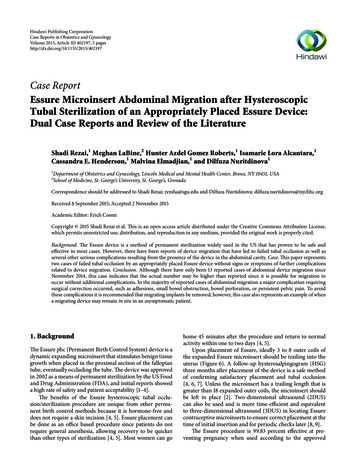
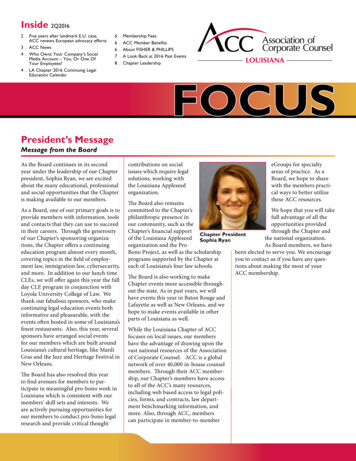
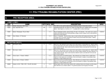
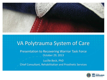
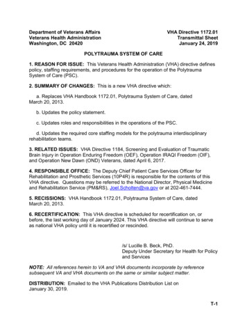
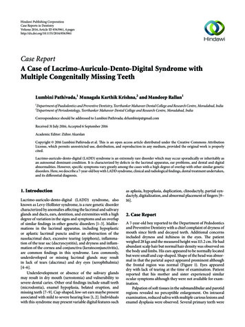
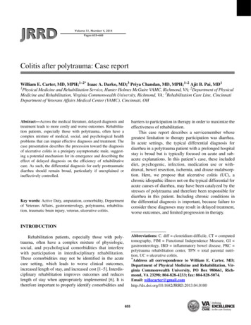
656JRRD, Volume 51, Number 4, 2014CASE: METHODS AND RESULTSPatient is a previously healthy and motivated 41year-old African-American male Active Duty servicemember who experienced a severe traumatic brain injurywith posttraumatic amnesia of 3 wk duration and multiple orthopedic injuries after massive trauma sustainedwhen his parachute malfunctioned during a trainingjump. Patient experienced aortic injury and subsequenthemorrhagic shock requiring massive transfusion andintubation. Acute hospitalization was complicated byhypertension, ventilator-associated pneumonia, bilaterallower-limb amputations, small bowel resection due toischemia, pelvic fractures, right elbow fracture, heterotopic ossification, total parental nutrition (TPN) use for3 wk, urinary retention, abdominal midline wound, andpain. Upon admission to the polytrauma rehabilitationcenter (PRC), his total Functional Independence Measure(FIM) score was 56.In the acute care setting, the patient had four loosestools per day with no documented diagnosis. Clostridiumdifficile (C. diff) toxin polymerase chain reaction assaywas negative on multiple occasions. These symptoms continued upon admission to the PRC. The primary differential diagnosis included withdrawal from 100 g fentanylpatch discontinued approximately 24 h prior to his rehabilitation admission, short bowel syndrome from 1 to 2 ft ofsmall bowel resection, and disuse from prolonged TPNuse. Infectious etiologies were lower on the differentialbased on absence of risk factors, fever, or leukocytosis.Pertinent medical history included a negative colonoscopyin 2009 and history of hemorrhoidectomy. Based on thedifferential diagnosis, he was initially managed with soluble fiber, colestipol, and loperamide, which achievedconstipation. Patient subsequently developed hematochezia along with increasing abdominal discomfort withoutrebound tenderness. On hospital day 10, gastroenterology(GI) specialists were consulted for evaluation of diarrhea,abdominal pain, and bright red blood per rectum. A computed tomography (CT) scan of the abdomen showed gallstones (Figure 1), and the GI specialists recommendedendoscopic retrograde cholangiopancreatography withsphincterotomy. Despite this procedure, the patient’ssymptoms continued. As evidenced by only a 3-pointimprovement in total FIM score from admission to hospital day 30 (FIM efficiency 0.10), therapy progress wasslow. Participation was severely limited by the diarrheaFigure 1.Computed tomography scans of abdomen and pelvis. (a) Normal finding sigmoid colon seen in patient. Artifact limited view.(b) Typical acute ulcerative colitis finding of thickened mucosaof sigmoid colon. Figure 1(b) is reprinted with permission fromHorton KM, Corl FM, Fishman EK. CT evaluation of the colon:Inflammatory disease. Radiographics. 2000;20(2):399–418.and the associated symptoms of pain, poor sleep, andfatigue. The bloody diarrhea progressed to become intractable with up to 14 stools per day, accompanied by severemalnutrition, despite augmenting the bowel regimen withiron, bismuth sulfate, and dietary modification to slowbowel propulsion. Abdominal imaging studies remainedunremarkable and C. diff assay was negatives times four.Fecal leukocytes were positive and C-reactive protein waselevated to 12 (normal: 3). Flexible sigmoidoscopy performed by the GI specialists on hospital day 30 showedtransmural inflammation (Figure 2), and biopsy returnedcryptitis and crypt abscess consistent with inflammatorybowel disease (IBD) (Figure 3). The patient was diagnosed with severe UC; placed on a nothing per mouth diet;and started on TPN, steroids, and mesalamine. Surgicalconsultation was obtained as a precaution to discuss anysurgical options. Over the ensuing days, the patient experienced progressive resolution as documented by clinical,colonoscopic, and pathologic evaluation. After diagnosisand treatment, he progressed well in therapy, tolerateda regular diet, was fitted with bilateral lower-limb prosthetics, and achieved a discharge FIM score of 112.DISCUSSIONUC, one of the two major disorders in IBD, is animmune-mediated chronic inflammatory condition characterized by relapsing and remitting episodes of inflammationlimited to the mucosal layer of the colon. It involves therectum and may extend proximally to the colon [7]. Typical
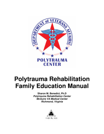
657CARTER et al. Colitis after polytraumaFigure 2.Sigmoidoscopy findings. Inflammatory pattern suggestive ofulcerative colitis.presentation includes rectal bleeding, frequent stools, andtenesmus with a mean onset of symptoms to diagnosis of10 mo [8].In UC, the bowel wall is thin or of normal thickness,but edema, the accumulation of fat, and hypertrophy ofthe muscle layer may give the impression of a thickenedbowel wall, especially seen in the terminal bowel region(Figure 1). CT scan will often show bowel wall thicknessof mean 7.8 mm [9–10]. Early disease manifests as hemorrhagic inflammation with loss of the normal vascularpattern, petechial hemorrhages, and bleeding. Edema ispresent, and large areas become denuded of mucosa.Undermining of the mucosa leads to the formation ofcrypt abscesses, which is a hallmark of the disease (Figure 3). While laboratory studies are useful for excludingother diagnoses and assessing disease severity, the diagnosis of UC is best made with endoscopy with biopsy.The diagnosis of UC in this patient was surprisingbecause he exhibited neither symptoms prior to injury norclear identifiable risk factors at the time of diagnosis. Hehad a negative colonoscopy several years earlier for hematochezia, with the colonscopy demonstrating internal hemorrhoids and polyps but no inflammation or other UCsigns. Risk factors known to contribute to UC includegenetic factors (e.g., first-degree relatives, Jewish descent)[11–12], autoimmune system reactions, environmental factors, nonsteroidal anti-inflammatory drug use [13], age(bimodal 15–25 and 55–65 yr) [14–15], low levels of antioxidants, psychological stress factors [16], and consumption of milk products. Although the patient receivedmultiple antibiotics, this UC induction mechanism is onlynoted in case studies of children [17]. In regards to subacute de novo UC presentations, increased incidence hasbeen noted after transplantation (a possible immunosuppression mechanism) [18], but no other such case presentations were noted in a literature search (Medline searches:“ulcerative colitis” AND polytrauma, “ulcerative colitis”Figure 3.Microscopic evaluation of colonic tissue. (a) Disease with relativesparing of submucosal. (b) Crypt abscess rectosigmoid consistent with ulcerative colitis (UC). (c) Crypt abscess and glanddestruction consistent with UC. (d) Normal colonic mucosa (notfrom patient). Figure 3(d) is reprinted under the Creative Commons License from Oranratanaphan S, Amatyakul P, Somran J,Thumumnuaysuk S. Colonic endometriosis mimicking sigmoidcancer: A case report. Gynecol Obstetric. 2011;1:101.AND “abdominal surgery”). This is despite the four proposed pathogenic causes of UC: dysbiosis, inflammation,impaired immunoregulation, and impaired mucosal functioning [19], all occurring in many settings of critical illness. There is no question that all four occur in the settingof a polytrauma involving bowel resection, infections, andabdominal wounds.In polytrauma, the initial insult, critical illness, subsequent surgeries, infections, and emotional burden allalter cytokines, potentiating medical pathology. Mechanistically, polytrauma produces a proinflammatory statefollowed by immunosuppressive states, mediated by systemic interleukins, tumor necrosis factor, and neuroendocrine changes, along with local tissue injury factors [20–21]. A large component of this immunodysregulation ismediated by T cells [22]. Using the well-studied stressulcer model, critical illness can produce ulcers throughincreased catecholamines, hypovolemia, and proinflammatory cytokines, leading to hypoperfusion and mucosalinjury [23]. It is proposed that stress not only causesimmediate pathology but can also produce more chronic
658JRRD, Volume 51, Number 4, 2014pathologic processes through induction of autoimmunediseases [24]. Moreover, although not clearly connectedwith the initial presentation of UC, these stress pathwaysare implicated to induce UC [25] and are known to exacerbate UC flares [26].In this case, diagnosis of UC was delayed until day30 of the rehabilitation admission despite loose stoolsbeing identified as pathologic during acute care stay.Sequellae included decreased therapy due to medicalworkup, malaise, weakness, and bowel accidents. Theabdominal pain combined with impaired absorptiondecreased nutrient intake, impairing wound and bonehealing and diminishing strength. Furthermore, pain andfrequent stools impaired sleep, resulted in polypharmacy,overburdened the nursing staff, and adversely affectedthe patient’s psychological well-being. As noted, therewas limited change in FIM score during the month priorto diagnosis and treatment despite relatively rapidimprovement thereafter in less than 2 mo (FIM efficiencyof 0.10 vs 0.88). At time of consultation, if colonscopyhad been performed, mild UC would have been the diagnosis; at time of diagnosis, the patient met 4 of 8 criteriafor severe UC despite missing data for other criteria(erythrocyte sedimentation rate was not checked, he wasmalnutritioned, and he was on a beta blocker potentiallymasking other criteria) (Table). Confounding factorsimpeding consideration of the diagnosis were numerous,from the past medical history of hemorrhoids, the multiple pathologies from the polytrauma, negative imaging,and confounding infections to those created idiopathically with medications and procedures. Only after symptoms worsened to a level beyond explanation wascolonoscopy reconsidered. Despite the delayed diagno-sis, this patient fortunately had a good outcome as evidenced by his discharge FIM score.CONCLUSIONSAlthough in the hospital setting, new onset symptoms are likely due to acute changes; in this case, lateconsideration in the differential diagnosis of an idiopathic chronic condition led to delayed diagnosis andprolonged rehabilitation stay. UC currently has unclearpathogenesis with typically prolonged symptomatologyprior to diagnosis. The presentation after polytrauma in apreviously asymptomatic male may support physiologicstress as an underlying mechanism for development ofUC. As such, UC should be considered in the differentialdiagnosis of early posttrauma diarrhea, particularly in thepresence of associated pain.ACKNOWLEDGMENTSAuthor Contributions:Drafting of manuscript: W. E. Carter, I. A. Darko, A. B. Pai.Critical revision of manuscript: W. E. Carter, P. Chandan.Financial Disclosures: The authors have declared that no competinginterests exist.Funding/Support: This material was based on work supported by theDepartment of Veterans Affairs, Veterans Health Administration,Office of Research and Development.Institutional Review: Permission from the veteran described in thiscase was obtained.Participant Follow-Up: The authors have informed the participant ofthe future publication of this study.Disclaimer: The contents of this article do not represent the views ofthe Department of Veterans Affairs or the U.S. Government.Table.Criteria for evaluating severity of ulcerative colitis (Truelove and Witts criteria) [27].VariableStools (number per day)Blood in StoolTemperature ( C)Pulse (bpm)HemoglobinErythrocyte Sedimentation Rate (mm/h)Colonic Features on RadiographyMild Disease 4IntermittentNormalNormalNormal 30—Clinical Signsbpm beats per minute.—Severe Disease 6Frequent 37.5 90 75% of normal 30Air, edematous wall, thumbprintingAbdominal tendernessFulminant Disease 10Continuous 37.5 90Transfusion required 30DilatationAbdominal distention and tenderness
659CARTER et al. Colitis after polytraumaREFERENCES1. Kobayashi L, Konstantinidis A, Shackelford S, Chan LS,Talving P, Inaba K, Demetriades D. Necrotizing soft tissueinfections: Delayed surgical treatment is associated withincreased number of surgical debridements and morbidity.J Trauma. 2011;71(5):1400–1405. 31820db8fd2. Liu V, Kipnis P, Rizk NW, Escobar GJ. Adverse outcomesassociated with delayed intensive care unit transfers in anintegrated healthcare system. J Hosp Med. g/10.1002/jhm.9643. Khashab MA, Tariq A, Tariq U, Kim K, Ponor L, LennonAM, Canto MI, Gurakar A, Yu Q, Dunbar K, Hutfless S,Kalloo AN, Singh VK. Delayed and unsuccessful endoscopic retrograde cholangiopancreatography are associatedwith worse outcomes in patients with acute cholangitis. ClinGastroenterol Hepatol. .org/10.1016/j.cgh.2012.03.0294. Adkinson JM, Shafqat MS, Eid SM, Miles MG. Delayeddiagnosis of hand injuries in polytrauma patients. AnnPlast Surg. 2012;69(4):442–45. e31824b26e75. Fairfax LM, Christmas AB, Deaugustinis M, Gordon L,Head K, Jacobs DG, Sing RF. Has the pendulum swung toofar? The impact of missed abdominal injuries in the era ofnonoperative management. Am Surg. 2009;75(7):558–63,discussion 563–64. [PMID:19655598]6. Khan S, Khan A, Feyz M. Decreased length of stay, costsavings and descriptive findings of enhanced patient careresulting from and integrated traumatic brain injury programme. Brain Inj. 2002;16(6):537–54. 101198627. Singh S, Graff LA, Bernstein CN. Do NSAIDs, antibiotics,infections, or stress trigger flares in IBD? Am J Gastroenterol. 2009;104(5):1298–1313, quiz 2009.158. Vatn MH, Jahnsen J, Benklev T, Moum B. Ulcerative colitisThe first attack: Diagnosis and outcome. Res Clin Forums.2000;22(2):31–40.9. Kim B, Barnett JL, Kleer CG, Appelman HD. Endoscopicand histological patchiness in treated ulcerative colitis. AmJ Gastroenterol. 1999;94(11):3258–62. 41.1999.01533.x10. Carucci LR, Levine MS. Radiographic imaging of inflammatory bowel disease. Gastroenterol Clin North Am. 2002;31(1):93–117, ix. 3(01)00007-311. Peeters M, Nevens H, Baert F, Hiele M, de Meyer AM, Vlietinck R, Rutgeerts P. Familial aggregation in Crohn’s disease: Increased age-adjusted risk and concordance in clinicalcharacteristics. Gastroenterology. org/10.1053/gast.1996.v111.pm878056212. Acheson ED. The distribution of ulcerative colitis andregional enteritis in United States veterans with particularreference to the Jewish religion. Gut. 0.1136/gut.1.4.29113. Felder JB, Korelitz BI, Rajapakse R, Schwarz S, HoratagisAP, Gleim G. Effects of nonsteroidal antiinflammatory drugson inflammatory bowel disease: A case-control study. Am JGastroenterol. 2000;95(8):1949–54. 41.2000.02262.x14. Jang ES, Lee DH, Kim J, Yang HJ, Lee SH, Park YS,Hwang JH, Kim JW, Jeong SH, Kim N, Jung HC, Song IS.Age as a clinical predictor of relapse after induction therapy in ulcerative colitis. Hepatogastroenterology. 2009;56(94–95):1304–9. [PMID:19950781]15. Ekbom A, Helmick C, Zack M, Adami HO. The epidemiology of inflammatory bowel disease: A large, populationbased study in Sweden. Gastroenterology. 1991;100(2):350–58. [PMID:1985033]16. Levenstein S, Prantera C, Varvo V, Scribano ML, AndreoliA, Luzi C, Arcà M, Berto E, Milite G, Marcheggiano A.Stress and exacerbation in ulcerative colitis: A prospectivestudy of patients enrolled in remission. Am J Gastroenterol.2000;95(5):1213–20. 41.2000.02012.x17. Shaw SY, Blanchard JF, Bernstein CN. Associationbetween the use of antibiotics in the first year of life andpediatric inflammatory bowel disease. Am J Gastroenterol.2010;105(12):2687–92. 39818. Wörns MA, Lohse AW, Neurath MF, Croxford A, Otto G,Kreft A, Galle PR, Kanzler S. Five cases of de novoinflammatory bowel disease after orthotopic liver transplantation. Am J Gastroenterol. .org/10.1111/j.1572-0241.2006.00624.x19. Osterman MT, Lichtenstein GR. Chapter 112: Ulcerativecolitis. In: DiMarino AJ, Coben R, Infantolino A, Sleisenger MH, editors. Sleisenger and Fordtran’s gastrointestinaland liver disease. 9th ed. P
polytrauma rehabilitation center, TPN total parental nutri-tion, UC ulcerative colitis. *Address all correspondence to William E. Carter, MD; Department of Physical Medicine and Rehabilitation, Vir-ginia Commonwealth University, PO Box 980661, Rich-mond, VA 23298; 804-